Hip dysplasia is a disease characterized by underdevelopment of the acetabulum and mismatch of articular surfaces. Due to the inability to cope with the load, the function of the limb on the affected side is impaired. Over time, the joint deteriorates. Large dogs are predisposed to the pathology.
Stages of hip dysplasia in dogs
The stages of dysplasia can be classified in several ways; for example, there is a method for assessing the degree of dysplasia according to Mitin, where the following radiological signs are taken into account to determine the stage:
- The Norberg angle is the angle between two straight lines: the one that connects the centers of the heads of both femoral bones, and the second tangent line that runs from the center of the femoral head along the anterior-outer edge of the acetabulum. Under normal conditions, this angle will be less than 105 degrees.
- The condition of the joint space should be narrow and uniform.
- Changes in the neck-shaft angle of more than 145 degrees are abnormal and lead to pathologies.
- The tangential angle is the angle between two straight lines: the first horizontal line goes through the anterior-outer edge of the articular cavity, the second straight line is a tangent, which seems to continue the cranial contour of the joint space. In the normal state, the second line runs below the first horizontal line, thus resulting in either a negative angle or zero when the lines coincide. If a positive angle is formed, then pathological processes occur.
- The index of insertion of the femoral head into the socket, that is, the amount covered by the part of the femoral head with the outer edge of the acetabulum, in relation to the radius of the femoral head. In normal condition, the index is smaller.
According to Mitin, there are six stages:
- A healthy joint with index a0 and a1, that is, ideal or no signs of dysplasia, is a “reserve”
- The stage of predisposition to hip dysplasia with index a2, that is, a still normal joint. In this case, one of the above five radiological signs was detected.
- Stage of predysplasia, that is, within acceptable limits with index b. Characterized by the detection of two radiological signs.
- The initial first stage of destructive changes is mild dysplasia with index c. Characterized by the detection of three radiological signs.
- The middle stage of pronounced destructive changes is moderate dysplasia with index d. Characterized by the detection of four radiological signs of joint subluxation.
- The last severe stage of destructive changes is severe dysplasia, index - e, characterized by the detection of four radiological signs, subluxation or dislocation of the joint, and the Norberg angle is less than 90 degrees.
Degrees of hip dysplasia in dogs
In Russia, a generally accepted international system is used, which distinguishes the following assessment:
- Grade A is a healthy joint without signs of dysplasia.
- Grade B - the joints are almost normal, there is no significant deviation.
- Grade C - mild degree of hip dysplasia.
- Grade D is an average level of dysplasia within acceptable limits.
- Grade E is a severe level of significant destruction.
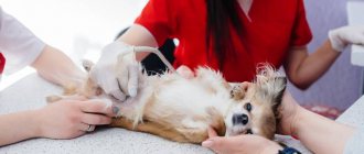
Resection of the femoral head: the surgical process and the first days after
The operation is highly invasive and is done under general anesthesia. The operation itself lasted about an hour and a half, and we waited another hour or so for the dog to come to his senses. After the operation and an hour-long wait (when the dog regained consciousness and could already move its paws a little and roll its eyes), we went home with the dog.
If you have a large dog, like ours, then be sure to take an old blanket or old blanket for such an operation, which will be strong enough to drag your dog, and also dense enough to absorb blood and other secretions that could take place.
The first night was very difficult, perhaps the most difficult in the entire time of having a dog. The dog tries to get up, encounters pain, squeals in bewilderment, and lies down. And so many, many, many times. And I feel very sorry for the dog.
Realizing that the dog could not lie down forever, we tried to position him ourselves. Through dog screams, whining and pain, we did it. After the first rise on three legs, the dog did not (still) know how to move on three legs: she tried to walk, but immediately whined and fell (we caught her and stood her up again). After 10-15 minutes I learned to more or less ride on three.
But the dog didn’t know how to lie down or sit up for about 4 days. My wife took a vacation and was able to follow the dog and help her lie down. He stood all the way until his paws began to shake, then we put him down. The dog couldn’t sit, so we kind of “hook” his paws to the side and immediately put him on his healthy side. When the dog woke up and tried to get up, they lifted it. It is worth noting that after a long and tiring period of standing, jumping and whining, the dog fell asleep instantly. And in general I slept a lot.
On the first evening (immediately after the operation) there was no talk of any walks - the dog moved within the confines of one room and simply could not move on. The first two days I went to the toilet as little as I could, on the floor, with a guilty face, standing on all three legs (the operated one was hanging lifelessly). They put up a huge basin, caught it, spilled it, wiped it off.
In fact, the dog went out for the first time about two days later, already outside. It’s unlikely that this is particularly important, but if your dog doesn’t shit in the first days, don’t worry as much as my wife. The clinic said that dogs stop going to the toilet, sometimes for five days.
The first three days we went to the clinic for dressings. There they injected Rimadil, Cifriaxon and Lincomycin. After 4 days they injected themselves with Cifriaxone, then with Norocarp tablets (because there was no Rimadyl), then Rimadyl (they found it).
Massage
There is an opinion that massage of the limb can speed up the recovery process. We massage about 2-3 times a day. The massage technique itself was taken from the book “Basic Facts about Physiotherapy for Dogs and Cats,” some of the pages of which we photographed in the clinic.
How to Identify Hip Dysplasia in Dogs
Diagnosis of hip dysplasia is possible only when the puppy gets older, because as the joints develop, manifestations of dysplasia become noticeable.
It is impossible to determine diseases at an early age, although the predisposition is already written in the dog’s genes. Dysplasia can be diagnosed after the first year of life, and for large breeds even after one and a half years of life. However, if the problems have obvious external manifestations, then it is worth showing the puppy to specialists even at an early age of 4 months.
You can identify dysplasia by the following most obvious external symptoms:
- Incorrect positioning of the limbs, swaying and instability are observed while walking
- Lameness.
- Apathy and reluctance to get up, it is easier for the dog to crawl to the goal.
- Rapid fatigue from active activities.
- Problems walking and standing on slippery surfaces.
- Rabbit running is when the limbs push off simultaneously, rather than one after another.
- Hypertrophy of the muscles of the forelimbs and pectoral muscles is possible, because most of the load falls on the front legs, as the animal tries to strain the hind pelvic muscles less
The most noticeable symptom that immediately catches your eye is the lameness of the animal. It can occur either due to pain in the joints or when it is difficult to move a limb forward. More often you can notice an initial lameness, which intensifies after a long period of sleep or lying at rest, but during a long period of activity the lameness completely disappears.
Monitor your pet closely in the first six months to a year of life, as timely treatment will help maintain your pet’s mobility and activity!
Risk group
Dysplasia of the hip joint and joint
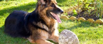
Predisposed breeds:
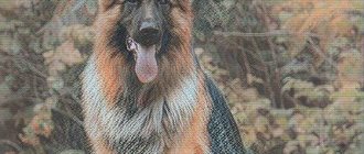
- German Shepherd;
- Labrador Retriever;
- Golden retriever;
- Saint Bernard;
- Rottweiler;
- basset hound;
- Bernese Mountain Dog;
- Newfoundland;
- and other large and giant breed dogs.
Risk factors are rapid increase in body weight, rapid growth, food with high energy value, and excessive exercise beyond age.
Retinal dysplasia
Predisposed breeds:
- Australian Shepherd;
- American Cocker Spaniel;
- English Springer Spaniel;
- collie;
- Labrador Retriever;
- Samoyed;
- Akita;
- bullmastiff;
- Yorkshire Terrier;
- rottweiler
Acquired infections may be risk factors.
Follicular dysplasia
Predisposed breeds:
| FD associated with color | FD black hair | FD unrelated to color |
|
|
|
Atrioventricular valve dysplasia
Predisposed breeds:
- Great Dane;
- German Shepherd;
- Labrador Retriever;
- Old English Sheepdog.
Causes of hip dysplasia in dogs
Often the cause of the development of the disease is a genetic predisposition. Errors in breeding work lead to the spread of pathology.
The main factors that contribute to the development of the disease:
- Overweight. Increased load on the articular components leads to disruption of normal functioning, and subsequently to dysplasia.
- Frequent injuries. Constant damage causes chronic inflammation and triggers destructive processes in the joint, ultimately leading to dysplasia.
- Unbalanced diet, use of low-quality dry food. The onset of the disease can be observed a short time after changing the diet. The key points are: excessive consumption of meat, which upsets the balance of calcium and phosphorus in the body, lack or excess of vitamin D in food.
As a result, a discrepancy between the sizes of the acetabulum and the articular part of the femur is formed. Over time, osteoarthritis develops, leading to joint destruction and dysfunction.
Features of nutrition of a sick animal
Most pets with dysplasia suffer from obesity, so they must be put on a low-calorie diet with a weekly fasting day. When reviewing your current diet, you must:
- Give up economy-class dry food or improve the quality of natural products.
- Exclude prohibited foods from the menu (sweets, smoked foods, pickles, canned food, flour).
- Make sure you have adequate protein intake.
- Reduce your usual portions of food to smaller amounts that allow you to lose excess weight.
With natural feeding, it is recommended to introduce dietary supplements into the diet, and with dry feeding, choose veterinary food with chondroprotectors.
Signs of hip dysplasia in dogs
The first manifestations of the disease can be noticed in animals at the age of 5-6 months.
These include:
- Frequently adopting a position lying on the stomach with the pelvic limbs spread to the sides. This sign occurs as a result of the puppy’s attempts to reduce stress and reduce pain.
- Rapid fatigue during physical activity. The animals are inactive; during long walks, they often take breaks and lie down. Shortness of breath may develop.
- "Rabbit Run". Due to impaired joint function, normal movement causes discomfort for animals. To reduce it, puppies push off simultaneously with both pelvic limbs when running.
- Minor lameness.
Hip dysplasia in dogs is subject to surgical and medical correction. In most cases, early intervention allows the animal to return to a normal lifestyle, but the individual is excluded from further reproduction due to the genetic inheritance of the pathology.
Breed Predisposition
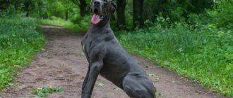
Any dog can develop dysplasia, but there is a certain predisposition. Some breeds get sick more often than others. Namely, large, massive and tall dogs, because their load on the musculoskeletal system is much more serious than that of medium or small dogs.
Dogs that have a lot of physical activity (work dogs, sled dogs) are also often affected. What kind of breeds are these? Great Danes, St. Bernards, Newfies, Shepherds, Rottweilers, Labradors and Golden Retrievers, Malamutes, divers and other representatives of the barking family.
Hip dysplasia in dogs symptoms
As the animal grows, the processes of joint destruction intensify and the symptoms of the disease increase.
Characteristic for adults:
- Increasing lameness. Due to the discrepancy between the sizes of the articular surfaces, lameness on the affected side increases with age.
- Difficulty getting up. After resting, it is difficult for the dog to get up, it tries to lean on something, and it is possible that it will fall when trying to stand on its paws.
- Unsteadiness of limbs. A later symptom indicating the destruction of most of the articular surfaces.
- X-shaped curvature of the pelvic limbs. The inability of the joints to cope with loads leads to curvature of the paws, which take the form of the letter X (closed in the center, spread apart around the periphery).
Hip dysplasia in dogs that appears after the age of 2 years is difficult to treat. Most often, dogs have problems with mobility for the rest of their lives.
Diagnostics
There is a misconception that hip and elbow dysplasia is diagnosed in dogs older than one year of age. In fact, this disease can be detected in a puppy as early as 4 months through x-ray examination.
To obtain the most accurate result, x-rays are taken in three to four projections under sedation of the animal. In the case of hip dysplasia, it is necessary to perform the Ortolani test, as well as identify the distraction index (DI), which allows one to determine “in numbers” how unstable the joint is.
Distraction index:
- less than 0.3 - the joint is stable, the likelihood of dysplasia is low;
- 0.3 — 0.7 - moderate instability, possible development of osteoarthritis in some dogs;
- more than 0.7 - unstable joint, development of severe osteoarthritis.
To exclude developing pathology, examination for retinal dysplasia should be carried out at the age of 6 - 9 weeks, and then at 6 - 9 months. Diagnosis is made by ophthalmoscopy.
Alopecia of colored hair with follicular dysplasia is diagnosed from 6 months to 2 years, a biopsy is taken, and the breed is looked at. Diagnosis of alopecia of black hair is the same as for alopecia of color, followed by histological examination.
In lipidosis pilaris, only the areas of brown hair growth on the face and limbs are affected. Airedale terriers and shorthaired dogs are often diagnosed with canine seasonal lateral alopecia, characterized by hyperpigmentation in a bilaterally symmetrical manner.
In cases of atrioventricular valve dysplasia, the animal undergoes echocardiography to identify abnormal development of the valve leaflets, tendon strings, or papillary muscles. Arrhythmia can be seen on the ECG. X-ray shows dilatation of the atria and/or ventricles.
Hip dysplasia in dogs treatment
There is no drug therapy with complete cure. After making a diagnosis, the veterinarian develops a treatment regimen with drugs that inhibit the development of the disease and relieve inflammation.
The treatment regimen includes:
- Chondroprotectors. They are taken orally or injected directly into the joint capsule by a doctor. The action is aimed primarily at preventing tissue destruction or slowing it down; restoring the original structure is almost impossible.
- Non-steroidal anti-inflammatory drugs. Prescribed for severe persistent pain. The carprofen they contain is highly selective and rarely causes complications even with prolonged use.
If a dog is diagnosed with hip dysplasia, the treatment of which with conservative methods is inappropriate, the question of surgical intervention is raised.
How long can dogs with hip dysplasia live?
This question naturally arises among owners immediately after the discovery of this diagnosis. We hasten to reassure you that dysplasia is not a fatal disease, but it can significantly worsen the quality of life of a pet. The outcome will depend on the time of detection of the disease and treatment. Of course, life expectancy will be influenced by factors such as the stage and degree of dysplasia, and the severity of its course.
Life expectancy will depend on owners and treatment. If the disease was detected in time, the pet was given proper treatment, and in subsequent years the owners carried out supportive therapy and fed and cared for the dog correctly, then the life expectancy can be 12-13 years. Without therapy and care, the dog will be limited in movement and will live much shorter. Be attentive to your pet and do not neglect treatment!
Prevention
Since dysplasia is hereditary, there is no way to prevent it in a puppy that has already been purchased. However, even before purchasing a dog whose breed is predisposed to THD and/or elbow joints, it is possible to determine the risk of its occurrence.
To do this, you need to carefully study the puppy's pedigree. It should include the results of tests for ancestral dysplasia. It is worth considering a purchase if at least one ancestor, even a great-grandmother or great-grandfather, has a degree of DTHD higher than grade “C”, and LS higher than 1.
If you asked yourself this question after purchasing a puppy, then your main task will be not to overfeed or overexert the puppy. At 4 months you can see your veterinarian and have an x-ray taken and a distraction index set.
Hip dysplasia in dogs surgery
Surgical intervention allows you to completely rid the animal of the manifestations of the disease. To do this, the affected joints are replaced with artificial ones. General endoprosthetics is indicated for severe forms of the disease. Widespread use of this type of intervention is hampered by its high cost.
Resection arthroplasty can reduce pain. During the operation, the head of the femur is removed. The result is no friction or pain. A side effect may be a change in joint mobility in advanced disease. This operation is a cheaper alternative to endoprosthetics.
Triple osteotomy surgery is also performed for hip dysplasia in dogs. During its course, the pelvic bones are dissected and special plates are placed at the sites of dissection, as a result of which the angle of the acetabulum changes and it more tightly covers the head of the femur. The pathological load on the joint is reduced, which leads to the disappearance of pain and restoration of function. This operation is not performed in severe forms of the disease, developed osteoarthritis or the presence of osteophytes.
What is this?
Joint dysplasia is the abnormal formation and development of joints, leading to impaired mobility and degenerative changes. In the initial stages, the disease entails deformation of the pet’s joint and then bone tissue.
An incorrectly formed or damaged joint, when rubbed, “erases” the cartilage tissue, causing severe pain. Gradually, the process affects the health and strength of the bones, preventing the dog from fully moving and leading an active lifestyle.
Note! According to statistics, dysplasia usually affects the hip joints. This is due to the fact that they bear the greatest load when running and jumping.
Drug treatment for hip dysplasia in dogs
As mentioned above, non-steroidal anti-inflammatory drugs are used for treatment. They have a symptomatic effect, that is, they help relieve pain, but do not eliminate changes in the joints.
The disadvantage of this type is that the painkiller causes the animal to actively use the damaged limb, thereby causing the development of joint damage. The dog does not feel pain and tension, so it strains the joint, which is not recommended.
Antioxidants, herbal preparations, viscoelastic drugs and even steroid hormones are also used. However, their use must be strictly coordinated with a doctor, as it can lead to undesirable side effects. And most importantly, the effectiveness of using these funds has not yet been proven.
There is also advisability in the use of chondroprotectors.
Detailed medical history
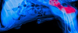
The dog's X-ray at that time looked like this:
Doctor Nikolaenko was offered the opportunity to perform juvenile symphysiodesis - a minimally invasive operation that made it possible to slightly change the growth of bones in order to make the dog’s skeleton more protected from dysplasia. The cost of the operation was around 5 thousand.
We did a blood test: it turned out that our phosphorus level was very high.
After consulting with probably a dozen veterinarians throughout Russia, we came to the conclusion that the operation is unlikely to help, however, there are practically no risks and we do not lose anything from it. The puppy was already old enough (5 months) to undergo such an operation, and juvenile symphysiodesis would hardly have been able to significantly change bone growth.
Some veterinarians expressed confidence that excess phosphorus in the dog’s body leads to improper development of the skeleton. As a solution, it was proposed to switch to food. We switched to Acana Puppy Large Breed, which we fed the puppy until he was a year old. Now we have switched to adult food from the same series - Acana Adult Large Breed. The phosphorus content in Acana is 1%.
However, we decided to try, since we had nothing special to lose (except for 5 thousand rubles).
The operation lasted a couple of hours. The dog recovered within a week. Since she took fairly strong painkillers (Previcox, 227 mg), she had no pain - neither from the operation nor from dysplasia. At the time of the operation, the puppy weighed 20 kg, 1/2 tablet of Previcox (227 mg) was given per day for a week.
It was decided to wait 3 months to see the result of the operation. During this time, we drank artoglycan in large quantities (spoiler: useless rubbish), and some other chondoprotectors.
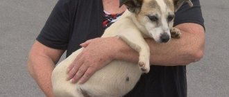
After 3 months, we took a repeat x-ray, which showed that the dysplasia had never gone away. The symptoms remained the same: the dog whined when getting up, walked crookedly and at random, and placed its paws poorly.
At the Lomakin clinic they threw up their hands: alas, alas, nothing can be done, we helped them in any way we could, all the best, good mood to you.
We went to the Ortho-Vet veterinary clinic, on Moskatova 16. There, first of all, they paid attention to the increased phosphorus and recommended to bring it down. As a way to reduce phosphorus, they suggested drinking Almagel, a remedy for heartburn and stomach ulcers. The principle was that the main active ingredient of Almagel is aluminum hydroxide, which, as it turned out, binds well to free phosphorus and removes it by the kidneys. We started taking Almagel. We drank about 8 ml of Almagel three times a day and gave it to the dog through a syringe.
In addition to Almagel, we were prescribed a course of the anti-inflammatory drug Rimadyl R. You can also use Previcox, but it is significantly more expensive. We paid about 1,000 rubles for a bottle of Rimadyl R (20 tablets).
There is a lot of fear on the Internet about Rimadyl, so you need to take it carefully. In our case, we drank it like this: for the first 7 days, 1 tablet in the morning, 1/2 tablet in the evening; over the next 2 weeks the dose was reduced - 1/2 tablet in the morning, 1/4 tablet in the evening. The veterinarian advised me to take Omez tablet first, then feed the dog, then give Rimadyl. But despite our warnings, 1.5 weeks after the start of treatment, side effects appeared: severe diarrhea. On the advice of the veterinarian, we paused for a week (continuing to give Omez twice a day), after which the course was successfully completed. It must be emphasized here that Rimadyl R also has an analgesic effect, so literally after a couple of tablets the dog becomes cheerful, but this improvement is imaginary, and the dog’s activity should be limited.
After Rimadyl, we injected the dog with Alflutop for 20 days. Alflutop is a chondroprotector, it was bought at a regular pharmacy, there were 10 ampoules of 1 ml in a box, it cost 1800 rubles per pack. They injected once a day in the evening, one at a time in the hind legs.
After 2-3 weeks of taking Almagel, phosphorus dropped, but not strongly enough. It was decided to double the doses of phosphorus taken. Another month with doubled expenses for almagel and as a result... off-scale phosphorus, the level of which is the highest in the entire history of observation! Despite all this, the dog continued to whine when standing up; All other symptoms of hip dysplasia were also observed.
Here they have already given up their hands in the second clinic: strange, strange, they said, well, let’s wait another six months, and then we’ll just do a resection of the femur (that is, we’ll cut off a piece of bone adjacent to the hip, to the edge of the hair dryer): there is no bone - there's nothing to hurt. We offered to do this right now, because we were very tired of watching the dog suffer, but the doctor convinced us to wait six months and take painkillers.
We had no choice but to wait. And then a thought came into my bright head: if we cannot effectively influence the output of phosphorus to the dog’s body, we can influence, so to speak, its input! Actually, we decided to study which foods contain the most phosphorus.
In addition to chocolate, which is prohibited for dogs, the greatest amount of phosphorus was contained in cheese, in all its types. Our dog loved cheese, and cheese was used as a treat for training at home. In fact, a complete ban was introduced on cheese and raw beef (which also contains a lot of phosphorus). Our dog practically did not eat other foods containing increased amounts of phosphorus.
After a week, the dog stopped whining when standing up.
After 2-3 weeks, the gait returned to normal.
Now (and about 2 months have passed since the beginning of the cheese embargo) the symptoms of hip dysplasia have completely disappeared. The dog not only returned to activity - she doubled, tripled it. There is no pain when standing up; when running, the dog moves its hind legs correctly; when standing, the dog places its hind legs parallel to each other; Visible dystrophy of the hip muscles disappeared, the dog’s legs became more fleshy and pumped up.
Soon we are going to do another blood test and, at one and a half years (in August), we will take another x-ray. In general, the dog is visually completely healthy; There is only some modesty in the size of the pelvis, most likely due to symphysiodesis; While other Rottweilers have a straight ass, ours has a neat little butt.
Consequences of dysplasia
Without timely treatment, the consequences of dysplasia can even become such a severe symptom as immobility of the animal. Also, without treatment, the dog may permanently limp or have difficulty walking. Any movement will cause severe pain, which means the dog will stop using its hind legs.
As a result, if left untreated in a severe stage, the dog can become aggressive and hostile towards people, as it constantly feels pain.
Timely detection of the disease and quality treatment will help restore your pet’s motor activity, guaranteeing him a long, happy and fulfilling life. Be careful and check your pet's health regularly!