An umbilical hernia in a puppy is the prolapse of internal organs through an opening into the cavity. Based on the location of the hernia and some of its other features, different types of pathology are distinguished. This is a common type of hernia. This pathological formation consists of an opening (hernial ring) and a sac containing the hernia: this is usually the omentum, but in the most severe cases it can be an intestinal loop.
Causes
Hernia is a strangulation of organs (usually the abdominal cavity) in a rupture of peritoneal tissue. That is, part of the intestines, uterus or bladder falls out into the resulting “hernial orifice”, which appears due to injury or as a congenital defect in puppies.
Diagram: structure of a dog’s umbilical hernia
This condition is classified according to several parameters; some types of hernia require immediate surgical intervention because they can lead to tissue necrosis and sepsis. Causes of pathology:
- Injuries, falls (directly related to the peritoneum or other place where the hernial tumor was found);
- Postoperative hernias (in case of suture dehiscence, improper care or restoration);
- The birth of large puppies (difficult childbirth is the most common cause of tissue ruptures);
- Chronic diseases that cause increased abdominal pressure (constipation, diarrhea, gas formation, volvulus, peritonitis, pancreatitis, gastritis);
- Age (as animals age, their cartilage, bones, and muscles weaken; in addition, due to decreased activity, excess weight and obesity appear);
- Heredity (if the parents have a hernia as a recurring problem, then there is a high probability that the puppies will have a similar disease);
- Predisposition (the appearance of hernial tumors is typical for Spaniels, German Shepherds, Poodles, Dachshunds, Bull Terriers);
- Excessive physical activity or its insufficiency (for each breed, a norm of daily physical activity is prescribed, which cannot be exceeded or underestimated; the puppy is prohibited from lifting heavy objects, jumping on steps, from high objects).
Which breeds are more susceptible
There are no differences between purebred and outbred cats and dogs. Both the highest-bred offspring of multiple prize-winners and “nobles” picked on the street can easily get the defect. Since the defect is caused by genetic pathologies, it can be found in kittens and puppies of any breed.
Symptoms and types of illness
The most important symptom is a characteristic protrusion of the organ in the form of a ball or swelling, most often on the stomach (but can also be on the side, neck, back and other places). Depending on the manifestation and quality of the neoplasm, hernia is divided into several types.
Congenital hernia
Heredity plays a major role in the occurrence of pathology: the inguinal canal is too wide, poor tissue regeneration (due to which the umbilical canal does not heal). Often, rupture of abdominal tissue occurs due to improper obstetric care, which results in unhealthy babies being born with various muscle and bone problems.
Acquired
Other forms of hernia appear during the dog's life for various reasons related to stress on the abdominal muscles due to illness or surgery. The risk group includes animals with chronic high blood pressure, heart defects, problems with blood vessels and blood flow . More often than others, female miniature breeds suffer from acquired hernia due to childbirth or a difficult pregnancy.
True
The most dangerous form of manifestation: the muscle ring diverges by more than 2 cm, and part of the organ falls out into the formed form. The hernial ball can reach the size of an orange. Over time, the cavity stretches, enlarges, and compresses the tissue of the uterus or intestines, which contributes to tissue death.
If such a formation is detected, the pet must be taken to a veterinarian for examination. Correct palpation will indicate the possibility or impossibility of reduction.
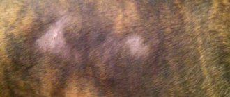
False
A form in which the separated muscle tissue is filled with fat rather than an organ. The ball is small in appearance (up to 2 cm in diameter), soft to the touch, and can roll under the skin or be smoothed out. Surgery on the animal is not necessary.
False hernia in the photo
Reducible
Only a veterinarian can classify the formation according to the degree of reducibility . If the organ can be returned to its place, the hernia is called reducible. Upon palpation, the ball is elastic, soft, easily enters the muscle ring and remains there while the animal is motionless. In this case, an operation is performed to eliminate the hernial orifice.
Irreversible or hard
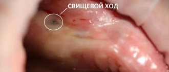
Such a hernia, rather, is a consequence of a truly reduced hernia, left without treatment. Appears when an organ is pinched in a muscular opening, tissue inflammation and the appearance of pus. Because of this, the cavity increases in size and cannot pass into the hernial orifice (it is hard, can bring pain and discomfort to the pet, and threatens health and life). Other symptoms also appear:
- Lethargy, lack of interest in life;
- Poor appetite;
- Temperature increase;
- When you tap on the cavity, a dull sound appears (like when playing a drum).
Why is it dangerous for dogs?
A strangulated hernia is fatal, and it is impossible to repair it at home. When pinched, pressure in the abdomen increases, blood flow is disrupted, and tissues of internal organs die and rot. The rotting process provokes the entry of pathogenic bacteria into the blood and blood poisoning.
Hernia repair in a dog
Features of the location of the hernia and its treatment
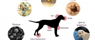
Bulges are also classified according to their location. Depending on the location, the pathology takes on different course patterns and side symptoms. To detect a hernia, ultrasound or x-rays are sometimes used if there are signs of muscle ring formation, but no swelling is visible.
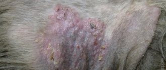
Hernia on the abdomen (Umbilical)
The most common manifestation, especially in newborns. Appears in the area of the umbilical canal and abdomen, part of the intestine falls into the cavity, which is why the hernia becomes rounded and becomes like a ball. The size of the neoplasm rarely exceeds 2-5 cm in diameter.
In adult animals, pathology occurs due to frequent constipation, increased gas formation, and bloating. When the disease is advanced, the dog’s activity decreases and appetite disappears. It is treated not only surgically, but also conservatively: bandages, bandages, massages.
Inguinal
More often observed in females. The uterus, bladder, and intestinal loops enter the hernial orifice. In addition to the appearance of a ball in the groin, there is an increase in basal body temperature, nausea, vomiting, depressed general condition, and increased heart rate and breathing.
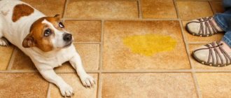
Males are characterized by pain impulses when palpating the testes, and for females - when palpating the loop or lower abdomen. If the bladder is pinched, anuria and involuntary urination occur.
Inguinal in small breeds
In small dogs, an inguinal hernia appears during pregnancy due to weak abdominal muscles. In this case, the uterus with the fetus descends into the ring, so the neoplasm increases as pregnancy progresses and the fertilized cell grows. This pathology is dangerous not only for the mother, but also for the unborn puppies.
Perineal
Rupture of the pelvic muscles. Those at risk are unneutered males, older dogs and breeds with short tails. Reason: constipation, prostatitis, inflammatory processes. The colon or rectum, prostate gland, and bladder prolapse into the hernial cavity.
A large ball-shaped lump appears around the anus or in the perineal area. The tumor is soft, causes severe pain when palpated, and makes defecation and urination difficult. If a rupture occurs, death is inevitable. For pregnant bitches, the appearance of a perineal hernia is equivalent to an abortion.
Intervertebral
A rare form, typical for older dogs that have been involved in transportation or sports all their lives. A hernia is characterized by protrusion of the vertebral cartilage into the formed fibrous ring of the spinal canal. The problem disrupts the functioning of several intervertebral discs.
A hernia is expressed by acute back pain, paralysis, lameness, and failure of the hind or forelimbs. The animal stops running, playing, and doesn’t want to go for walks. For an accurate diagnosis, an x-ray is needed.
Diaphragmatic
From the name it is clear that such a hernia occurs due to a violation of the integrity of the diaphragm. Because of this, organs from the abdominal cavity rise into the intercostal or thoracic space. This pathology is accompanied by pulmonary edema and disturbances in the functioning of the heart. Characteristic symptoms appear:
- Blue discoloration of mucous membranes;
- Oxygen starvation;
- Anemia;
- Arrhythmia;
- Nausea and vomiting;
- Bulging in the chest area.
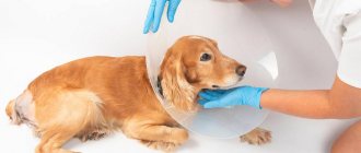
Surgical hernia removal
There are two options for treating pathology: conservative (massages, bandages) and surgical. The first is carried out in the case of false and reducible hernias (less often with umbilical hernias in puppies). Animals with diaphragmatic, intervertebral, perineal, and inguinal problems require surgery. Umbilical tumors in adult animals are also treated surgically.
The animal is operated on under anesthesia in a supine position. The skin is cut, the hernial sac is separated from the peritoneal tissue, the organ is set (if the process of tissue death has not begun), the cavity is truncated and the window is sutured. If the formation is filled with pus and the decomposition process has begun, the bag is completely removed. The last step of the operation is suturing and antiseptic treatment.
Scheme: removal of a hernia in a dog
The animal is prescribed anti-inflammatory, immunomodulating, analgesic drugs (sometimes laxatives or fastening and symptomatic). The dog is also prescribed a diet.
Rehabilitation takes place over 10-14 days, during which time daily treatment of the stitches with antiseptics, wearing a bandage or bandage, and maintaining rest are required.
Briefly about the main thing
- Hernias can be congenital - due to improper development of the fetus, or more often hereditary, and acquired - after injury or surgery.
- There are umbilical, inguinal, perineal, diaphragmatic, intervertebral and hiatal hernias, which mainly differ from each other in their location.
- Most often, the only effective treatment is surgery.
- Hernias are diagnosed not only visually, but using special research methods - radiography, ultrasound, endoscopy, etc.
- After surgery, it is important to limit the dog’s movements and provide a special diet.
Preventing the development of a hernia in a pet
Hernial neoplasms are not so common; pathology can be easily avoided if you monitor the pet’s condition, examine its body, respond to changes in mood, and periodically show it to the veterinarian. To prevent the appearance of a muscle tear, it is enough to follow the rules:
- It is necessary to promptly treat problems of the digestive system and other disorders in the functioning of the body, normalize nutrition, and monitor the process of bowel movements;
- It is better to sterilize females not intended for bearing and giving birth to offspring (the same applies to males);
- After injuries or falls, it is important to have the animal examined in a clinic;
- The main condition for keeping a dog is the required amount of physical activity, proper training, regular walking (to avoid obesity or excessive strain on the muscles);
- To operate on a pet (for any reason), select a trusted clinic, and during the recovery period you need to strictly follow the veterinarian’s instructions and take care of the sutures;
- Elderly animals are fed special foods with a high calcium content;
- Dogs are purchased only from trusted breeders. It is important to first get acquainted with the puppy’s parents and find out about their chronic diseases.
To avoid the appearance of a hernia in puppies, a specialist obstetrician is invited during childbirth to help the female without injuring the offspring.
Any hernial neoplasm needs to be examined by a doctor. Therefore, if a lump (reducible or hard) appears, it is necessary to contact your local veterinarian and undergo examination, and then rehabilitation.
4 / 5 ( 3 voices)
Caring for a sick animal
After the operation, the veterinarian prescribes anti-inflammatory antibiotics and mild painkillers in the form of injections. Rehabilitation after removal is quick.
You can speed up the healing of the suture with special ointments that disinfect, relieve swelling and inflammation.
In some cases, immunostimulants are prescribed to quickly restore health.
There can be no relapse of the disease after surgery. However, for preventative purposes, it is better to visit a veterinarian regularly for examination.
A number of vitamins and medications that support intestinal microflora must be included in the dog’s diet during treatment and rehabilitation.