But there are parasites that do not cause itching themselves, but can lead to the development of a bacterial skin infection, which also results in itching. These parasites include the causative agent of demodicosis - the subcutaneous mite of the genus Demodex. In fact, almost every dog is a carrier of demodicosis, receiving the mite at birth from its mother. Normally, Demodex mites do not cause any clinical signs in a healthy dog. But if the body’s immune system malfunctions, then the development of the disease demodicosis with clinical signs begins. But even such animals today can be easily helped by using modern drugs for treatment. After all, the most important thing in the fight against any parasites is timely treatment of your pet against them.
Demodectic mange in dogs – what is it?
The causative agent of demodicosis is a subcutaneous microscopic parasite, the mite Demodex canis. It feeds on dead skin cells (epidermis), the secretion of the animal's sebaceous glands and is localized in the hair follicles. A dog's healthy immune system can inhibit the development of these mites, resulting in the dog and the mite living peacefully together. In a young animal with normal immunity, demodex does not develop - the body's immune forces successfully restrain it. However, dogs under one year of age are at risk (since their immunity is not yet strong), as well as older animals (over 7-9 years old).
Description of the disease
Demodex canis are microscopic mites, shaped like a very elongated oval of light gray color. Adults have 4 pairs of legs, which are located in the upper part of the body.
In large cities, from 38 to 67% of dogs are carriers of Demodex. Short-haired purebred animals are most susceptible, since their sebaceous glands are more developed than those of long-haired animals. The disease is a severe pathological process that develops in the sebaceous glands, hair follicles and is manifested by acute dermatitis, hyperkeratosis and rapid exhaustion of the pet.
Demodicosis is a dangerous disease and extremely difficult to treat, although at an early stage of the pathology, when the animal is only bothered by ordinary itching, it is difficult to imagine. Therefore, if you suspect your pet has “red scabies,” you should seek the help of a specialist as soon as possible for a full diagnosis.
Forms of demodicosis
Depending on how the disease progresses and how it spreads, two forms of demodicosis are distinguished:
- Localized. It is more common in young dogs (under 2 years of age). The main symptom of the local form is the development of alopecia (foci without hair) in limited areas of the animal’s body. There may be 3-5 such lesions, but most of the body is not affected. The lesions do not exceed 3-4 cm in size. The localized form can go away on its own without therapy.
- Generalized. In this case, the lesions are large, there are many of them, and almost the entire surface of the body can be affected. The generalized form of demodicosis is observed in older animals and is often associated with severe stress or exhaustion, leading to weakened immunity. In young individuals, a generalized form of the disease also occurs as a result of the development of a local one. The generalized form occurs with complications in the form of a secondary infection, since areas affected by demodicosis are very vulnerable - inflamed skin is an excellent place for the development of bacteria and fungi.
Diagnostics
The diagnosis is made comprehensively, taking into account medical history (complaints from the owner, medical history), physical examination, and laboratory tests. The main method of confirming the diagnosis is microscopy of skin scrapings. Scrapings are necessary from all affected areas of the body. The scraping should be deep enough, carried out with a scalpel until the first drops of blood appear, since the mite sits in the deep layers of the skin (hair follicle). Trichoscopy (examination of plucked hairs) or tape test (taking material for examination using a narrow strip of tape) may also be useful. If there are entire pustules on the body, it is imperative to conduct a microscopy of their contents. To make a diagnosis, you need to detect a large number of mites at different stages of their development. The discovery of just one tick may be an incidental finding, but still should not be completely ignored. In such cases, it is recommended to repeat scrapings after some time (2-3 weeks) to clarify the diagnosis. If otodemodecosis is suspected, then microscopy of the contents of the external auditory canals is performed. In especially doubtful cases, a skin biopsy with histological examination may be suggested. Also, in doubtful cases, the doctor may suggest a trial treatment, even if the diagnosis could not be confirmed at the initial appointment.
Juvenile demodicosis
Puppies experience so-called juvenile demodicosis. It usually occurs before one year of age and is caused by a hereditary defect in the immune system, causing it to become unable to control the number of mites. In this case, the number of mites increases and they begin to cause disease. Subcutaneous mites easily multiply in the epidermis of young animals, causing baldness and inflammation.
As a rule, juvenile demodicosis occurs easily and without complications. It responds well to treatment, for which it is necessary to contact a veterinary clinic for a diagnosis and treatment.
But in some cases, local juvenile demodicosis can progress and become a generalized form. In this case, it is important to contact the clinic in time for timely treatment. It is very important to understand that since generalized juvenile demodicosis is a hereditary disease, a dog that has had it cannot be used for breeding even after complete recovery.
A dog is considered recovered from juvenile generalized demodicosis if it does not experience a relapse (re-development of the disease) within a year.
Briefly about the main thing
- Canine demodicosis is a common disease caused by the Demodex canis mite, which penetrates the hair follicle and destroys it. The disease is characterized by severe skin lesions, complicated by other dermatitis, leading to exhaustion of the dog and even death.
- Dogs become infected from sick individuals, most often at a young age. There is an assumption that demodexes normally live on almost all dogs, but do not manifest themselves in any way as long as the animal’s immunity is normal.
- There are the following forms of the disease: juvenile demodicosis, adult demodicosis, localized, generalized, scaly, pustular and mixed.
- Treatment should be prescribed exclusively by a doctor, since specific therapy is toxic to dogs and a clear dose calculation is required for each individual case.
Have we answered your question fully enough? If not, post your question in the comments below and our veterinarian will answer it.
Did you like the article? Share it with your friends on social media. networks. This will help them get useful information and support our project.
How can a pet become infected?
As we have already said, Demodex mites are conditionally pathogenic. Subcutaneous mites are transmitted from mother to puppies during feeding in the first days after birth. With the exception of the indicated route of initial transmission, the disease itself is not considered infectious. This means that a small number of mites on a dog's skin is normal. Pathology when the animal’s immune system is unable to restrain the growth of their population and the disease progresses.
Normally, the immune system is able to control the number of mites in the skin, so healthy animals and humans do not develop the disease. If there are serious disturbances in the functioning of the immune system, the body loses the ability to control the number of subcutaneous mites and a disease called demodicosis occurs.
Thus, demodicosis in dogs is not contagious, that is, it is not transmitted from dog to dog, since each animal has its “own” ticks, and whether the disease occurs or not depends on the animal’s immunity. In addition, mites parasitize only in the skin and do not affect internal organs.
Danger to humans
Demodex is a strictly specific parasite, that is, a species that parasitizes dogs, but cannot parasitize humans. And, as noted above, Demodex is a normal inhabitant of animal skin. It multiplies, causing disease, only in the conditions of a specific organism (due to decreased immunity or a genetic defect) and, accordingly, is not contagious.
Symptoms
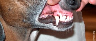
The first signs of the disease are usually alopecia - bald spots, most often in the head and paws. Itching, as a rule, is not pronounced, and appears only when a bacterial infection is attached, which is not uncommon with demodicosis. In this case, the dog constantly tries to scratch itself and this significantly reduces its quality of life. To prevent teeth from scratching the affected areas, veterinarians recommend wearing a protective collar on the animal. Since most drugs that relieve itching are contraindicated for demodicosis.
In advanced cases, the animal may become completely bald, a severe purulent or fungal infection develops, and the general condition of the animal becomes quite serious.
Clinical signs
With a mild form of the disease, the only signs may be slight redness, comedones and peeling. Alopecia and/or hypotrichosis are possible. In more severe forms of the disease, peeling of the skin, crusts, furunculosis, fistulas, areas of ulceration, hyperpigmentation, unpleasant odor and other lesions are noted.
In most cases, the process begins with the muzzle and forelimbs and can then spread to other areas of the body.
Generalized demodicosis is almost always accompanied by a secondary bacterial infection, and lymphadenopathy, lethargy, and fever may also occur.
If otodemodecosis is suspected, a microscopic examination of earwax is performed. As a rule, otodemodecosis is accompanied by bilateral ceruminous otitis.
Pododemodecosis is a mite infestation of the skin of the paws. Sometimes these lesions are independent, and sometimes they are part of a generalized process. In cases of podomodexosis, the skin of the fingers may be thickened, erythematous, and hyperpigmented, with significant tenderness in these areas.
Treatment of demodicosis in dogs
For a local form of demodicosis, the treatment regimen consists of treating the dog with special drugs that destroy subcutaneous mites. These may be in the form of drops or tablets.
In case of severe generalized demodicosis, the veterinarian may add points such as:
- daily treatment of affected skin areas with antiseptics and softening ointments;
- bathing with dermatological shampoos (with chlorhexidine, antiseborrheic, keratolytic);
- the use of antibacterial and antifungal drugs in the presence of a secondary skin infection;
- balanced diet, vitamin and mineral supplements.
Treatment of generalized demodicosis usually takes quite a long time, and can be stopped only after two series of scrapings with an interval of one month do not reveal a single subcutaneous mite, either alive or dead, in any of the scrapings.
Usually the patient's external recovery occurs before treatment can be stopped. If treatment is stopped prematurely, relapse (recurrence of the disease) is very likely. Treatment may take 2-5 months or sometimes longer, depending on the drug chosen.
The main treatment for any form of demodicosis is drugs that stop the activity of the subcutaneous mite. Today, one of the most effective methods of treating demodicosis is the use of drugs from the isoxazoline group, which include Simparica tablets.
When treating demodicosis, the course of taking the drug is 2 months. Tablets are given twice with an interval of 30 days. Simparica is used for adult dogs and for puppies from 8 weeks, and is active not only against demodicosis, but also against fleas, 9 types of ixodid ticks, otodectosis (ear scabies) and sarcoptic mange (scabies).
The tablet is easy to give - it has a liver taste that is attractive to dogs, so dogs eat it as a treat.
Breed predisposition to the disease
Demodectic mange is more common in purebred dogs, and certain breeds are affected more often than others. According to some studies, these are the Shar Pei, West Highland White Terrier, Scottish Terrier, English Bulldog, Boston Terrier, Great Dane, Weimar Pointer, Airedale Terrier, Alaskan Malamute and Afghan Hound. The diagnosis of demodicosis is often made in dogs of other breeds (for example: Doberman Pinscher), but the incidence of the disease in dogs of these breeds is incomparably lower than in those that have already been noted.
Other predisposing factors include: short hair, poor nutrition, estrus, childbirth, stress, endoparasites. As well as diseases that weaken the body. However, as foreign authors point out, most of these factors are very difficult to assess, and many of them are unlikely to be so significant. After all, the vast majority of cases of the disease occur in purebred dogs. At the same time, they are well maintained, receive excellent nutrition, and are otherwise in good condition.
Therefore, for some time the question arose as to which of the reasons are really decisive for the development of this disease.
A number of studies were conducted aimed at studying the virulence of some strains of the pathogen, but as a result, the theory about the existence of some special types of pathogen that necessarily cause the development of demodicosis in dogs was rejected.
At the same time, the development of demodicosis was noted in adult dogs undergoing immunosuppressive therapy, or having cancer, or suffering from serious metabolic disorders.
It has been suggested that the main reason for the formation of demodicosis may be immune disorders in dogs.
Various parts of the immune system were studied, and on this basis it was found that in dogs with a generalized form of demodicosis, the system of neutrophil phagocytes and the complement system are not depleted and are quite active, and humoral factors are not subject to any significant changes.
In particular, histological examination of the bone marrow, lymph nodes, spleen, as well as blood tests revealed that the number of B lymphocytes is normal, or even increased. When these animals are immunized against Aleutian mink disease, or canine distemper, or viral hepatitis, the amount of antibodies produced is also normal. What is important to understand is that animals suffering from a generalized form of demodicosis have no contraindications to vaccinations.
It has been established that in dogs with generalized demodicosis, there are disturbances at the level of T-cell immunity.
This is confirmed by studies that indirectly indicate the activity of T-cell function, such as the reaction of blast transformation of lymphocytes, as well as the reaction of changes in the migration of leukocytes using mitogens: phytohemagglutinin (PHA), concanavalin A (ConA). These methods are relatively reliable. This is probably why, until recently, it was not completely clear at what level immunosuppression occurs in demodicosis.
Recent studies based on the determination of lymphocyte subpopulations have confirmed the theory that the “breakdown” of the immune system occurs at the level of cellular immunity. In particular, a decrease in the production of interleukin-2 (IL-2) and a lack of expression of the T-cell receptor (TCR) for this type of interleukin were found. Scientists have suggested that dogs with demodicosis have a reduced T-helper response, which, in turn, appears to be determined by heredity.
The statement about the defect at the level of T-lymphocytes is confirmed, in particular, by the results of intradermal studies with an antigen obtained from a tick, the causative agent of demodicosis. Thus, in healthy dogs and dogs in which demodicosis spontaneously resolves, the result of the test for skin hypersensitivity is adequately delayed, while in dogs with a chronic disease it is not.
Indeed, the relatively increased activity of plasma cells, an indirect increase in the level of immunoglobulins in the blood of dogs with demodicosis, and a decrease in the level of IL-2 production probably indicate a defect at the level of T-helper type 1 (Th-1).
It is Th-1 cytokine production that determines the development and formation of cellular immunity. The cytokine series produced (Th-2) reduces the development of cellular reactions. In particular, IL-10 prevents the activation of macrophages, which leads to marked immunosuppression. (see: “Visual immunology”, p. 30, author: Burmester et al., M. 2007).
Further study of this issue, from the point of view of immunology, using modern research methods (including flow cytometry), will subsequently allow us to have an undoubted idea of what specific mechanisms in the immune system are affected in canine demodicosis.
Clinical forms of demodicosis
In general, there are two main clinical forms of demodicosis: localized and generalized demodicosis. The juvenile form of the disease is also distinguished, since this is important from the point of view of prognosis and the choice of approaches in subsequent therapy. Localized demodicosis.
Localized demodicosis occurs as: one to several small, circumscribed, erythematous, scaly, nonpruritic or pruritic alopecias, most commonly on the face or forelegs.
The most common place for their appearance is the muzzle, especially in the area around the eyes and in the corners of the mouth. The next most common place for scaly spots to appear is the front paws. Less often, 1-2 spots are found on the body or on the hind legs. Most cases of the disease occur in the first 3-6 months of life and are cured without the use of therapeutic measures. True localized demodicosis only in rare cases turns into a generalized form. The fur on the affected areas begins to grow back within 30 days.
Affected areas may appear and disappear over several months. Relapses are rare because the skin appears to become less favorable for mites to multiply rapidly, or the host's immunocompetence returns to normal.
It is believed that if we are talking about the presence of 1 to 5 spots on the skin of a young animal, then this is a juvenile localized form of demodicosis and does not require therapy. If the number of these spots is greater, or during the process of observing the animal, new lesions appear, then a diagnosis of a generalized form of demodicosis should be made and therapy should be resorted to.
It should also be noted that this rule does not apply to adult animals, and if a localized form of demodicosis is detected in adults, then in this regard, you should think about treating demodicosis without much delay.
Generalized demodicosis is usually recognized in dogs with a large number of affected areas. In this case, for example, a certain part of the body may be affected: for example, the muzzle; or two or more paws.
| Alopecia and pyoderma: diffuse spread | Alopecia, excoriation and pyoderma on the dog's head | Alopecia and excoriation on the face | Alopecia and pyoderma of the finger area |
Generalized demodicosis usually begins at an early age: from 3 to 18 months. If appropriate therapy is not carried out on time, then, as a rule, the disease accompanies the animal into adulthood.
True late acquired generalized demodicosis is also distinguished. It is rare and in such cases it should be said that the dog has been carrying and controlling demodectic mites as part of its skin flora for many years. Until resistance to the disease decreased and the number of ticks suddenly increased thousands of times. In this case, it should be borne in mind that there is some internal disease that has caused immunosuppression.
Among the general health disorders in dogs with late acquired demodicosis are hypothyroidism, natural or iatrogenic hyperadrenocorticism, leishmaniasis and malignant neoplasia, which leads to the development of secondary demodicosis. In these cases, the dog’s condition must be carefully monitored and research conducted to exclude primary causes that can lead to demodicosis. If it is not possible to detect the cause of the development of demodicosis, then in this case, the likelihood of treatment success decreases.
Generalized demodicosis manifests itself in the presence of numerous affected areas on the head, paws and torso. These areas increase in size, some of them connect and form entire diffuse areas of lesions.
Usually we are talking about the development of folliculitis. When secondary pyoderma appears as a complicating factor in the affected areas of the skin, deep folliculitis usually occurs, with exudate being released and thick crusts forming on the skin.
In some dogs, the affected areas have an atypical appearance, such as nodules, so the possibility of demodicosis should always be kept in mind if a specialist is faced with the presence of any nodules on the dog’s skin. In particular, an English bulldog may have this type of nodular form of the disease.
Bacteria, actively multiplying, complicate the course of demodicosis in dogs. Staphylacoccus intermedius is the most common bacterium complicating generalized demodicosis. Pseudomonas aeruginosa causes serious pyogenic complications and is particularly resistant to antibiotics, especially when it comes to demodectic pododermatitis. Proteus mirabilis is another serious bacterial agent, the presence of which may additionally cause itching, as a clinical manifestation of pyoderma against the background of generalized demodicosis.
After several months, the chronically affected skin becomes covered with purulent, hemorrhagic and follicular-furunculous bald spots with the presence of crusts. The skin of the abdomen is affected to a lesser extent, probably due to the fact that there are fewer hair follicles there.
Demodectic pododermatitis. This form of demodicosis can manifest itself only on the plantar part of the paws of dogs, with a complete absence of affected areas on the animal’s body. At the same time, the history shows whether the dog previously suffered from demodicosis, which remained only in the form of bald spots on the lower part of the paws, or whether the paws are the only affected part of the body. In the affected areas in the digital and interdigital areas, secondary pyoderma especially often develops. In some animals, demodectic pododermatitis can take a chronic form, which is extremely resistant to therapy. Large dogs, such as Great Danes, Newfoundlands, St. Bernards, and English Shepherds, are particularly affected by pain and swelling.
Establishing diagnosis
Properly taken and described skin scrapings can help make the diagnosis of demodicosis. The affected skin should be squeezed firmly to dislodge the mites from the hair follicles, and skin scrapings should be deep and extensive. Extremely sensitive areas should be avoided as the bleeding this will cause will make the results difficult to interpret. The diagnosis is made either when a large number of ticks is detected, or when there is an increased ratio of their immature forms (embryos, larvae, and nymphs) to adult individuals. The discovery of an occasional adult tick on skin scrapings should also not be ignored. The dog should have other areas scraped before rejecting the diagnosis of demodicosis.
Skin scraping is apparently a simple laboratory procedure with a clear result. Adequate skin scrapings are mandatory in all cases with a combination of canine pyoderma and seborrhea. When skin scrapings are negative in a Shar Pei or in a dog with fibrous lesions, especially in the interdigital area, a skin sample should be biopsied before the diagnosis of demodicosis can be ruled out.
Sometimes, as an additional test, hair removal is carried out by plucking in those areas where high-quality scraping cannot be carried out, for example, in the area between the fingers. Subsequent trichoscopy will help in making a diagnosis.
Differential diagnosis
Because skin scrapings reveal the presence of mites in the vast majority of cases of demodicosis, this disease is difficult to confuse with other skin diseases.
Demodicosis should be differentiated from other diseases that can cause folliculitis. The most common among them are pyoderma itself and dermatomycosis. In general, the likelihood of demodicosis should be suspected in every case of folliculitis.
Superficial excoriations in young dogs sometimes resemble erythematous areas of localized demodicosis. Acne, as well as early areas of juvenile cellulitis on the face of young dogs, can also sometimes resemble demodicosis, accompanied by the presence of pustular lesions.
With contact dermatitis, erythematous papules appear, which are also sometimes similar to demodicosis.
Therapy
• Localized demodicosis . It is a mild condition that resolves without medical intervention in 6-8 weeks, but may wax and wane in a localized area over several months. There is no difference in the rate of recovery between dogs receiving and not receiving drugs. There is no evidence that treatment of localized demodicosis prevents the development of the general form of the disease.
At a follow-up visit after 4 weeks, the specialist will be able to determine whether there are signs of generalized demodicosis. Skin scraping at the beginning of the spread of localized demodicosis often reveals a large number of living adult mites and their immature forms. After four weeks of observation, skin scrapings from healing areas should contain fewer mites and fewer immature forms. If the lesion spreads and the number of mites (including the ratio of immature forms to adults) is high, the condition may progress to generalized demodicosis.
• Generalized demodicosis. With treatment, most cases, perhaps almost 90%, can be completely cured, but the treatment process can take almost a year. A common problem in the treatment of demodicosis is premature cessation of therapy, since the clinical form of the disease may disappear before all mites are destroyed.
It is unjustified to euthanize dogs, especially at the age of 6 to 12 months, because they have a severe form of general demodicosis, since some of them can generally recover on their own (according to some data, up to 50%) with control of pyoderma and seborrhea and with good state of health. However, it is always better to prescribe therapy, especially since it is currently quite accessible and generally safe.
Before prescribing any treatment for demodicosis, if necessary, adult dogs that are suspected of having problems with their general health should be examined. Since their disease can be provoked by some kind of systemic disorder, and when the cause is eliminated, such a dog either recovers on its own or responds better to treatment.
Dogs with demodicosis should have regular examinations and skin scrapings, usually every 2-4 weeks. To determine the effectiveness of treatment, it is best to always take skin scrapings from the same areas and record the results in the patient's chart.
Pyoderma and seborrhea, noted in dogs with demodicosis, are the result of mite infestation, and cannot be cured until they are completely exterminated. The choice and duration of antibiotic use depends on each specific case.
Treatment against demodicosis must be continued for an additional 30 days or more after skin scrapings are negative. Symptoms disappear in dogs several weeks before all parasites are destroyed. A parasite cure means that the dog's skin scrapings are free of live or dead mites at any stage of development. Only after scrapings from at least 4-6 areas give a negative result can we talk about getting rid of parasites. The set of areas depends on the specific case, but it must include an area on the muzzle and on the front paw.
A number of drugs are used to treat demodicosis
The drug Amitraz is a diamide, N'-(2,4-dimethylphenyl)-N'-(((2,4-dimethylphenyl)imino)methyl)-N-methylmethanidamide. It has a number of commercial names (for example: Mitaban; Ectodex (Ectodex Dog Wash) and Taktic); and contains different concentrations of the active substance. Therefore, with regard to dilution of the drug, in each specific case, you should be guided by the attached instructions for use.
The recommended frequency of use may vary: once every 7 or 14 days. To achieve maximum results, you must follow the following rules:
1. Dogs with long to medium coats are cut short to allow the aqueous solution to better contact the skin and penetrate the hair follicles.
2. All scabs are removed. In some cases, a sedative or pain reliever is necessary because some scabs adhere tightly to the skin and are painful to remove without an anesthetic. The use of sedatives that are α-adrenergic agonists should be avoided. (eg, xylazine) as synergistic toxicity may occur.
3. The entire dog is washed with a medicated shampoo such as Doctor or Peroxiderm to kill bacteria and remove scales and exudate. Despite the fact that the skin may seem rough and irritated after the described procedures, contact of the drug with the affected skin will be optimal. The dog is carefully dried with a towel. Alternatively, a preparatory wash of the dog can be done the day before treatment.
4. The amitraz solution is best applied with a sponge. This solution should be applied to the entire body: both healthy and affected areas of the skin. Although the solution is not irritating, people applying amitraz should wear protective gloves and work in a well-ventilated area. Amitraz causes short-term sedation for 12-24 hours, especially after the first use. And in some dogs, at the initial stage, itching may develop against the background of treatments. Other side effects are rare and include allergic reactions (urticaria or redness), skin irritation and various systemic signs of allergy. Severe forms of the reaction due to intoxication can be treated with yohimbine or atipamesol. In case of severe side effects, the dosage of the drug is usually reduced during subsequent applications to the skin. In rare cases, as a reaction to amitraz solution, dogs may experience severe weakness, ataxia, and drowsiness. If it is necessary to continue amitraz therapy, yohimbine is additionally used, which prevents or significantly reduces the severity of these undesirable effects. In some people, contact with amitraz may cause skin dermatitis, migraine-like headaches, or asthma attacks. When infected with pododermatitis, the paws can be immersed in a small bath of amitraz solution and lightly massaged to enhance penetration of the solution. There is no need to rinse the paws or body. The drug should remain on the skin for 2 weeks.
Although approximately half of the drug remains in the skin for 2 weeks, some may wash off if the dog gets wet or swims. In this case, you can re-apply ahead of schedule.
In the early 1990s, scientists conducted pilot studies on the effectiveness of oral ivermectin or milbemycin in hopes of finding a therapeutic alternative for dogs intolerant or unresponsive to amitraz. The results of these studies were so successful that now the treatment of generalized demodicosis with the help of these drugs is common practice.
Milbemycin can be used in dogs that are sensitive to ivermectin, but unfortunately it is not commercially available in our country. Milbecin is used at a dose of 2 mg/kg daily. The duration of treatment depends on the specific situation, and can range, according to the authors, from 60 to 300 days.
Moxidectin, a type of milbemycin, can also be used to treat demodicosis in dogs at a dosage of 0.2 to 0.4 mg/kg.
Ivermectin is used in a dose of 0.45 to 0.6 mg/kg, and its effectiveness is also high. Temporary side effects with ivermectin may include lethargy, anorexia, ataxia and stupor. These manifestations disappear with drug withdrawal and subsequent dose adjustment. Ivermectin is highly toxic to the following breeds: Collie, Australian Shepherd, Sheltie, Miniature Australian Shepherd, Silky Windhound, Longhaired Whippet, German Shepherd, Border Collie, Bobtail, English Shepherd, Mac Nab; and for their mixed breeds (before prescribing ivermectin, it is necessary to use a genetic test for the presence of a defect at the level of the MDR-1 gene)
Today, drugs from the isoxazoline group have appeared on the market (Bravecto (Intervet), Frontline Nexgard, Nexgard Spectra (Merial)). Their safety and effectiveness in the treatment of demodicosis have been proven.
The period of therapy for demodicosis is usually carried out until the presence of two negative series of scrapings carried out with an interval of 30 days.
If a dog relapses within the first 3 months, it can likely be treated with more aggressive therapy using the same drug. If the dog relapses again after the second course of treatment, or if the first one occurs 9 months or more after stopping therapy, it is unlikely that further treatment with the same medication will help the dog. If the dog was initially treated with milbemycin, additional treatment may be given with ivermectin and vice versa.
Dogs with negative skin scrapings cannot be considered recovered for at least 12 months after cessation of treatment. During this observation period, any alopecia that appears should be scraped. You should also avoid using any immunosuppressive drugs for this animal for a year.
Left - Before treatment. Right - After treatment.
Prevention measures
Generalized demodicosis is a hereditary disease of young dogs. Until the mode of inheritance is established, preventive measures cannot be taken if infected dogs and littermates are used for breeding.
The only preventive measure is considered to be sterilization of sick dogs with a generalized form of demodicosis.
Abroad, dermatologists do not treat dogs for generalized demodicosis if these dogs are intended for breeding. And they believe that if everyone follows this policy, the disease will be eradicated (see: “SMALL ANIMAL DERMATOLOGY”: Scott, Miller, Griffin, 457-474).
Prevention
Parasite prevention is important for dogs of all ages. But taking into account the fact that demodicosis is normally present in the skin of healthy dogs, and the manifestation of the disease is associated with problems with the immune system, the measures to prevent this disease differ from other parasitic diseases. And may include the following recommendations:
- Do not allow dogs with a history of clinical demodicosis to be bred;
- To prevent relapses of the disease (exacerbations) in dogs that have already had clinical signs of the disease in the past, use prophylactic drugs, for example, Simparica;
- For animals ever suffering from demodicosis, avoid taking drugs that reduce immunity;
- Monitor compliance with the conditions of keeping the animal, adhere to balanced feeding, and, if possible, avoid situations that are stressful for the dog.
Simparic tablets are well suited as a preventive agent for dogs with a history (life history) of clinical manifestations of demodicosis. One tablet of Simparica lasts 5 weeks (35 days) and protects against most external parasites in dogs.
Prevention is always easier and cheaper than treatment. It is she who is the key to the health of your pets and your peace of mind.
Rehabilitation period
The recovery period begins with the selection of proper nutrition and living conditions. Medicines are administered to restore immunity. For this, various options are used, depending on the severity of the disease. To strengthen a weakened body, glutarginic acid, karsil and essentiale are used, which restore liver function. To maintain kidney activity, droppers with glucose solution are used.
The skin is treated with wound-healing drugs, such as panthenol ointment and others. The choice here is up to the veterinarian and depends on the condition of the skin at the moment. The course of treatment for this disease requires constant monitoring by a veterinarian, since the direction and choice of drugs for treatment is individual for each dog. In severe advanced cases, for example, it is necessary to use medications that improve tissue repair and metabolism. For these purposes, the drugs erbisol and bioglobin have been developed.
Demodectic mange is very dangerous for a dog's health. Even after complete recovery, signs of its former presence remain. Therefore, it is very important to take preventive measures against the spread of ticks, and at the slightest sign of infection, immediately contact a veterinarian and prevent the development of the disease.
Find out more about skin diseases in our review (lots of photos): Skin diseases of dogs: how they manifest themselves and how to treat them
Complications and consequences
Against the background of weakened immunity, demodicosis is almost always accompanied by fungal and microbial infections. This complicates the course of the disease. It takes more time, effort and medications to carry out complex treatment.
In addition, demodicosis may be accompanied by conjunctivitis and dermatitis.
In the generalized course of the disease, damage to the gastrointestinal tract and liver may occur, and problems with the endocrine system may arise.
Vitamins
This is where expert opinions differ. Some believe that vitamins for demodicosis create favorable conditions for the reproduction of mites. Others unequivocally claim that vitamins strengthen the immune system and enhance the body’s ability to resist parasites. I believe that everything is individual. And such a wonderful supplement as fish oil is more likely to do more good than harm.
In any case, before giving an animal any drug, you need to make sure that the pet is not allergic to it.