Surgical treatment of dislocations. Performing the most complex traumatological operations at the clinic. Daily intake of animals with orthopedic pathologies.
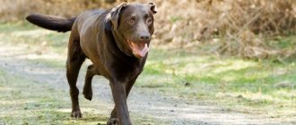
Dislocation in a dog is a fairly common phenomenon that almost every owner of a four-legged friend can encounter. Such an injury can occur as a result of falls or a blow from an unsuccessful jump, or on a slippery surface, as well as during active games with other pets.
In order to understand how to act when a dog sprains, read the following information about the types, symptoms and treatment options for this injury.
First of all, what is a dislocated joint in a dog? A dislocated joint in a dog is an anatomical displacement of bone structures, accompanied by damage to the integrity of tissues and destruction of cartilage, tendons and ligaments of the joints, as well as blood vessels.
How to determine a dislocation in a dog?
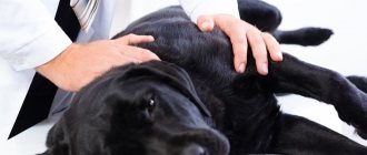
In order to diagnose a dislocated joint in a dog, you should examine your pet for a number of symptoms. If they are present, then you should definitely take your pet to the nearest trusted veterinary clinic to confirm the diagnosis using x-rays or other visual diagnostic methods. Our veterinary clinic is located in Moscow, and we are always ready to help you and your pet.
The symptoms of a dislocation will be noticeable immediately, as the pet will have difficulty moving. The owner may notice the following symptoms of a dislocation in a dog:
- the dog stops using the sore paw and does not step on it,
- when a front paw is dislocated, the animal will tuck it under itself so as not to strain it,
- with a hip dislocation, the animal has difficulty standing up, does not use the damaged paw, edema or swelling in the pelvic area may also be noticeable,
- the animal does not allow you to touch the sore spot, swelling or does not allow you to straighten your tucked paw, does not make contact. In case of severe pain, the animal may behave aggressively and not even allow you to approach it,
- sometimes the dislocation can even be heard, as there may be a grinding or clicking sound when trying to move the damaged paw, or a slight sound of bone rubbing against the joint,
- dislocation can lead to inflammatory processes, when the temperature may rise, and this, in turn, leads to apathy and loss of appetite,
Factors influencing the rate of bone fusion
The healing of a broken bone depends on a number of factors that either speed it up or hinder it. The regeneration process itself is individual for each patient.
First aid is critical to the speed of healing. With an open fracture, it is important to prevent infection from entering the wound, because inflammation and suppuration will slow down the regeneration process.
Healing occurs faster when small bones are broken.
In open fractures, callus forms much longer if a wound infection develops, which is accompanied by bone sequestration and post-traumatic osteomyelitis. That is why, if the fracture is treated incorrectly, the formation of callus slows down or does not occur at all. In such situations, fractures that do not heal for a long time, characterized by slow consolidation, as well as false joints appear:
- If patients suffer from hypovitaminosis and vitamin deficiency (osteomalacia in pregnant women, rickets, scurvy).
- If there are disturbances in the activity of the parathyroid glands (decreased calcium concentration in the blood) and adrenal hyperfunction.
- The presence of concomitant diseases occurring in the chronic stage, as well as inflammatory processes. Any pathological processes in the body significantly delay the recovery period after a fracture.
- The presence of excess body weight negatively affects the healing process of bone tissue.
- Metabolic disorder.
- Failure to comply with the terms of wearing a plaster cast. Many cases of bone tissue taking too long to heal are due to the fact that a person does not want to walk in a cast for a long time and removes it before the time prescribed by the doctor. The fused area of the bone is under pressure and displacement occurs.
How quickly bones heal depends on factors such as the need to install an implant. This occurs in cases where there are too many bone fragments, they are very small, and it is not possible to reassemble them.
What to do if your dog has a sprain?
As soon as you have discovered the above symptoms, you yourself have seen the process of injury, for example, from a blow, or you have noticed how the animal has difficulty moving and is clearly experiencing severe pain, then you are faced with the question of providing first aid.
After detecting obvious symptoms of a dislocation, you should contact a professional veterinary clinic, where you will undergo an X-ray examination of the joint, which will be performed in the lateral and direct projections.
Only after an x-ray will the doctor be able to make an accurate diagnosis and determine the type of dislocation, as well as prescribe subsequent treatment.
Attention! Under no circumstances should you try to straighten the paw yourself, as you can only harm the animal and increase ruptures and damage. Try not to let your pet lean on the affected limb, as putting pressure on the damaged joint will only worsen the condition.
Treatment will depend on the type of dislocation. Let us further consider the main ones - a dislocation of the paw: front or rear, hip dislocation in a dog, dislocation of the patella, dislocation of the femur and dislocation of the shoulder joint. Each of them has its own characteristics and this must be taken into account.
Prevention
If there are congenital pathologies, lameness is difficult to prevent. As for external factors, the owner is able to protect his four-legged friend from them.
It is enough to follow the basic rules:
choose suitable places for walking with minimal risk of bites and injuries;- do not overload the dog with physical activity during exercise, selecting exercises that are feasible for the animal;
- Carry out daily examinations of your pet to promptly identify injury or disease.
A balanced diet plays an important role in a dog’s health. The mineral components included in the feed strengthen bones and blood vessels and make muscle tissue elastic. This helps prevent the animal from developing problems with its limbs.
Dislocation of a paw in a dog: types and consequences
This is one of the most commonly diagnosed types of injury in active pets.
It is a displacement of bones or joints without compromising their integrity. In this case, soft joint tissues are damaged - these are ligaments, tendons, adjacent muscles and blood vessels, which entails dysfunction of the entire limb.
Dislocation of the paw can be classified according to one or another nature of the phenomenon. So there are several varieties
- Congenital type - established during the development of the puppy before its birth, the baby will be viable, but it is quite difficult to completely cure a dislocation in adulthood
- Traumatic - this is one of the most common types of paw dislocation in dogs, as it subsequently appears due to injuries from a fall, pinching of the limbs by some heavy object or a collision with a heavy object at high speed, and also often in car injuries
- The pathological appearance occurs as a reaction to the occurrence of various pathologies of the musculoskeletal system, when the cartilaginous bone tissue is greatly thinned
- The paralytic type of dislocation appears as a result of atrophy of the muscle group that supports the joint
- Non-reducible and habitual ones appear with repeated dislocations, when once-stretched ligaments or muscles weakly support the joint. This leads to constantly occurring repeated dislocations, that is, the usual type or an unreducible old one, when new tissues form between the articular heads, which interfere with the restoration of the joint.
It is also necessary to distinguish between types and the timing of the appearance of the first symptoms, that is, how much time has passed from the onset of injury to the detection of symptoms and diagnosis. So there are three varieties - fresh, stale and old blow.
- A fresh dislocation is detected if you notice it within three days of receiving the injury.
- A stale appearance is a period of three to fourteen days when, over a sufficiently long period, the dog does not immediately show symptoms
- And an old dislocation is from two weeks or more, usually the dog begins to limp heavily and the symptoms will be clearly visible.
Of course, the sooner a dislocation is detected, the easier it will be to treat it and the easier it will be for the pet itself. Failure to provide assistance may result in dire consequences.
It is important to immediately visit a specialist for an x-ray after detecting symptoms in order to receive a diagnosis and the necessary treatment, since the consequences of an untreated dislocation will cause severe pain in the animal. The dog will have difficulty moving, which means it will be more difficult for it to move around for food or other natural needs. Severe pain initially leads to apathy and a reluctance to get up for a walk or food, and over a long period it leads to severe aggression and the fact that the pet does not obey the owner. A long period of failure to provide assistance for a dislocation most often leads to lameness, since the articular bones do not fuse properly. The pet begins to limp and his gait is greatly distorted.
Another option for late treatment is swelling and inflammation that can occur in areas of joint damage, which leads to horrific consequences for inflammation. This can go so far that the pet may lose a leg due to the necessary amputation due to gangrene.
Also, if the paw is not used for a long time, the muscles begin to atrophy and paralysis occurs.
All these horrific consequences can be avoided if treatment is started on time.
Dislocation of the front and rear paws
A dislocation of a dog's front paw can be determined because the dog is pulling it close to its body to make movement as easy as possible and not strain the affected paw. It is impossible to determine the exact cause without contacting a specialist and undergoing a number of tests in addition to x-rays, since the cause could be, for example, arthritis, muscle weakness or another disease as a side effect. How to treat a dislocated paw will depend on the reason that caused the dislocation of the joint.
It is important to accurately determine the specifics of the disease in order to be sure that the treatment received is correct.
All that is needed from the owner is to take the pet to a veterinary clinic for an examination and until this moment do not allow the dog to lean on its paw, I try to use it and overcome the pain. If you called a doctor to your home, then ensure that your pets are at rest and apply something cold to the damaged area, for example, a frozen item from the freezer
Symptoms of pathology
Lameness in dogs is a change in gait, which is accompanied by a shortening of the step, partial or complete exclusion of the limbs from the walking process, when the animal only leans lightly on it, or even tucks in when moving. It is not an independent disease, but a symptom of pathologies that are associated with pain, discomfort or muscle weakness in the limbs.
The disorder can manifest itself spontaneously, after prolonged physical exertion or a state of rest, and is sometimes accompanied by additional symptoms - swelling, muscle atrophy, general malaise (loss of appetite, lethargy, apathy, etc.). Features of lameness and accompanying manifestations play an important role in making a diagnosis, so they need to be given special attention.
Lameness is not an independent disease, but a symptom of pathologies
Hip dislocation in a dog
Hip dislocations are quite common due to the complex multi-axial structure of the hip joint itself.
In most cases (more than 95%), complete traumatic dislocation of the head of the hip joint occurs in dogs, and incomplete dislocation - that is, subluxation - is less common in dogs with joint dysplasia.
Traumatic dislocation of the hip joint leads to a complete inability to lean on the damaged limb, and the pelvic part becomes swollen and asymmetry.
A definitive diagnosis of hip dislocation can only be made after an X-ray examination.
Treatment of a dislocated hip joint should be done as quickly as possible after discovery of the disease by repositioning the dislocated joint or surgery. The most optimal period is the first hours after the injury; if you urgently consult a doctor during the first 24 hours after the injury, the specialist will be able to help you and correct the dislocation without surgery.
However, at a later stage, reduction of hip dislocation using the closed reduction method is not always successful, and relapse may occur when the femur comes out of the acetabulum again.
Therefore, the surgical method of treating this injury is most often used. The doctor will determine the method of recovery based on: the age of the animal, its weight, the characteristics of the breed, the severity of the dislocation and the appearance of associated injuries, and so on.
Medical practice recommends the use of four types of intervention and treatment for hip dislocation.
The first method of suturing the joint capsule is the so-called capsulorrhaphy. This option is necessary when there is a tear in the joint capsule, which stabilizes the hip joint. The method results in open reduction by tightly suturing the joint capsule, after which the animal requires strict rest for a period of two weeks.
The following methods are the round ligament replacement method and the transarticular stabilization method, in which the damaged hip joint is fixed in the correct anatomical position using a metal pin, which fixes the composition to the floor of the acetabulum. After this, the animal requires strict rest for a period of up to three weeks, and the metal pin is removed when this period is reached.
When replacing the round ligament, instead of a wire, a graft is inserted into the position of the round ligament, and the remnants of the round ligament are removed, along with which the joint capsule is sutured. The method also requires restricting the animal's movement for a period of two weeks.
The last surgical method for treating hip dislocation is removal of the femoral head, that is, the method of resection arthroplasty.
Recovery in the post-rehabilitation period requires first strict rest for up to 2-3 weeks, and then rehabilitation procedures such as physiotherapy and swimming. A small amount of exercise will help to gradually restore the animal's mobility.
Risk factors
Many factors can increase your risk of developing DVT, which include:
- Age. At age 60, the risk of DVT increases, although it can happen at any age.
- Sitting for long periods of time, such as while driving or flying. When your legs remain motionless for several hours, your calf muscles do not contract. Muscle contractions promote blood circulation.
- Prolonged bed rest, such as during a long hospital stay or paralysis. Blood clots can form in the calves if the calf muscles are not used for a long time.
- Trauma or surgery. Injury to the veins or surgery may increase the risk of blood clots.
- Pregnancy. Pregnancy increases pressure in the veins of the pelvis and legs. Women with an inherited bleeding disorder are at particular risk. The risk of blood clots from pregnancy can last up to six weeks after the baby is born.
- Birth control pills (oral contraceptives) or hormone replacement therapy. Both factors can increase the blood's ability to clot.
- Exposure to drugs or chemicals. Certain drugs can cause blood clots. Consult your physician before use.
- Overweight or obese. Excess weight increases pressure in the veins of the pelvis and legs.
- Smoking. Smoking affects clotting and circulation, which may increase the risk of DVT.
- Cancer. Some forms of cancer increase levels of substances in the blood that cause blood clotting. Some forms of cancer treatment also increase the risk of blood clots.
- Heart failure. Increases the risk of developing deep vein thrombosis and pulmonary embolism. Because people with heart failure have limited heart and lung function, symptoms caused by even a small pulmonary embolism are more noticeable.
- Inflammatory bowel diseases. Bowel diseases such as Crohn's disease or ulcerative colitis increase the risk of DVT.
- Personal or family history of DVT or PE. If you or someone in your family has had one or both of these, you may be at greater risk of developing DVT.
- Genetics. Some people inherit genetic risk factors or disorders, such as factor V Leiden, that make their blood clot more easily. The hereditary disease itself may not cause blood clots unless it is combined with one or more other risk factors.
- Risk factor unknown. Sometimes a blood clot in a vein can occur without an obvious underlying risk factor. This is called unprovoked VTE.
Patella luxation
This type of dislocation most often occurs in small and dwarf breeds of dogs, such as terriers and Spitz, poodles, as they have a genetic predisposition to such a dislocation.
Dislocation of the calyx as a result of injury occurs in larger breeds of dogs. Also, the displacement of the kneecap is often associated with age-related changes, since closer to old age the muscles of the body weaken and this type of injury is possible.
When a dog's kneecap dislocates, it can move both outward and inward - if the kneecap moves toward the abdomen, then this type is called a lateral dislocation, and in the opposite direction from the abdomen, it is a medial dislocation.
The last type - medial dislocation - is more common, small breeds of dogs are most susceptible to it, and for dwarf breeds it poses the greatest threat, as it can lead to irreversible consequences and the dog will lose mobility.
Classification
In order to diagnose a dislocation and determine treatment, it should be taken into account that doctors distinguish 4 degrees of severity of patella dislocation in dogs:
- The first degree is the easiest; after a knee injury, the cup will return to its place without consequences
- The second degree is characterized by an unnatural position when bending, and may fall into place in rare cases
- The third degree occurs during movement when bending and extending the paw; it requires surgical intervention, as it gets into place for a short time and loses its position again
- Fourth degree - with it the kneecap cannot return to its normal state and, accordingly, is always in a state of dislocation
Prolonged lack of treatment can lead to a chronic form of chromatia.
The symptoms of a luxated kneecap are as follows:
- The first stage is when the knee does not bend because the cup is not in its natural position, but it can be realigned. this condition rarely worsens and can be treated non-surgically
- At the second stage, the cartilage is severely damaged due to the sliding of the cup outside the joint, which leads to severe changes and the possible need for surgical intervention
- The third stage is a persistent dislocation, and without surgical intervention it is impossible to return the cup to its natural state, since even with successful reduction a relapse occurs and the cup flies out again
- At the fourth stage, the stability of the dislocation is also repeated, and even surgical intervention does not guarantee a 100% result
A luxated kneecap can be identified by the following external signs:
- the dog jumps or seems to shake off its paw during walks and exercise, but this does not bring it discomfort
- after a long rest, when the dog has not stood up for a long period, difficulty may be noticeable when trying to stand up; it is quite difficult to lean on its paws. After a long period of activity, nothing like this is observed.
- the dog moves its paw with less speed and range of motion
- the animal tries to constantly hold its paw in a bent state or press it to itself
- In the last severe stages, the joint becomes audible, since with any movement it crunches and clicks are heard
Diagnosis of a luxated patella occurs as follows:
It is necessary to take the pet to a veterinary clinic or invite a specialist to your home, after which the doctor will determine the position of the kneecap and determine whether it can be set without surgery.
Next, an x-ray must be taken in two positions: straight and lateral, which show the degree of dislocation and the presence of extraneous damage.
It is important to consider that exclusively medicinal treatment is based on the use of painkillers, and this is not always the right solution, since the dog stops experiencing pain and begins to actively use the damaged paw again. This is strictly contraindicated, as more damage occurs and the animal only harms itself.
Surgery for medial patellar luxation usually occurs in one of two versions:
The first method is plastic surgery, that is, removal of part of the cancellous bone, onto which fibrocartilage subsequently grows again.
The second is three-dimensional chondroplasty. This method is only suitable for pets up to six months old.
Since the first degree is not so dangerous, and when the kneecap is reset, the threat of re-dislocation is much less, you should be careful about the second and third degrees of dislocation.
With surgical intervention at these levels, a higher percentage of success and the absence of re-dislocation can be guaranteed. To do this, you should help your pet and carry out physical therapy with him, carefully monitoring the amount of exercise.
Shoulder dislocation
This is a pathological displacement of the scapula relative to the shoulder joint. The most common cause of dislocation is injury after jumping, fighting, etc., that is, injury to the shoulder girdle occurs due to the activity of mobility of the forelimbs.
Since the shoulder complex works in several planes at once, that is, it produces flexion-extension of the limb, rotation and adduction-abduction forward-backward, it is not surprising that the risk of damaging the joint increases with the number of movements.
Shoulder dislocation in dogs most often occurs at the site of a strong blow to the shoulder girdle, or due to excess load.
Subluxation of the shoulder joint is also highlighted, this is the moment when the sockets and heads of the bone have already moved away from each other, but are still in contact, thus, the head of the articular bone falls out of the socket, but then returns to its place. Such subluxation can lead to the development of dislocation.