Spinal diseases also occur in wild animals, but in cases where people have interfered with nature’s plan, the frequency of pathology increases as changes are introduced and radicalized. Dogs that look minimally similar to their wild ancestors are more susceptible to spinal and joint damage than others. Most often, intervertebral hernias occur.
Causes of the disease
The spine is made up of many individual bone fragments called vertebrae. For mobility, they are connected to each other by elastic and dense cartilaginous formations - discs. They serve as shock absorbers for shocks and shocks when walking and running, allowing the spine to gently bend and return to its original position.
With injuries, diseases, genetic predisposition, or as the body ages, intervertebral discs can “shrink out,” become deformed, and become displaced. When moving, they can be pinched by the vertebrae, pressing on the nerve endings and putting pressure on the spinal cord. This can cause not only severe pain and problems with movement, but also lead to partial or complete paralysis.
Doctor of Veterinary Sciences Kozlov N.A.
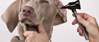
- Peculiarities of symptoms in dogs with discopathy in the thoracic and lumbar spine.
What is discopathy/disc herniation in dogs?
One of the most pressing problems of modern veterinary medicine for small animals is intervertebral disc herniation/discopathy/disc pathology or disc disease in dogs, which is associated with their wide distribution. Probably, the more correct term is disc pathology, but in Russian-language literature the combination “disc herniation” in dogs has become firmly established.
Studying discopathy is undoubtedly important, since the pathology is common in dogs. Intervertebral disc herniation in dogs is one of the most common diseases characterized by neurological disorders, and, at the same time, the most common spinal pathology in dogs (Braund, 1993; Sharp NJH, Wheeler SJ, 2005). Degenerative changes can affect any intervertebral disc between C2 and S1 and, as a result, lead to the formation of a herniated disc. According to a number of researchers, disc herniation (with clinical manifestations of varying degrees) accounts for 3...5% of all diseases in dogs (Hoerlein BF, 1987, Bubenik L., 2005). Among dachshunds, this pathology occurs in 25% of animals (Hoerlein BF, 1987). According to our data, in Moscow and the Moscow region, discopathy accounts for 65...70% of all neurological pathologies. Despite the fact that a large number of new surgical techniques have been proposed in veterinary vertebrology, the results of treatment of animals with degenerative diseases of the spine (disc herniation) are too early to be considered satisfactory.
There are 3 types of discopathy (disc herniation in dogs) or intervertebral disc pathologies.
Type I discopathy in dogs
In chondrodystrophic breeds (dachshunds, French bulldogs, pugs), pathologies are caused by chondroid metamorphosis (cartilaginous changes) of the nucleus pulposus and, as a rule, the formation of a Hansen type 1 disc herniation or disc extrusion at the age of 2-7 years. In this case, parts of the nucleus pulposus, having shifted into the spinal canal, cause compression (compression) of the spinal cord.
Discopathy type II in dogs
In dogs of non-chondrodystrophic breeds, age-related degenerative changes occur, which leads to destruction of the annulus fibrosus and calcification of the nucleus pulposus, which, in turn, contributes to the formation of Hansen type 2 disc herniation (disc protrusion), respectively. With this discopathy, the altered fibrous ring affects the contents of the spinal canal: the spinal cord, spinal nerve roots, etc. From personal experience, it should be noted the occurrence of type 1 hernias in large breeds of dogs, in particular in Rottweilers and German shepherds. Typically develops between the ages of 6-9 years. There are exceptions; several dogs were operated on at the age of 5-8 months.
Type III discopathy in dogs
Separately, there are type III disc herniations associated with acute damage to the spinal disc. In the English literature, this type of disc lesion corresponds to the term “acute non-compressive nucleus pulposus extrusion (ANNPE)” or “disc explosion”. Such lesions are characterized by structural changes in the spinal cord (myelopathy), lack of compression and a small volume of hernial material, which represents elements of the nucleus pulposus. More often, pathology is noted in dogs of decorative breeds.
It's important to remember that
The success of conservative and surgical treatment (i.e. restoration of dogs) for all types of hernias/discopathies is determined by correct and timely diagnosis, treatment and rehabilitation of the patient in the postoperative period.
Symptoms of disc herniations depending on the part of the spine.
Features of the clinical picture in dogs with hernias in the cervical spine.
In most dogs with hernias in the cervical spine, we observed a combination of the following signs: lameness in one or both thoracic limbs and impaired proprioception of the limbs. In the future, as symptoms progress, impaired proprioception in the pelvic limbs may be added. The following signs may be observed: the dog's head is always lowered down (ventroflexion), hemiparesis (paresis of 1 limb), spontaneous whining, twitching of 1 or several muscles in the scapula area. The only sign that we observed in all animals with cervical hernias was a neurological deficit in the thoracic limbs or one of them (“radicular” symptoms). The most severe condition for dogs with hernias in the cervical spine is tetraplegia - paralysis of 4 limbs, the animal is completely paralyzed and unable to move.
Features of the clinical picture in dogs with hernias in the thoracic and lumbar spine
These hernias are characterized by severe clinical manifestations affecting the pelvic limbs. The leading symptoms observed in most patients with hernias in the thoracolumbar region are pain in the back and neurological deficit of the pelvic limbs. In most animals, kyphosis was determined, which, as a rule, correlates with pain; the stronger the pain, the more pronounced the kyphotic hump. Paradoxical reactions are often observed: with strong finger pressure on the spinous processes in the area of the hernia, the animal does not react in any way, and with a light touch it shows severe pain. Also, such animals often had a tense abdominal wall. Therefore, in such dogs, neurological problems must be differentiated from abdominal pathologies.
Features of the clinical picture in dogs with hernias in the lumbosacral spine
Herniation between L7-S1 can be manifested by impaired movement of the pelvic limbs, lameness, and neurological disorders of the SUI type. The most common symptom observed with this pathology was pain in the L7-S1 area and pain with hyperextension of the pelvic limbs. In some cases, it was more pronounced when this area was lightly touched than when pressure was applied strongly to the spinous processes. This sign could also be noted with hernias in the thoracolumbar region. Pain as the only symptom in sick animals was extremely rare. As a rule, it was accompanied by a deficit of proprioception and paresis or paralysis of the pelvic limbs, tail, as well as urinary and fecal incontinence, which is associated with dysfunction of the pudendal and pelvic nerves. In this case, incontinence was characterized by leakage of urine from the very beginning of the development of the pathological process without “overexposure” of urine. As a rule, the anal reflex is reduced. In some cases I encountered the syndrome of “pseudohyperreflexia”. It manifested itself as follows: a false strengthening of the knee reflex occurs, due to the fact that the tone of the muscles innervated by the sciatic nerve decreases. These muscles counteract the muscles involved in the knee reflex. Sometimes it manifests itself as follows: when testing the knee reflex, the reflex appears on the unexamined limb.
Diagnosis of discopathy in dogs
Diagnosis of discopathy in dogs consists of several stages:
- Clinical examination and neurological examination;
- X-ray examination if necessary;
- MRI or CT.
To determine the neurological status of the animals, reflexes on the thoracic and pelvic limbs, panniculitis (also called the skin strip reflex), and proprioception were determined. It should be remembered that the degree or severity of the process is determined, as a rule, by the presence of deep pain sensitivity (DPS). Its absence is a negative prognostic criterion; other reflexes determine to a greater extent the localization of the process. Of course, in the presence of a hernia in the thoracolumbar region and the absence of a knee-jerk reflex, we can talk about a severe course of the process and probable descending myelomalacia, and not about the localization of the disc pathology.
- Definition of proprioception
- Knee Reflex Testing Technique
- Biceps/triceps reflex
- Flexion reflex
- Definition of HBC
- CT or MRI
These techniques make it possible to make a final diagnosis and establish the exact location of the disc herniation. The question of choosing a technique, either CT or MRI, is controversial. For myself, I defined this choice as follows: if you need to determine the presence of a hernia, then CT is preferable, and if the task is to determine mainly changes in the spinal cord, then it is better to choose MRI. At the present time, with the availability of MRI in veterinary medicine with a field power of 1 T, I consider it more appropriate to conduct MRI diagnostics.
Treatment of discopathy in dogs
1. Conservative treatment is based on limiting the pet’s mobility;
2. Hormonal or non-steroidal anti-inflammatory drugs;
3. Acetylcholinesterase inhibitors. Previously, the drug Prozerin was used in veterinary medicine. Now the drug Neuromedin has become more widespread - a dose of 0.05 mg / kg - 2 times a day.
4. Vitamins of group B, the most widely used preparations are B1, B2 and B6, sometimes B12. Complex drugs are also used: Combilipen, Milgamma. Dosage:
B1 - 3 mg/kg subcutaneously, 1 time per day
B2- 3 mg/kg, 1 time per day
B6- 1.5 mg/kg, 1 time per day
4. Drugs of various groups. In some cases, for discopathy in dogs, drugs such as Cerebrolysin, nootropil, vinpocetine, etc. are used. It should be remembered that a scientifically proven result from such drugs has not been established! As a rule, the improvement associated with their use is associated with limited mobility, and not with the use of the drug!
The site contains articles devoted to the treatment of dogs with intervertebral disc herniations:
To summarize the treatment, in my opinion, the indication for conservative treatment is the presence of the initial stages of neurological disorders in the animal. Indication for surgical treatment is relapse (repetition) of symptoms or their aggravation, absence of HBC.
Prognosis for discopathy in dogs
Depends on the stage of neurological disorders before surgery, the doctor’s experience, the age of the animal, the presence of concomitant pathologies (diabetes, renal failure), etc. Based on my own experience, based on more than 3000 operations, the likelihood of recovery in dogs with type 1 hernias is in the presence of HBC it is 97%.
Main symptoms
Signs of a hernia are:
- Pain in different parts of the back.
- Weakness.
- Gait disturbances, stiffness. The animal tries in every possible way to limit its own mobility.
- Paresis and paralysis.
- Complete or partial loss of sensation.
- Involuntary urination and/or defecation.
The appearance of such signs should certainly alarm dog owners and force them to visit a veterinary clinic. This needs to be done as early as possible, because in the early stages the problem can be dealt with much easier and faster.
How to massage a hernia without causing harm
General principles
Massage for a spinal hernia can be done immediately after eliminating the main symptoms of an exacerbation.
Here are 5 basic principles of the correct procedure:
- You should not use shock techniques, as they can provoke an aggravation.
- You cannot apply mechanical pressure to the spine - all manipulations are carried out in the paravertebral region.
- The massage should be relaxing, soothing, the patient should experience only a pleasant feeling of relaxation.
- Any increase in pain resulting from the influence of a chiropractor is a sufficient reason to terminate the session.
- It is necessary to massage the back and the affected limb, because as a result of compression of the nerve roots, pain, numbness, and decreased muscle strength occur not only in the back, but also in the limb on the affected side.
Five massage techniques
- Using light circular movements of your palms in the direction from the lower back towards the shoulders and from the paravertebral region to the lateral surfaces of the body, massage the back on the left and then on the right side.
- Using the palms of your hands with light pressure on your back, perform several vertical movements along the spine from the lower back to the neck.
- Visually divide the left and right halves of the back into two more halves with longitudinal lines. Using your fingertips, use gentle pressure in a circular motion to work your back muscles along these mental lines to promote relaxation. Repeat until you feel less muscle tension in your back.
- Perform stroking, rubbing, kneading with your palms: straight and circular movements from the center of the back to the lateral surfaces of the back and slightly upward. This will improve venous drainage from recently cramped muscles, which will reduce the pain and fatigue associated with GP.
- Using your palm with your fingers spread apart, with light force, perform several movements from the bottom up (from the lower back to the shoulder area) on each side of the spinal column.
Best Eyelid surgery for Shar Pei
For a massage course, use cream or massage oil with a calming effect.
You can do this massage every day. This is an excellent remedy for improving overall well-being: it reduces pain and muscle stiffness, restores skin sensitivity and muscle strength in the limbs. And massage for a vertebral hernia in combination with physical therapy helps to form a good muscle corset and prevent exacerbations of HP or reduce the degree of their manifestations.
Treatment method and prognosis
In the initial stages and with minor damage, conservative treatment may be prescribed, consisting of limiting the animal’s mobility and using specific medications. These are painkillers and anti-inflammatory drugs of non-steroidal origin.
In severe cases, surgery is necessary. Spinal surgery is complex, painstaking, dangerous and expensive. It requires the highest qualifications of the surgeon.
During the operation, tissue is removed that puts pressure on the spinal cord and nerve endings, causing severe pain and impaired mobility. In rare situations, implantation of an artificial intervertebral disc is used. This is expensive and not always possible, since it requires special equipment and a high level of training and experience of the veterinarian.
If sensitivity in the animal’s limbs is not completely lost, the prognosis is positive. But the owners will need to put in a lot of effort and spend money on treatment and restoration measures.
Prevention
An effective conservative treatment plan or surgery allows the dog to make a full recovery. The main condition is to promptly seek help from a veterinary clinic. Recurrences are rare and usually only in dachshunds, with the hernia developing in another area (for example, in the cervical spine).
To avoid the formation of a hernia, it is enough to follow useful recommendations from experienced veterinarians:
- Avoid intense physical activity . They must be optimally calculated for a specific breed of dog, taking into account the weight of the animal, as well as previous diseases and injuries;
- Provide a proper and balanced diet. This is especially true for older dogs who need to strengthen their musculoskeletal system. Their diet should contain a large number of foods high in calcium;
- Attend regular checkups. Make an appointment with a veterinarian every six months to rule out the presence of a hernia and other serious diseases, as well as to detect them in a timely manner and begin treatment;
- Losing excess weight. Don't let your pet become obese. If this situation has already developed, stick to a special diet, since extra pounds create a huge load on the spine.
Health to you and your pet!
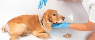
What to do at home
At home, the animal needs to be provided with the most calm and quiet place where it will not be disturbed and forced to get up often. At first, you need to not allow the dog to move a lot. It is strictly forbidden to run, jump, walk on steps or on very rough terrain.
It is best if the sick dog is kept in a special small enclosure, or in a room in an apartment. At first, he himself will not have the strength or desire to frolic, but as the pain decreases, activity will increase. During this period, it is important not to allow the dog to make sudden movements. The rehabilitation period depends on the severity of the pathology and can last up to one and a half to two months.
Life prognosis for dogs with discopathy
The life prognosis for dogs with this diagnosis is very good. Veterinarians assure that timely started therapeutic treatment “puts about 70% of sick animals back on their paws” without surgery. The main thing is to strictly follow the doctor’s recommendations and take medications according to the schedule. If the animal is overweight, then it is necessary to get rid of it, as this leads to a relapse of the disease.
To prevent relapses, it is useful to do physiotherapeutic procedures, massage and make your pet swim, as this forms a good muscular frame in the animal.
A healthy spine means a vigorous, active and cheerful dog!
Authors of the articles: Belanta Clinic team
Classification
Degenerative disc disease Hansen type 1 or IVDD 1 usually occurs due to cartilaginous degeneration of the disc. They are more common in chondrodystrophoid dog breeds (dachshunds, Pekingese, pugs, shih tzus, beagles, etc.), but sometimes in others. This pathology is accompanied by excruciating pain.
Hansen type 2 or IVDD 2 intervertebral disc disease occurs due to fibrous disc degeneration. Protrusion of the nucleus pulposus occurs through part of the fibrous rings, while the remaining fibrous rings remain intact, hypertrophy and protrude into the spinal canal. This pathology occurs more often in aging dogs of non-chondrodystrophoid dog breeds and is accompanied by less severe pain.
Types of intervertebral disc diseases.
Surgical methods
Depending on the type of roof, veterinarians use several methods of surgical treatment.
Methods of surgical treatment of hernia:
- The Hering-Sedamgrotsky method differs in that during the operation the hernial sac is immersed into the abdominal cavity through the hernial ring, after which knotted sutures made of silk threads are applied to the edges of the ring.
- The Sapozhnikov method involves repositioning the contents of the hernial sac into the abdominal cavity, after which the hernial sac is twisted around the longitudinal axis 2-3 times, stitched with catgut and inserted into the hernial ring. The quack rings are sewn up with knotted sutures.
- Olivekov method . In this technique, the hernial sac is grabbed with special tweezers, rotated 360 degrees along the longitudinal axis, and a silk ligature is placed on top. In this case, the ends of the ligature are passed through the edges of the hernial ring using a needle. After that, the ends of the ligature are tightly pulled together until the ring is completely closed and two additional interrupted sutures are applied.
- Tarasevich's technique is used for operations on wide hernial rings, abscess or necrosis of the hernial sac. The operation begins by exposing and dissecting the hernial sac, then it is excised, and its contents are inserted into the peritoneal cavity. The hernial ring is sutured with loop-shaped sutures under finger control. In case of abscesses or necrosis, the hernial sac is amputated.