The skeletal system in the animal's body forms the skeleton. This is a frame for movement, muscle attachment, and also support. The size of the skeleton and the shape of its bones determine the size of the dog, body type, constitution, and body proportions.
Bone is a very durable and strong, but living organ. It grows, changes its structure, is restored and destroyed, is a calcium depot, reacts to changes in internal and external environmental conditions, is supplied with blood and innervated.
| BONE STRENGTH | |
| Compound | Structure |
| Inorganic substances - calcium | Compact bone tissue (substance) - bone plates consisting of tubes (osteons) tightly adjacent to each other |
| Organic substances – collagen | Spongy bone tissue (substance) – in the bone it is located under a compact substance, bone plates are located at a great distance from each other, these cells contain bone marrow |
Depending on the structure of the bones, they can be flat, mixed, or tubular. The bone is covered on top with periosteum, due to which it can grow in thickness. Tubular bones have 2 ends - the epiphyses and the body of the bone (diaphysis). In growing animals, tubular bones have cartilaginous plates in the area of the epiphyses, which ensure the growth of the bone in length.
During the process of embryogenesis (fetal development) and growth of a puppy, the skeleton goes through three stages of development:
- membranous (formed by connective tissue);
- cartilaginous;
- bone.
During the period of the membranous skeleton in embryogenesis, a notochord is formed, from which the skull and spine are subsequently formed; the limbs appear later. At birth, bones are partially constructed from cartilaginous tissue and contain ossification centers. After complete ossification of the cartilaginous layers in the bones, the dog’s growth stops.
The skeleton is divided into two sections: axial and peripheral.
Dog skeleton: structure of the skull, spine, limbs
To study the anatomy of a dog, it is worth knowing the skeletal structure of this animal. So, the skeleton is the basis of the body, which consists of the spinal column, skull, ribs and limbs. All the bones of these formations make up the dog’s skeletal system. The degree of development of a dog’s skeletal system plays a significant role in its life. The entire musculoskeletal system of a dog includes, in addition to the skeletal system, also joints with ligaments and muscles with tendons. Young animals have more elastic bones than old ones, because with age the bones become brittle and lose their strength. How metabolic processes occur in a dog’s body can be judged by the degree of development of bones in the area of the metacarpus, metatarsus, hock and carpal joints, as well as by the condition of the teeth.
Dog skull: structure
The skull consists of cranial (cerebral) bones (forming the cranial or cerebral cavity) and facial bones. The brain section of the skull is formed by unpaired and paired bones. The facial part is wedge-shaped, the lower jaw is attached to the skull. The facial part of the skull also has unpaired and paired bones.
All the bones of the medulla are connected by cartilage or connective tissue in the form of sutures. In older dogs, the sutures become stiff. Only the lower jaw is connected by a movable joint, thanks to which the dog chews food.
Paired dog skull bones:
Unpaired skull bones:
Thus, the dog’s skull consists of 20 paired and 7 unpaired bones.
Side view of dog skull:
Dog's dental system and bite
Consists of incisors, clicks, false roots, molars, premolars and molars (42 teeth in total). The breed standard determines the correct bite of a dog. The bite may be:
Interracial differences in the structure of dog head elements
Dogs of different breeds have heads that look different, mainly due to the ratio (percentage), that is, the ratio of the skull to the muzzle. The length and width of the skull to the muzzle is also important (for example, in the case of knots the width is 3/5 of the length of the entire head), as is the width and length of the muzzle itself (for example, very short in relation to the skull in the Bulldog and Pekingese, and also very long and slender in greyhounds).
The muzzle is also wide at the base and tapers slightly towards the nose. The upper part of the muzzle of most breeds is flat and straight, that is, without convexities (rarely found, for example, in the Bull Terrier). When determining the characteristics of rocks, the angle formed by the stop with the front angle of the head is also taken into account. In some breeds the nuchal swelling is not easily visible, while in some it is noticeable (in some cases with slender heads). The nostrils should be large and allow the dog to breathe freely. The upper lips are moderately drooping in some breeds and very drooping in others, but always meet at the front to form an inverted "V" shape. The ears may be erect (such as in a German Shepherd), semi-erect (such as in a Terrier), or completely drooping (such as in a Dachshund). Ears can also be long or short and set high or low. In some breeds in some countries it is legal to copy or cut the ears to give them the shape of short and erect ears.
Front and rear limbs
The dog's forelimb is formed by an obliquely set shoulder blade, which connects to the humerus with the help of the glenohumeral joint. The elbow joint is the connection of the forearm (from the radius and ulna) and the humerus. The carpal joint consists of 7 carpal bones, which are connected to the metacarpus (5 bones). The metacarpus goes into 4 three-phalangeal and 1 two-phalangeal fingers. The fingers have non-retractable, strong claws.
The dog's hind limb is represented by the hip joint, which connects the limb itself to the pelvis. The hind limb is formed by the femur, which is connected to the lower leg with the help of the knee joint (2 bones - the tibia and the tibia). On the other side, the tibia is connected to the tarsus using the hock joint. The tarsus consists of tarsal bones, among which the powerful heel bone stands out. 4 (occasionally 5) metatarsal bones are attached to the tarsus. They form a metatarsus, which then turns into 4 3-phalanx fingers with claws.
The forelimb is connected to the spine with the help of powerful shoulder muscles, and the upper edges of the shoulder blades slightly exceed the protruding processes of the thoracic vertebrae - this is how the withers are formed. Dog height: the length of a perpendicular line extending from the edges of the shoulder blades to the ground. Each dog breed has its own height according to the standard.
The degree of development of a dog’s skeletal system plays a significant role in its life. The entire musculoskeletal system of a dog includes, in addition to the skeletal system, also joints with ligaments and muscles with tendons. Young animals have more elastic bones than old ones, because with age the bones become brittle and lose their strength. How metabolic processes occur in a dog’s body can be judged by the degree of development of bones in the area of the metacarpus, metatarsus, hock and carpal joints, as well as by the condition of the teeth.
Source
Dog spine: anatomy
The spine consists of the following bones:
- 7 – cervical
- 13 – thoracic vertebrae
- 7 – lumbar
- 3 – sacral
- 20-23 – tail
The first cervical vertebra is called the atlas, and the second is called the epistropheus. Both of them are somewhat different in structure from the rest of the vertebrae of the dog's column. Thanks to them, the mobility of the animal's head is ensured. The ribs are attached to the thoracic vertebrae through the joints, which form the dog's chest.
The tail vertebrae are very mobile, but in some dogs, according to the standard, the tail must be docked.
Scull
The dog’s skull is light, with a developed brain part; depending on the breed, the shape of the skull can vary greatly, with 3 main types:
The bones of the skull form the walls of the brain, oral and nasal cavities to house and protect soft organs (brain, sensory organs, initial parts of the respiratory and digestive systems). The bones of the head serve as extensive surfaces for attaching the chewing and facial muscles.
The head skeleton can be morphologically divided into:
Functionally, the head skeleton can be divided into two sections:
Anatomy of a dog's skull
Skull and teeth. The connection of the skull bones is movable. It is this that gives the pet the ability to chew, gnaw, and so on.
The dental formula includes: canines, incisors, molars and premolars. Bite is influenced by breed and breed standards.
Forms of bite in dogs
- Scissor-shaped. Here the lower ones seem to be under the upper incisors, and also have a tight connection with each other.
- Pincer bite – This form of bite occurs when the incisors close together.
- Straight. The incisors stand on top of each other.
- Snack. The lower jaw protrudes forward and the teeth do not align.
Brain section of the skull
The medulla of the skull contains the cranial cavity, which contains the brain. It includes 3 paired (frontal, parietal, temporal) and 5 unpaired (occipital, interparietal, sphenoid, pterygoid and ethmoid) bones.
Frontal bone
Parietal bone
Interparietal bone
Occipital bone
Sphenoid bone
Pterygoid bone
The pterygoid bone (os pterygoideum) is a thin short bone plate fused with the pterygoid process of the basisphenoid and with the perpendicular plate of the palatine bone. Takes part in the formation of the lateral wall of the choanae. It has a short hook on the bottom edge.
Ethmoid bone
Temporal bone
general characteristics
To make it easier to understand the anatomy of a dog, the dog’s body parts are conventionally divided into four main components, namely:
Unfolded skeleton of a dog
The external characteristics of the dog, its body parts and their location, which are inherent in the breed and gender, are called the exterior and look like the signature of the breed.
Note! In addition to the general idea, it would not hurt for the dog owner to study the textbook, authored by Volmerhaus and Frewein, which provides a diagram and location of all the animal’s organs. The practical textbook is regarded as a guide for dog breeders.
Skeleton or skeletal system
Of course, the study of dog anatomy begins with the structure of the dog’s skeleton. The skeleton of a dog is the basis on which all the internal organs and muscles of the dog exist. Each component of the skeleton requires special consideration.
Dog skull: structure
There are facial and brain parts, which contain paired and unpaired bones.
Expanded dog skull with explanations
Important! The dog's skull consists of 27 bones, which are connected by cartilage tissue. The older the pet, the more the cartilage becomes ossified, and only the lower jaw retains mobility of the head.
There are 3 types of dog skulls:
For your information! The third type makes up approximately 75% of all breeds.
The differences become visible in the structure of the front part. So, for example, in brachycephalic breeds the muzzle has a flattened shape, and the jaw protrudes forward. This structure of the dog affects its health. In veterinary medicine, the main cause is related to the respiratory tract.
Limb structure
A dog's paws have a complex structure. So:
Important! The owner should monitor the condition of the pet's nails especially carefully. So, too long nails interfere with walking and can even deform the skeleton.
Beautiful dog paws enlarged
Body structure
The dog's body includes the vertebral section and ribs, which are connected to each other. A skeleton is attached to all this. The dog's spine consists of four components:
Spine structure
The pet's body consists of a torso axis and ribs that form the skeleton. The spine is formed from several sections:
At the base of the tail there are 20-23 movable vertebrae. It is the ribs that protect the heart and lungs and, depending on the type of breed, have different curvature. The lumbar vertebrae are large, due to which muscles and tendons are securely attached to them.
For your information! Many dog breeders are often interested in how many ribs a dog has. The chest consists of 13 pairs of ribs.
Structure of teeth
The teeth are used to grind the food received and protect the owner. Dogs are born without teeth. They erupt only at the age of 2-3 weeks. The tooth that appears during this period is temporary. It falls out at the age of 4-5 months, and only after that permanent ones begin to appear.
A set of teeth is formed by:
General view of the teeth of an adult dog
1.1.1. OSTEOLOGY
The first important section is OSTEOLOGY
- Dedicated to the study of bone structure. Bones are located inside the body, they act as a frame and levers for the attachment of muscles, form body cavities, and serve as a place for bone marrow.
The totality of a dog's bones forms its skeleton (Fig. 1). It is a system of bones and cartilage interconnected in a certain way, has longitudinal and lateral symmetry and consists of 289–292 bones, the number of which varies due to the different number of caudal vertebrae.
Rice. 1. Skeleton of a dog
1. Muzzle. 2. Frontal part of the skull. 3. Lower jaw. 4. First cervical vertebra (atlas). 5. Cervical vertebrae. 6. Shoulder blade. 7. Humerus. 8. Bones of the forearm. 9. Carpal joint. 10. Metacarpal bones. 11. Phalanges of fingers. 12. Claws. 13. Thoracic vertebrae. 14. Lumbar vertebrae. 15. Sacral vertebrae. 16. Caudal vertebrae. 17. Ribs. 18. Sternum. 19. Pelvic bones. 20. Femur. 21. Knee joint. 22. Shin. 23. Hock joint. 24. Metatarsal bones
The axial skeleton consists of 109 bones, and the peripheral skeleton - of 180. The male dog has an additional bone - the bone of the penis.
The bones of puppies and young dogs are very elastic due to the large amount of moisture and organic substances - collagen fibers. With age, bones, losing water and these organic components, become more fragile and brittle.
The structure of bones varies by type.
PLATE bones - bones of the head, ribs and pelvic bones.
Spongy bones - bones of the wrist, tarsus, etc.
AIR bones (with cavities filled with air) - frontal and maxillary bones.
MIXED bones - vertebrae.
TUBULAR bones are some limb bones that have a complex structure, characterized by the presence of a middle part (diaphysis) and its two ends (epiphyses). The structural features of tubular bones give them increased strength under vertical loads, however, under sharp diagonal loads, these bones suffer from fragility, which is a common cause of their fractures.
It should be noted that the tubular bones contain yellow bone marrow. The first parts of the dog embryo to appear are the vertebral column and ribs (2–3 weeks of pregnancy), then the bones of the limbs are formed, and the bones of the head are formed last.
The source of bone growth is the periosteum, which covers all bones from the outside, while its inner layer is bone-forming, while the outer layer has a protective function. The process of ossification in dogs is very complex, ensuring long-term growth and plasticity of bones, and is completed completely by about two years.
AXIAL SKELETON consists of:
• bones of the brain and facial parts;
• bones of the spinal column, divided into cervical, thoracic, lumbar, sacral and caudal;
• thirteen pairs of ribs;
• sternum.
The dog's SKULL consists of 29 bones, firmly connected to each other by sutures, and 3 movably connected bones: the paired mandibular and hyoid. During an external examination of a dog’s skull, the expert has to deal with only a few of them (see Fig. 2).
THE SPINAL COLUMN forms the longitudinal axis of the body. In front it connects to the skull, in the back it ends at the tail and consists of a total of 50–55 individual vertebrae.
Rice. 2. Dog skull
1. Occipital bone. 2. Parietal bone. 3. Frontal bone. 4. Lacrimal bones. 5. Nasal bone. 6. Upper jaw. 7. Incisive bone. 8. Temporal bone. 9. Zygomatic bone. 10. Lower jaw
The spinal column is divided into 4 sections (see Fig. 3).
Rice. 3. Sections of the spinal column
1. Cervical (7 vertebrae). 2. Thoracic (13 vertebrae are normal, sometimes 12, rarely 14). 3. Lumbar (7 vertebrae are normal, rarely - 6). 4. Sacral (3 vertebrae). 5. Caudal (20–23 vertebrae)
Each vertebra (Fig. 4) consists of a body, an arch and processes. The vertebral arches form the spinal canal in which the spinal cord is located. Paired transverse processes extend laterally from each arch, to which the ribs and muscles of the spinal column are attached. A spinous process extends upward from each arch.
Sense organs or analyzers
Perhaps every dog owner knows that such animals hear better than any person. And the dog’s sense of smell is at the highest level. But you need to have a more detailed understanding of each sense organ.
Ear structure
The sound reaches the pet through the outer ear, and then it moves to the middle ear and ends up in the inner ear. The outer one consists of the auricle, thanks to which he hears even the quietest sounds. Following the auricle is the auditory canal, which consists of several parts:
The eardrum plays an important role in the hearing system, thanks to which acoustic waves are heard. The middle ear contains the auditory bones of the dog, thanks to which the eardrums and inner ear are attached to the membrane.
For your information! The inner ear provides auditory receptors and contains the vestibular apparatus.
Close-up view of a dog's ear, implementation of auditory activity
Structure of the nose
The nose allows the dog to have a connection with the outside world. The animal reacts sharply to the smell, which allows it to be used not only for peaceful, but also for official purposes. The animal remembers the smell of its owner and recognizes it, even when for some reason it cannot see it.
Important! There are 125 million olfactory receptors in animals, but only 5 million in humans.
The dog senses smells through his nostrils. The pet receives a large percentage of air through the side cutouts on the nose. There is also mucus in the nose, which is why it remains moist.
It consists of the external nose and the nasal cavity, which includes several parts. The upper one is the repository of olfactory reflexes. The lower one serves as a connecting link through which air enters and passes to the nasopharynx, where it warms up.
Internal organs
Of course, a dog's anatomy is not limited to the skeleton and sensory organs. Internal organs occupy a special place in the anatomy of a dog.
Expanded image of a dog's internal organs
Digestive system
It originates from the mouth, where food enters. Afterwards, the food moves through the esophagus and reaches the stomach, where the incoming food is digested. Gastric juice and enzymes grind food into a homogeneous mass called zymus.
Next, the small intestine comes into play, interacting with the pancreas, duodenum and liver. The walls of the small intestine transmit incoming substances into the blood.
Next in line is the large intestine, where the digested food goes. By the time it enters the large intestine, all useful vitamins, minerals and other substances have already been received. Feces are formed from the remains of waste food.
Circulatory system
The main circulatory organ in dogs, like in humans, is the heart. Through the arteries, blood enters each internal organ and circulates through the veins to the heart. The heart is located between the 3rd and 6th ribs in front of the diaphragm.
The heart consists of four chambers and is divided into the right part, where venous blood circulates, and the left part, where arterial blood flows. The parts of the heart are in turn divided into the atrium and the ventricle. The walls of the heart consist of an inner layer - the endocardium, an outer layer - the epicardium and the cardiac muscle of the myocardium.
Important! The size and rate of its contractions depend on the breed, sex and age of the pet.
Respiratory system
The respiratory organs provide the supply of oxygen. The respiratory system consists of two sections:
The physiology in this case is as follows: air enters through the nostrils, and their size depends on the breed of the dog. In the nasopharynx, the incoming air undergoes a warming process, and the nasal glands act in this process as a filter that rejects dirt and dust. The incoming air then continues to move through the larynx. After this comes the trachea. The air then reaches the lungs, which contain blood vessels.
For your information! On average, a dog takes 10-30 deep breaths per minute. Small breeds breathe more frequently than larger breeds. The frequency of inhalations may increase depending on the emotions that the animal experiences, and this indicator may look different.
Urinary system
The dog's urinary organs are designed to empty the body and maintain an acceptable level of water-salt balance. The kidneys produce hormones that control hematopoietin and renin. That is why disruption of the urinary system leads to a number of diseases and often death.
Excretory system
The activity of the excretory system depends on the kidneys. The kidneys work with the bladder through the ureters and end in the urethra. The main task of the excretory system is to remove urine from the body.
The kidneys are equipped with nephrons, which are surrounded by blood vessels. The older the animal, the more often problems with this internal organ may occur.
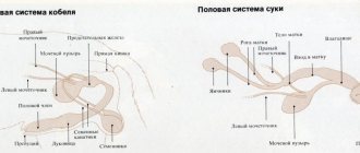
Reproductive system
The reproductive system is intertwined with the excretory system. In male dogs, the urinary tract is the vas deferens. In addition to the testes, male dogs have a prostate, which provides life to sperm.
The reproductive organ of females is the uterus. The dog's uterus is equipped with horns to which the ovaries, fallopian tubes and vagina are attached. Bitches go into heat twice a year, but it still depends on the type of breed.
Note! In northern breeds, estrus occurs once a year and lasts 28 days. Once finished, the female can be bred.
Muscular system and skin
The muscular system is responsible for the activity of the dog. There are three types of muscles:
The muscles are enveloped in nerve endings that transmit signals to the dog’s brain.
The skin, like the muscle system, has a number of functions. It reacts to any changes occurring in the pet and regulates body temperature. It is permeated with vessels and glands, and on the surface layer there are hair roots.
Knowing the anatomy of a pet will make life easier for its owner, because it is not always possible to understand the reasons for the anxiety of your four-legged friend. And so, having an idea of the work of internal organs, you can provide him with timely assistance.
Source
Muscles and skin
The muscle system ensures the dog's mobility. There are 3 types of muscles in the body:
- smooth – located on the walls of all blood vessels;
- striated – attached to the skeleton; dogs have more than 180 single and group muscles of this type;
- cardiac.
All muscle fibers are penetrated by a huge number of nerve endings that transmit impulses to the brain. When contracting, heat is released, which is why in hot weather during activity the body overheats.
Skin provides many functions in a dog's body. It plays the role of a natural sensor that reacts to any external changes, and also protects muscles from injury and regulates body temperature. All layers of the dermis are penetrated by blood and lymphatic vessels, sebaceous and hepatoid glands, and on the surface layer - the epidermis - there are hair roots. The type of coat, its color and structure are determined by breed standards.
Photo: wikimedia.org
Dog skeleton
The skeletal system in the animal's body forms the skeleton. This is a frame for movement, muscle attachment, and also support. The size of the skeleton and the shape of its bones determine the size of the dog, body type, constitution, and body proportions.
Bone is a very durable and strong, but living organ. It grows, changes its structure, is restored and destroyed, is a calcium depot, reacts to changes in internal and external environmental conditions, is supplied with blood and innervated.
| BONE STRENGTH | |
| Compound | Structure |
| Inorganic substances - calcium | Compact bone tissue (substance) - bone plates consisting of tubes (osteons) tightly adjacent to each other |
| Organic substances – collagen | Spongy bone tissue (substance) – in the bone it is located under a compact substance, bone plates are located at a great distance from each other, these cells contain bone marrow |
Depending on the structure of the bones, they can be flat, mixed, or tubular. The bone is covered on top with periosteum, due to which it can grow in thickness. Tubular bones have 2 ends - the epiphyses and the body of the bone (diaphysis). In growing animals, tubular bones have cartilaginous plates in the area of the epiphyses, which ensure the growth of the bone in length.
During the process of embryogenesis (fetal development) and growth of a puppy, the skeleton goes through three stages of development:
During the period of the membranous skeleton in embryogenesis, a notochord is formed, from which the skull and spine are subsequently formed; the limbs appear later. At birth, bones are partially constructed from cartilaginous tissue and contain ossification centers. After complete ossification of the cartilaginous layers in the bones, the dog’s growth stops.
The skeleton is divided into two sections: axial and peripheral.
Skeleton of a dog's torso
In the spine, there is a distinction between the spinal column (spine), which is formed by the vertebral bodies, and the spinal canal, which is formed by the connection of the vertebral arches.
| DEPARTMENTS OF THE SPINE | ||
| Departments | Number of vertebrae | Structural features |
| Cervical | 7 | The first is the atlas: it consists of two arches and forms a movable connection with the occipital bone. The second is epistrophy, the spinous process in the form of a ridge |
| Chest | 13 | Developed spinous processes. Places for attachment of ribs on the vertebral body |
| Lumbar | 6-8 | Developed transverse processes |
| Sacral | 3 | They have fused into one bone - the sacrum. The transverse processes formed the wing of the sacrum |
| Tail | 20-23 | Reduced, in the latter there is no spinal canal |
The thoracic spine, ribs, and sternum (breastbone) form the rib cage.
The peripheral skeleton (the skeleton of the thoracic and pelvic limbs) provides support and passive movements.
The skeleton of the thoracic limb is the shoulder girdle and the free thoracic limb.
The free thoracic limb is the shoulder, forearm, wrist, metacarpus and fingers.
The humerus is a long tubular bone with a body and two epiphyses.
The forearm is formed by two bones - the radius and ulna, the radius is located in front of the radius and is shorter. The ulna in the upper (proximal) section has the ulnar tubercle and coronoid processes.
The bones of the wrist, metacarpus and fingers form the front paw.
The skeleton of the pelvic limb is the pelvic girdle and the free pelvic limb. In addition to support and passive movements, the pelvic limbs are involved in moving the body forward.
Sections of the free pelvic limb: thigh, lower leg, tarsus, metatarsus, fingers.
How does the internal skeleton of a dog work?
Upper spine (neck). It consists of seven vertebral bones. The very first one is called “Atlas” (translated from Latin “Atlas”). It differs from the others in its ring-shaped shape and ensures vertical mobility of the head. The second vertebra is called “Epistrophy” and is responsible for the horizontal movements of the animal’s head.
The ribs are attached to the transverse processes of the vertebrae of this section. The spinous processes of vertebrae 1 to 10 are directed towards the tail, but the eleventh is called the diaphragmatic one. Its spinous process is directed upward. These same processes from the 12th to 13th vertebrae are directed towards the head of the animal.
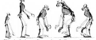
Comparison of human and dog skeletons
Low back or lumbar region. These vertebrae are oval. Their processes are long, flat, ribbon-shaped, transverse - the costal articulars are superbly developed.
The spinous processes of the lumbar vertebrae are directed towards the head. The length of each (up to the fifth) gradually increases, and then immediately shortens. The sacrum is the fusion of three or four sacral vertebrae into one bone. The main function of this part of the spine is to firmly connect the spinal column with the hind limbs.
In females, the sacrum is longer and wider than in males. Such sizes are due to the reproductive function of females. In this part of the spine, the spinal processes merge into the crest of the same name.
Tail. The first four vertebrae are well developed. They are endowed with all the corresponding characteristics, like ordinary vertebrae. Further, the vertebrae of the caudal region serve only to attach the muscles that allow movement of the tail.
In case of spinal injuries or congenital pathologies, the pet is given an MRI and treatment is prescribed. The shoulder girdle includes the scapula bones and rudiments of the clavicle. The scapula bone is attached to the dog's body near the first pair of ribs. Thanks to this belt, the forelimbs are attached to the skeleton.
Comparison of animal limb bones
Limbs. Dogs only have four paws.
The pectoral girdle of the limbs consists of:
- The shoulder, which is made up of the humerus.
- Forearm, includes the ulna and radius bones.
- Brush. It consists of seven carpal bones, five metacarpal bones and phalanges. The dog has five toes, which consist of three phalanges.
The pelvic limb girdle includes:
- Pelvic bones (iliac, pubic, ischial).
- The hips consist of the femur and the kneecap.
- The tibia includes the tibia and fibula.
- Stop. It consists of seven tarsal bones and five metatarsal bones. The phalanges of the fingers and their structure are the same as the thoracic region.
Dog pelvic bone