Sometimes owners of pet dogs face a serious problem - the dog’s paws are lost. This often occurs due to an accident or other accident, but some breeds have a genetic tendency to develop hind limb paralysis.
Most often, misfortune happens suddenly and greatly shocks the owners. Therefore, they must know what to do if the dog’s hind legs fail.
Causes (etiology)
One of the main causes of paresis of the hind limbs is a violation of the conductive function of the spinal cord due to its compression by elements of a prolapsed intervertebral disc (hernia) in the lumen of the spinal canal.
Rice. 1. CT image in a soft tissue window. Dachshund dog, female, age 6 years. Hernia (loss) of the disc between the 9th and 10th thoracic vertebrae. In the photo in the sagittal (median) projection we see the presence of a white mass in the lumen of the spinal canal. This is a herniated disc. This hernia is sequestered, i.e. elements of the disc, mixed with blood, fell into the lumen of the SC canal and migrated cranially (towards the head) along the SC canal. Medical history: in the evening the dog’s hind limbs began to tangle (paresis), lethargy, and in the morning the dog was unable to get up (degree 3 neurological deficit), urinary incontinence. Clinical examination revealed a grade 4 neurological deficit, i.e. the dog had no superficial pain sensitivity
Rice. 2. CT image in a soft tissue window. This is the same dog. Axial (transverse) projection. A hernia is visible in the lumen of the spinal canal, which compresses the spinal cord, causing disruption of its function. After examination, this animal was successfully operated on. The rehabilitation was successful. The functions of the hind limbs were completely restored
What to do if your dog's back legs fail
First of all, go to the vet.
When considering possible pathologies, treatment is completely different, and independent therapy will lead to dire consequences.
The veterinarian will prescribe an x-ray of the pelvic limbs and spine, which will show pathologies of the paws and spine.
Only an experienced veterinarian can treat the disease.
Clinical picture
Based on clinical symptoms, 6 neurological syndromes (stages) are distinguished, corresponding to the degrees of myelopathy (spinal cord damage):
- Pain syndrome: the animal is inactive, lethargic, constrained, and cannot jump onto elevated objects. One of the main signs of the presence of a hernia in the thoracolumbar region is hyperesthesia (increased sensitivity to irritants), hypertonicity (tension) of the muscles of the back and abdominal wall, hunched back (forced kyphosis). And in the cervical region - a forced position of the neck (head in a half-lowered position) and sharp pain with squealing, twitching of the head and neck.
- Decreased proprioceptive sensitivity, ataxia, dysmetria, paresis, but the animal can stand up and move independently. May present with or without pain.
- The paresis is severe, the animal cannot stand up and move independently, but sensitivity is completely preserved (see video No. 1);
- Paralysis - voluntary movements are absent, superficial pain reactions are reduced or absent, conscious reaction to deep pain is preserved. A “seal” positioning of the limbs is possible;
- Severe paralysis (plegia) - superficial and deep pain reactions are absent. “Seal” positioning of the limbs;
- After the dog reaches stage 5 neurological disorders, the process of myelomalacia begins to progress. (See video #2).
If animals have a 4-5 degree neurological deficit, an emergency CT or MRI examination and subsequent (based on the examination results) surgical intervention are necessary because time passes by minutes, and the faster we decompress the spinal cord (surgical decompression), the greater the chance of recovery neurological status.
Rice. 3. Intraoperative. Left hemilaminectomy. We see a sequestered encircling prolapse (hernia, prolapse) of the T13-L1 disc in a French bulldog dog. Interesting fact: this type of hernia does not respond to conservative therapy, because the hernia encircles the spinal cord and, in the process of involution (resorption of sequestration blood elements and formation of fibrin), compresses the spinal cord around the circumference, like a belt. Computed tomography is most suitable for visualizing such hernias, followed by MRI.
Rice. 4. Intraoperative. Left hemilaminectomy. After removing part of the sequestrum, we observe a bluish-colored area of the spinal cord, which indicates significant compression. As a consequence: impaired conduction function - paresis, paralysis. This dog was operated on on time (the first day). The rehabilitation was successful. Walks and runs
Treatment
Of course, in order to help your pet, you need to know exactly the cause of the disease, and for this you need to contact a veterinary clinic. It is advisable to get an appointment immediately with a doctor specializing in neurology. Even simple lameness or difficulty getting up can be a reason to visit the veterinarian. Do not think that this is a short-term phenomenon that will go away on its own. It’s good if so, but this could also be the first sign of very serious illnesses.
If your pet is injured, jumps unsuccessfully, or pulls a muscle, do not delay going to the veterinarian. Only competent treatment can save the dog from subsequent negative manifestations. It is strictly forbidden to use painkillers without a doctor's recommendation. Moreover, the pain will limit the animal’s movement, which means the risk of even greater injury will be eliminated.
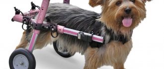
Remember that only a timely visit to a specialist and a competent approach to treatment will help to fully get the dog back on its feet. Otherwise, help may be ineffective and then your pet will only have one sentence - a stroller. Depending on the diagnosis and cause of the disease, the veterinarian may prescribe treatment with medications, massage, certain physical activities, diet, etc.
First aid for a pet
Regardless of the nature of the injury, its extent or signs, it is important to get your pet to the clinic as soon as possible. At the same time, you cannot force him to walk if his motor function is still possible. Pick up the dog or place it in the car and take it to the veterinarian. The specialist must establish the integrity of paw sensitivity, check for pain, the presence of injuries and pathologies. The doctor may also take blood and urine tests for additional information.
If your dog's back legs are giving out, you should restrain him on a strong, hard surface. Any medications, including painkillers, cannot be given. It is important to carefully deliver the dog to the veterinary clinic as quickly as possible without unnecessary shaking.
Treatment. Algorithm of actions in the event of a neurological syndrome caused by IVD herniation
So, the dog developed a neurological syndrome of 1-3 degrees (see Clinical picture). After a neurological examination, corticosteroid hormones (dexamethasone, methylprednisolone, hydrocortisone, etc.), B vitamins and symptomatic treatment (H2-histamine receptor blockers, laxatives, etc.) in therapeutic doses are prescribed. Further, the sequence of actions depends on the dynamics of the increase or decrease in the degree of neurological deficit during treatment with anti-inflammatory drugs:
Scheme 1
Dexamethasone (concentration 4 mg/ml) solution – 0.1 ml/kg (0.4 mg/kg) body weight 2 times a day (first day), then 0.05 ml/kg (0.2 mg/kg) – the next 3-5 days intramuscularly; Mannitol (20% solution) – 5 ml/kg (1g/kg) – 2 times a day intravenously drip for 1-3 days; Ranitidine (tab.) 0.15 g or Zantac 0.025 mg/ml (injectable ranitidine) – ¼ tab. per 10 kg of body weight or 0.7 ml per 10 kg of body weight 2 times a day intramuscularly; Neosmectin (3.76 g packet dissolved in 50 ml of water) - ¼ per 10 kg of body weight 3 times a day orally; Milgamma (2.0 ml) solution – 1.0 ml per 15 kg of body weight 1 time per day 10 injections; For constipation: Glycerin suppositories ½ per 10 kg of body weight 2-4 times a day. It is necessary to empty the bladder: catheterization or squeezing through the abdominal wall at least 3 times a day.
Scheme 2
Methylprednisolone (4 and 16 mg tablets) – 2 mg/kg per day, divided into 2 doses (first day), then 1 mg/kg, divided into 2 doses – the next 3-5 days, orally
In the acute period, with increasing neurological deficit, passing to 3-4-5 degrees:
Methylprednisolone (powder 250 mg + 4 ml solvent) – 5 mg/kg 2 times a day (first, second days), then 2.5 mg/kg for 3-5 days, intramuscularly and intravenously;
Mannitol (20%) – 5 ml/kg (1g/kg) – 2 times a day intravenously drip for 1-2 days; Ranitidine (tab.) 0.15 g or Zantac 0.025 mg/ml (injectable ranitidine) – ¼ tab. per 7 kg of body weight or 0.7 ml per 10 kg of body weight 2 times a day intramuscularly; Neosmectin (3.76 g packet dissolved in 50 ml of water) - ¼ per 10 kg of body weight 3 times a day orally; Milgamma (2.0 ml) solution – 1.0 ml per 15 kg of body weight 1 time per day, 10 injections.
Why do I not recommend using loading doses of corticosteroids?
Because in my clinical practice, quite often, I encountered animals with myelomalacia (irreversible destruction of the spinal cord) after the use of corticosteroids at a rate of 10-30 mg/kg body weight (methylprednisolone and analogs). In addition, from my clinical experience I know that large doses of 10-30 mg/kg cannot solve those problems that effective doses (0.5-5 mg/kg) do not solve.
Glucocorticoids have mineralocorticoid activity: they retain sodium and water in the body by increasing reabsorption in the renal tubules, and stimulate the excretion of potassium. Fluorinated glucocorticoids (triamcinolone, dexamethasone, betamethasone) have virtually no mineralocorticoid activity .
Edema of the spinal cord and brain is better relieved by dexamethasone, therefore, in my opinion, of the two treatment regimens above, it is better to choose the first one (regimen 1).
When prescribing glucocorticoids, it is necessary to take into account their equivalent doses: for the anti-inflammatory effect: 5 mg of prednisolone corresponds to 25 mg of cortisone, 20 mg of hydrocortisone, 4 mg of methylprednisolone, 4 mg of triamcinolone, 0.75 mg of dexamethasone, 0.75 mg of betamethasone.
Symptoms
Usually, owners quickly notice that the animal is not in order. If there was an injury, then the dog most often simply stops getting up on its paws immediately or the next day.
If the disease develops gradually, symptoms may more often manifest themselves in behavior:
- The dog is reluctant or takes a long time to get up after sleep
- A previously active and playful dog ceases to be interested in walks, favorite toys, and outdoor games.
- Change in gait: the dog drags its paws, walks more slowly, or its gait loses its natural harmony and grace
- Sometimes the dog may yelp or whine. This happens when stroking, leaning on a sore paw, or walking.
- The dog is slouching, hunching over
- In severe cases, involuntary urination may additionally occur.
1-2 degree of neurological deficit. The animal can move independently
- in the case of an increase in neurological deficit to grade 3-4-5 within 12-24 hours during therapy, examination (CT, MRI) followed by surgical intervention is recommended;
- If the neurological condition improves within 12-24 hours against the background of anti-inflammatory therapy, we continue to monitor the animal for 3-5-7 days. Then we cancel anti-inflammatory therapy and conduct a neurological examination after 24-48 hours. If pain and neurological deficit reappear, we perform a CT or MRI examination.
Caring for an old dog
Be sure to regularly take your elderly pet to the veterinarian. Do not wait until the emerging disease enters the phase of irreversible changes. Brush your dog's teeth regularly with a special toothpaste for animals. Diseases of the gums and teeth are the scourge of older dogs.
Old animals need frequent bathing and regular brushing. When brushing your pet, feel the skin to detect growths or tumors in time. Check your dog's eyes and ears regularly.
Do not change the long-established way of life of your pet - this causes stress in an elderly animal, which will lead to illness and a shortened life. Let the dog live in its usual place, eat its usual food. An elderly dog needs increased attention and affection from its owners. Physical activity should be feasible for the animal and not cause severe fatigue and severe shortness of breath.
What breeds are at risk?
Some breeds are more likely to lose their paws than others. This is explained by strict selection, forced changes in the skeleton for the sake of working or decorative qualities.
Typically, leg failure occurs in young and adult pets between 3 and 8 years of age, and less commonly in puppies.
Arthrosis or spondylosis makes itself felt in older dogs - at 11 - 14 years old and older.
- dogs with a long body - dachshunds, basset hounds;
- brachycephalics - pugs, bulldogs (English and French), Pekingese, Brabançons, boxers, sharpeis;
- large breeds - St. Bernards, Great Danes, Shepherds, Labradors, Alabais.
Measures to prevent paresis of the hind legs
To prevent paralysis of the limbs, adhere to the following recommendations:
- X-ray examination of breeds that are at risk is carried out once a year;
- periodically give chondoprotectors to puppies and adult dogs of large breeds;
- make sure that dachshunds, basset dogs and large pets do not jump from heights until they are 12 months old;
- up to six months, all puppies are carried in their arms while going down the stairs;
- control loads - both insufficient and excessive are harmful;
- monitor your pet's weight;
- make up a complete diet;
- avoid potentially traumatic situations;
- do not leave the dog lying or sleeping in a draft so that it does not get a cold in the spine.
It is impossible to completely protect a dog from hind leg failure. But these measures will reduce the risk of paralysis significantly.
It is important to carefully select a puppy: during the examination, you need to pay attention to how it moves, runs, and plays.
You need to purchase a baby from a trusted breeder, who will present a pedigree, passports and a medical certificate of the baby and his parents.
How to avoid problems during puppyhood
You can minimize the risk of limb problems in puppies by following some care rules:
- The pet must not stand on its hind legs, sit in an upright position, go up and down stairs, or jump from a height;
- you need to make sure that there is a flat, non-slip surface where the puppy plays;
- the diet should include cottage cheese and other fermented milk products;
- It is necessary to give your pet supplements containing calcium, glucosamine and chondroitin.
Regular walks and moderate physical activity are also necessary.
Features of therapy
The exact treatment depends on the cause of the disease. To eliminate spasmodic conditions, nosh-pa and other antispasmodics are prescribed. If the animal experiences pain, a course of analgesics is prescribed in the form of injections. At the same time, the neurologist can offer a course of B vitamins, which restore the normal functioning of nerve fibers.
In the treatment of paralysis, blocking individual nerves using novocaine injections has a good effect. The affected muscles can be warmed up, and massage treatments are prescribed. Please note: novocaine blockade is an intervention that is carried out only in a clinic under the supervision of an experienced veterinary neurologist!


