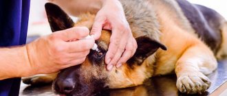
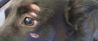
Eye diseases in dogs are among the most common. The condition of the eyes and auxiliary organs is a clear diagnostic sign. Thus, inflammation of the conjunctiva accompanies many infectious diseases.
For diagnosis, examination, special devices and tests are used. But owners can also identify eye diseases in a dog - the presence of discharge, swelling, the dog vigorously rubs its muzzle with its paw, the eye does not open.
Scheme for studying eye diseases in dogs
The ophthalmological examination should be methodical and complete. If you do not complete it completely and do not specifically look for the cause of the disease, you will not be able to identify it.
- Vision and symmetry of the eyes are determined at a distance (including strabismus, which evaluates the condition of the III, IV, VI pairs of cranial nerves).
- The blinking and protective reflex evaluates the state of the II, V, VII pairs of cranial nerves.
- Accessory organs of the eye: examine the eyelids, third eyelid, conjunctiva, eyelashes.
- Turn off the light and use the ophthalmoscope. The pupillary reflex (PR) is checked, and then the cornea and anterior chamber are examined on one and then on the other side.
- Pupillary reflex.
- Cornea: vessels, pigmentation, edema, lipid/calcium deposition, scars, ulcers.
- Anterior chamber: check the anterior chamber for the presence and size of pathological changes. Then the iris is examined. Do not forget to check the aqueous humor for the presence of pathological inclusions.
- The pupils are dilated, and then the lens and fundus of the eye are examined. Initially, according to indications, a Schirmer tear test is performed. The Schirmer tear test should be performed before any drops are administered to the eye. The pupil is not dilated if the animal has glaucoma or signs of lens luxation (aphakic disc or vitreous in the anterior chamber). If glaucoma is suspected, intraocular pressure is measured. In general, if the eye is not painful and the pupil is not impaired, then the pupils can be dilated.
- Other tests: While waiting for pupil dilation, other tests may be performed, such as fluorescein testing, cytology, tear duct catheterization, and obstacle avoidance test.
- Fundus examination: check for opalescence. Then a retinal examination is performed.
Character
German Shepherds are smart, confident, fearless and know the limits of their territory. Any aggression directed towards the owner or other family members will be regarded by them as a signal to attack the offender. The dog will protect the people she loves to the last drop of blood. However, being well developed physically, she has every chance of defeating attackers.
Adults are distinguished by a calm and balanced character and can be trained much faster than representatives of other service breeds. It is no coincidence that they are actively used in the army and police.
At a young age, a dog may show disobedience and an attempt to dominate, but with proper training and moderate methods of physical influence, these qualities can be easily suppressed. The dog recognizes the authority of the owner and will completely obey him.
German Shepherds get along well with children and tolerate childish pranks stoically. However, you should not abuse the patience of animals. Children need to be taught that a dog is a member of the family and should be treated with care.
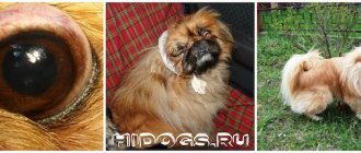
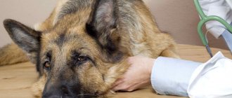
The dog lost his sight
Blindness Caused by Cataracts in a Labrador Retriever
Blind eyes may appear completely healthy; in such cases, additional tests are necessary. Most ophthalmic disorders are not pathognomonic of any etiological agent. Even if the structure causing blindness is identified, additional tests are needed to clarify the ethnology.
The first step in diagnosing possible vision loss is confirmation of blindness. This requires observations of the animal’s movement in the examination room and its overcoming of obstacles. From a distance, you can evaluate the symmetry of the dog’s orbits, vision and the condition of the II, III, IV, VI pairs of cranial nerves. Assessment of visual function includes:
- whether the animal can maneuver around the room or whether it moves cautiously and bumps into objects;
- it looks at the objects it hears;
- vision can be assessed with further examination.
Video - loss of vision in a Chihuahua dog
Functional tests and tests
Throw a cotton ball in front of the dog and see if it follows the ball as it falls. You can test different fields of vision by throwing a cotton ball from behind the animal in different directions. If necessary, night and day vision can be tested by walking the animal through obstacles at the end of the examination.
Obstacle test - the animal is put through obstacles in both a lighted and dark room. If an animal has signs of nyctalopia (night blindness), it may develop progressive retinal atrophy - inherited in an autosomal recessive manner, characterized by an increase in tapetum reflectivity, attenuation of retinal vessels, a decrease in non-tapetal pigmentation, night blindness with progressive day blindness and the formation cataracts.
To avoid air currents, place a plexiglass plate in front of the animal or make vertical movements with your hand. Vision is also assessed by visual reactions and postures. You can place the animal in front of the table and encourage it to move towards the table. If it sees a table, it will approach it and place its forelimbs on the table.
Origin of the breed
The German Shepherd, considered the national symbol of Germany, is one of the most famous and ancient breeds. It is believed that its ancestors were northern wolves. As a separate breed, “Germans” appeared at the end of the 19th century and earned a reputation as excellent livestock guards and detection dogs.
A huge contribution to the breeding of the breed was made by captain Max Emil von Stefanitz, who later became the founder of the German Shepherd Lovers Club. Painstaking selection work began with herding dogs from the Southern and Central regions of Germany, as a result of which an average type was obtained. This was the dog Greif, the first to be included in the pedigree of the breed.
He was distinguished by his quick reaction, carried out commands better than others and showed good results in grazing livestock. However, due to the dirty coat color, the dog did not fit the definition of a national symbol.
The official date of appearance of the first true German Shepherd is considered to be April 1889. The male Horand von Grafat had impressive dimensions, similar to those of a wolf, and an aesthetic appearance, as well as high quality characteristics. In the same year, the dog, which won an exhibition in Germany, was recognized as the progenitor of the breed. Subsequently, as a result of crossing, several lines of German shepherds were born that fully met the breed's qualities.
The Second World War practically nullified all the work of breeders, and the breed was threatened with complete degeneration. Fortunately, von Stephanitz's followers did not allow this to happen. Today, German Shepherds are very popular and are one of the three smartest breeds.
Inflammation of the conjunctiva
Eye discharge, either serous or purulent, indicates irritation (eg, viral, bacterial, injury, etc.). They can also be caused by blocked tear ducts. Please note: purulent discharge is not pathognomonic of a bacterial infection. Any chemotactic stimulus to neutrophils can cause purulent discharge.
Conjunctivitis in a dog
The most common cause of conjunctivitis is mechanical injuries, irritations of various types caused by foreign bodies, acids, alkalis and gases, pyogenic microorganisms that have entered the conjunctival sac, as well as complications of blepharitis and keratitis.
Read more about conjunctivitis in dogs in this article.
Mucus discharge may indicate keratoconjunctivitis sicca (KKS).
Eyelid diseases in dogs
Blepharitis (inflammation of the eyelid)
| Description | Treatment |
| Staphylococci, the most common infectious pathogens, can cause blepharitis, either through direct infection or due to hypersensitivity. In puppies, a hypersensitive reaction most commonly occurs, characterized by multifocal, often large abscesses along the eyelid margin with severe swelling. | Treatment consists of applying warm compresses several times a day and appropriate systemic antibiotic therapy. Concurrent therapy with oral corticosteroids is often necessary, starting with 1.1 mg/kg prednisone 2 times a day for 10-14 days, gradually reducing the dosage. |
Agenesis (underdevelopment) of the eyelids
| Description | Treatment |
| Puppies are born with underdeveloped eyelids, and for this reason their eyes can be open immediately after birth. In most cases, the absence of part of the eyelid is bilateral. If there is a significant lack of eyelid tissue, the function of closing the palpebral fissure and irritation of the cornea are likely. | In case of significant defects, the animal should be protected from exposure to bright light. In case of significant eye defects, surgical treatment (reconstructive blepharoplasty) is performed to reduce the palpebral fissure defect and restore proper eyelash function. |
Inversion, inversion
| Description | Treatment |
| Pay attention to any asymmetry in the length or position of the edges of the eyelids and eyeballs. Volvulus is differentiated from spastic blepharospasm caused by pain. To do this, a local anesthetic and iridocycloplegic are injected into the eye. The blepharospasm will disappear. | A reliable method should be considered surgical intervention, which must be rushed if the eyelashes wrapped inside irritate the cornea and conjunctiva. The most common operation for pathology is to excise folds of skin. |
Chalazion
| Description | Treatment |
| Formation in the thickness of the eyelid, caused by inflammation of the meibomian glands and compaction of the secretory substance in the gland. It is painless and may be visible on the conjunctiva of the eyelid as a yellow lesion. | Curretage can be performed under local anesthesia, then the eyelid is everted and the contents of the chalazion are removed using a chalazion curette or other instrument. In this case, a short course of corticosteroids and antibiotics is indicated. |
Stye on a dog's eye
| Description | Treatment |
| Dogs commonly have a bacterial infection of the meibomian glands. A stye is a focal abscess in the sebaceous gland, usually caused by a staphylococcal infection. | Should be treated with hot compresses and systemic or local antibiotics. Surgical excision/drainage may be required. |
Benign and malignant neoplasms of the eyelids
| Description | Treatment |
| Neoplasms are observed at different ages. They are divided into two main groups: benign and malignant. Benign ones include warts, papillomas, fibromas, skin horns and cysts. Malignant ones include sarcomas, melanosarcoma and carcinoma. | Radiation therapy, repeat cryosurgery or laser surgery, or wide excision, which often requires skin grafting. In the case of multifocal lymphoma, the eyelids may also be affected, in which case appropriate drug treatment is indicated. |
Main symptoms
Inflammation of the cornea of any etiology and degree is characterized by the appearance of general symptoms, united by the concept of corneal syndrome:
- cutting and burning pain in the eye;
- intense lacrimation;
- blepharospasm (an involuntary reaction of closing the eyelids even without the presence of irritants);
- intolerance to bright light;
- sensation of the presence of a foreign body under the eyelid;
- decrease in visual acuity while maintaining the ability to distinguish the outlines and colors of objects, their location in space.
The appearance of these phenomena is due to irritation of the receptors on the cornea by the infiltrate secreted by the tissues in response to damage. It also reduces the transparency of the eye shell. With non-infectious and viral inflammation, the exudate is colorless or grayish, since lymphoid agents predominate in it. Infectious keratitis is accompanied by the release of yellow or greenish exudate. This staining is due to the accumulation of leukocytes and purulent discharge in it.
As the disease progresses, a superficial erosion may form at the site of the inflammatory focus, which, if untreated, transforms into a deep ulcer. Externally, such formations look like a localized defect with a rounded or slightly uneven edge, a gray bottom and an accumulation of exudate in it. After healing, a cloudy spot remains at the site of the ulcer - a thorn.
Important! Along with inflammation of the cornea, 50-70% of patients experience concomitant inflammatory processes: conjunctivitis, scleritis, uveitis and others.
Pathologies of eyelashes
Pathologies of eyelash growth: magnifying devices are used to identify pathologies of eyelash growth.
- Districhiasis: Abnormally positioned eyelashes growing from the meibomian gland duct (eyelid margin). Looks like a double row of eyelashes, where one or more eyelashes grow from the openings of the meibomian glands and are directed towards the eyeball.
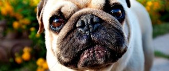
- Trichiasis: A condition where the hair grows normally, but is perpendicular to the eye and touches it (for example, folds on the face). If pigmentation in the medial corner of the eye is detected in brachycephalic breeds of dogs with strongly pronounced folds on the muzzle, one should not think that this is associated with trichiasis; it may be due to lagophthalmos.
- Eyelash ectopia: eyelashes growing from the meibomian gland and perforating the conjunctiva of the eyelid. Slightly invert the eyelid or palpate the conjunctiva of the eyelid to detect ectopic eyelashes.
Districhiasis
Trichiasis
Ectopia
Possible complications
With mild keratitis, traces of infiltration disappear completely after 2-4 weeks from the start of treatment. Complications threaten central and paracentral inflammation of the cornea, which occurs in severe form. These types of diseases most often cause a decrease in the transparency of the cornea due to the formation of scars on it. Pathologies accompanied by an ulcerative process are complicated by the spread of infection to the contents of the eyeball. They, in turn, can result in phlegmon and atrophy of the eye and optic nerve, sepsis and other life-threatening conditions.
Corneal diseases
The surface of the cornea should be smooth and shiny. Using a transilluminator, the cornea is examined for the presence of blood vessels, pigmentation and other opacities.
Possible pathological changes in the cornea in a dog:
- Changed blood vessels are a nonspecific sign of the disease. The surface vessels are long, thin and branched, similar to tree branches. They indicate a superficial pathological process. The deep vessels are short and not branched. They talk about the presence of intraocular pathology (for example, glaucoma, scleritis, anterior uveitis).
- Pigmentation indicates corneal irritation. Its location indicates the source of irritation. Pigmentation is a secondary disease. Its etiological factor is identified.
- Deposits of lipids and calcium in the corneal stroma indicate corneal dystrophy. Steroid use may worsen the disease. It is necessary to differentiate from cicatricial changes.
- Ulcers: for better diagnosis of corneal ulcers, a fluorescein test is performed (more details in the article about Corneal Ulcers in Dogs).
- Corneal precipitates are accumulations of cells attached to the endothelium of the cornea. They may be very small and diffusely distributed, or they may be large, isolated masses called sebaceous precipitates.
- Edema.
- Scarring.
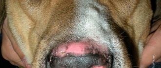
Uveodermatologic syndrome
Uveodermatologic syndrome, also called Vogt-Koyanagi-Harada syndrome-like syndrome (VKH-like syndrome), is considered an autoimmune disease in which melanocytes become target cells. During particularly severe bilateral anterior uveitis, posterior or panuveitis, poliosis and vitiligo of the eyelids and skin around the eyes and mouth are also observed. Skin disorders often cause severe itching, crusting, ulceration, and excoriation.
The surface and mucocutaneous border of the eyelids are affected, this manifests itself in the form of blisters, ulcers, crusts and alopecia. A skin biopsy is taken to confirm the diagnosis, and oral immunosuppressive doses of corticosteroids are often necessary. For relapses, azathioprine may be required.
Although this condition can occur in any breed of dog, Akitainus are the most predisposed.
Initial treatment consists of oral immunosuppressive doses of corticosteroids, then reduce the dosage to the minimum that can control symptoms. This disease is often difficult to treat and requires high doses of steroids, which causes many side effects. The addition of azathioprine (Imuran, Burough-Wellcome) is often necessary. After controlling the condition, both poliosis and vitiligo disappear. Autoimmune dermatoses (pemphigus foliaceus, pemphigus erythematous, systemic lupus erythematosus, discoid lupus erythematosus) may appear on the eyelids and around the eyes even before the appearance of generalized symptoms.
Pathological accumulations
The anterior chamber is the area delimited by the cornea, iris and anterior lens capsule. The anterior chamber and lens should be transparent. When examining the anterior chamber, the cavity is first illuminated from the front. Then they examine from the side for the presence of synechiae, neoplasms on the iris and clouding of the aqueous humor. When examining a dog, you can detect various pathological inclusions in the eye, such as pus, fats, parasites, tumors, and fluid accumulation.
Hypopyon is an accumulation of pus or leukocytes in the anterior chamber. Neutrophils accumulate in response to any chemotactic stimulus and are usually not caused by an infectious process. In cases where there is a deep corneal ulcer, neutrophils migrate from the iris rather than from the cornea.
Hyphema - blood in the anterior chamber
Pet owners may notice hyphema. Because it is a form of anterior uveitis, episcleral injection and anterior chamber hyperemia are often observed in these animals. Hyphema has many causes, the most common being trauma. A single hemorrhage resolves quickly. Recurrent bleeding, which is a consequence of retinal detachment, can lead to secondary glaucoma or synechia. If examination of the retina is not possible due to hyphema, then ultrasound of the eye is used to identify retinal detachment.
Hemorrhage into the chamber of the eye
It is necessary to identify and treat the primary disease, while simultaneously providing local eye treatment, as for anterior uveitis. If bleeding recurs, intraocular pressure should be constantly measured and secondary glaucoma treated.
Cloudiness of aqueous humor
The anterior chamber normally contains very little protein. When the aqueous humor-blood barrier is destroyed, as in anterior uveitis or iritis, protein leaves the uveal vessels into the aqueous humor.
When there is an abnormally large amount of protein in the aqueous humor, the light is partially scattered so that if you shine a narrow beam of light into a dog's eye, you can see the beam passing through the anterior chamber. This phenomenon is called aqueous opacification and indicates anterior uveitis. In order to see cloudiness of the aqueous humor, you must:
- a slit lighting head is placed on the ophthalmoscope and held less than 1 cm from the eye;
- turn off the lights in the room;
- then the anterior chamber is examined.
Normally, a beam is visible passing through the cornea and lens, but not the aqueous humor. When the aqueous humor becomes cloudy, a ray is visible passing through the aqueous humor.
Breed standard
In breeding the German Shepherd breed, the standards developed by Emil von Stephanitz apply. To briefly describe the “Germans”, they are strong dogs of medium height with a powerful and well-developed muscular corset.
The weight of an adult male varies from 35 to 40 kilograms, height (at the withers) - 60-66 centimeters. Bitches are smaller: weight - 25-32 kg, rarely reaching 60 cm at the withers.
Description of the standard:
- The head is wedge-shaped, convex forehead without grooves. Lips are dark, dry, tight-fitting. The bridge of the nose is straight.
- The jaws are well developed, powerful, with a scissor bite.
- The ears are of medium size, straight, erect, with sharp tips, the distance between them is average. Black lobes.
- The eyes are almond-shaped, slightly slanted, and small. The iris is dark.
- The sternum is pronounced, long, moderately wide. A special requirement for the hips: they should not be thick or barrel-shaped.
- The neck is strong, strong, muscular, without a layer of skin and fat.
- Back: Well developed, strong croup, different in length, smoothly blending into the base of the tail.
- The forelimbs are straight, parallel to each other.
- The paws are tightly packed, round in shape, with hard pads and dark claws.
- The tail is long, ending at the level of the hocks, the lower part is covered with hair. When moving, the tail rises slightly, in a calm position it hangs gently and is slightly curved.
The skin fits tightly to the body and does not form folds. Wool has an undercoat and comes in three types:
- long, soft;
- long hard;
- short hard.
The color can vary from pure black to a weakened and darkened zonular.
Reasons for disqualification are deformation of the ears and tail, white coat color, light eyes, broken or drooping ears, excessive nervousness, manifestations of cowardice or aggression.
Iris and pupil
Iris pathologies: look for changes in iris color, edema, vascular injection, residual pupillary membrane, dyscoria (change in pupil shape) or cysts/tumors. Cysts, unlike tumors, transmit a beam of light directed at them.
Pupil dilation – necessary for further diagnosis
For further examination of the pupil, it is necessary to dilate it. If an animal needs a Schirmer tear test (STT), the TTS is performed first. Do not dilate the pupil if the animal has glaucoma or lens subluxation. In general, if the eyes do not cause discomfort and the visual acuity is normal, the animal does not have glaucoma. If there is any doubt, measure the intraocular pressure first!
To dilate the pupil, one or two drops (make sure to inject the same number of drops into both eyes) of 1% tropicamide are injected into the conjunctival sac/cornea of each eye. Typically, maximum pupil dilation occurs within 15-20 minutes. While waiting for the pupil to dilate, additional tests such as fluorescein testing, Schirmer tear test, cytology and culture can be performed.
Iridocyclitis and iritis
These are inflammatory diseases of the anterior vascular tract. Isolated inflammation of the iris is relatively rare. Depending on the nature of the exudate, it can be serous, serous-fibrinous, fibrinous, purulent and hemorrhagic.
Causes:
- penetrating wounds;
- inflammatory processes in the cornea;
- dog distemper;
- infectious hepatitis.
It manifests itself as pain in the eye and soreness of the eyeball on palpation. The iris is always swollen, may be greenish or rusty in color, its pattern is unclear, the pupil is narrowed, and posterior synechiae often occur. With serous iritis and iridocyclitis, clouding of the moisture of the anterior chamber is noted; with fibrinous and purulent iritis, white and white-yellow exudate settles at the bottom of the anterior chamber (hypopiomas).
Fibrin is often deposited on the inner surface of the cornea in the form of precipitates. Intraocular pressure is often reduced. Visual acuity decreases. With hemorrhagic iritis, blood appears in the anterior chamber.
Treatment should be aimed primarily at eliminating the cause of the disease. For this purpose, use a 1% solution of atropine 4-6 times a day or GLP with atropine 1 time a day. The following mixture is administered subconjunctivally once every 3-4 days:
- 1 ml of 0.5% novocaine solution;
- 0.2 ml of 1% atropine solution;
- 0.1-0.2 ml hydrocortisone.
For purulent iridocyclitis, 10-20 thousand units of a broad-spectrum antibiotic are added to the mixture. Prescribe eye ointments with antibiotics, as well as orally - butadiene 0.15 g or reopirin 2.5 g 3 times a day for 10 days, 10% calcium chloride solution one tablespoon 3 times a day, diphenhydramine 0.03-0.05 g 2-3 times a day, multivitamins, sulfonamide drugs.
For hemorrhagic iridocyclitis, subconjunctival injections of fibrinolysin 80-1000 units are used every other day. When treating the consequences of iridocyclitis, tissue preparations are used (vitreous body, FiBS, placenta suspension).
Fundus and retina
Fundus reflection. With the transilluminator located approximately an arm's length away from the animal and the light turned off, direct a beam of light into the animal's eye and look at the reflection of the tapetum. This is the most effective way to detect aphakic crescent and impaired transparency of the intraocular media (for example, incipient cataracts). This is also the best way to differentiate lens nucleus sclerosis from cataracts.
Retinal examination: indirect ophthalmoscopy provides a wider field of view and is the best method for examining patients with fundus pathologies.
- Optic disc
- Vessels: are they pathologically tortuous, is hypertension suspected?
- Retina: Is the retina in the center? If not, the animal may have a retinal detachment.
Retinal disinsertion
It can be partial or complete. It occurs in older animals, as well as in a number of diseases.
- Etiology. It occurs as a result of a decrease in the volume of the vitreous body due to its atrophy, with severe traumatic injuries, a large accumulation of exudate, and hemorrhage into the space between the retina and the choroid.
- Symptoms. The animal's vision suddenly deteriorates or blindness occurs. The pupils are dilated and the reaction to light is slow. Ophthalmoscopy reveals various disturbances in the fundus of the eye and protrusion of the retina in the form of a white bubble.
- Treatment. With complete retinal detachment, treatment is useless. In case of partial detachment, 0.1-0.2 ml of hydrocortisone with novocaine is injected subconjunctivally after 3-4 days; 0.3-0.5 ml of dexazone daily. A 1% solution of atropine and a 2% solution of dionine are instilled into the conjunctival sac.
Our services in ophthalmology
The administration of CELT JSC regularly updates the price list posted on the clinic’s website. However, in order to avoid possible misunderstandings, we ask you to clarify the cost of services by phone: +7
| Service name | Price in rubles |
| Appointment with an ophthalmologist (primary) | 3 900 |
| Comprehensive OCT examination of the retina (one eye) | 3 500 |
| Ultrasound scanning of the anterior segment of the eye | 1 000 |
| Revision of the vitreous cavity | 44 000 — 70 000 |
All services
Make an appointment through the application or by calling +7 +7 We work every day:
- Monday—Friday: 8.00—20.00
- Saturday: 8.00–18.00
- Sunday is a day off
The nearest metro and MCC stations to the clinic:
- Highway of Enthusiasts or Perovo
- Partisan
- Enthusiast Highway
Driving directions