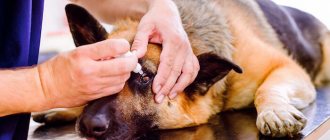
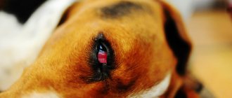
Dog owners sometimes notice that the hair around their pet's eyes begins to thin, fall out, and the skin becomes inflamed. The animal may scratch the sore spot, and the eyes may become watery, festered, swollen, and look very inflamed.
If such signs appear, owners should immediately take their dog to the veterinarian, because this condition can have many causes. Among them there are quite dangerous and unpleasant for the dog.
Causes of the disease
There are many reasons why hair may fall out around the eyes. Some of them are associated with allergic reactions. They can be triggered by the food you eat. In most cases, the cause lies in a food allergen or an excess of proteins in the foods the dog is fed. More often this happens when feeding the animal natural, homemade food.
Loving owners without a second thought put plenty of chicken, fresh cottage cheese, eggs, and liver into their pet’s bowl, not realizing that they can provoke unwanted health problems.
The same thing happens when you wash your dog too often and use too aggressive or “human” cosmetics that are not suitable for the dog. This can be soaps with fragrances, shower gels, shampoos and even perfumes and deodorants.
Animals with hypersensitive skin and coat may react to them. Since the skin around the eyes is the thinnest and most delicate, it is the first to become a “victim of allergies.”
In young dogs, juvenile blepharitis can cause hair loss. With it, the eyes begin to water, ache and become inflamed, suppuration appears, and then hair is lost. Another disease can provoke the same manifestations - meibomitis, or inflammation of the meibomian gland. It is located in the thickness of the eyelid and manifests itself as an internal “stye”.
Old age, metabolic disorders, hormonal problems, the presence of helminths and skin parasites, such as mites, can also provoke baldness.
Neoplasia – tumors of the eyelids of dogs
Most common neoplasms (adenomas, papillomas, melanomas) on the eyelids of dogs are benign and are eliminated using resection, cryosurgery and laser ablation. All removed tissues must be subjected to histopathological examination, since malignant degeneration of neoplasms is possible.
The following malignant eyelid tumors have been noted:
- squamous cell carcinoma;
- basal cell carcinoma;
- adenocarcinoma;
- mast cell tumor;
- malignant melanoma;
- hemangiosarcoma;
- myoblastoma.
The most common tumor is squamous cell carcinoma, which tends to be locally invasive, with frequent relapses and metastases. Their treatment consists of radiation therapy, repeat cryosurgery or laser surgery, or wide excision, which often requires grafting of skin flaps. In the case of multifocal lymphoma, the eyelids may also be affected, in which case appropriate drug treatment is indicated.
Eyelid tumors in dogs
In practice, up to one-fourth of the eyelid is removed using conventional wedge resection, after which only a simple suture is required. However, there are exceptions to the rule, and dogs with longer eyelids or tighter periocular skin will have more tissue removed. To remove this swelling, a complete “pentagonal” resection is performed, and since the upper eyelid is long, an initial suture can be placed.
After the eyelid gap narrows as a result of shortening the upper eyelid, provided the tissue is elastic, they can be compared, and thanks to the gradual stretching of the tissues, by the time the sutures are removed, the eyelids take on a more normal and quite acceptable appearance. Over time, further transformation and restoration of the original form will occur.
Aseptic pyogranuloma
The eyelids may be affected by aseptic granuloma of unknown etiology. This apparent neoplasm may be extensive, multiple and ulcerated. Relapses may occur. On histopathological examination, this disorder appears as diffuse or nodular granulomatous or pyogranulomatous inflammation.
There is no evidence of neoplasia and no etiological factors have been identified. The results of bacterial and fungal cultivation are negative. Most dogs do not respond to systemic antibiotics, but have responded well to immunosuppressive doses of oral corticosteroids.
Which breeds are more susceptible
All breeds suffer from the problem of hair loss, but due to the specific nature of their coat, long-haired breeds are most affected by infections and parasites, especially those with tufts of hair on the face, which can irritate the eyes. Inflammation of the eyes due to this can be so severe that it will lead to loss of hair around them.
More often than others, dog breeds that have fairly large and deep skin folds experience problems with their fur, for example, pugs, sharpeis, boxers, chow-chows, mastiffs and bullmastiffs, bulldogs, and chihuahuas.
Prevention
Regarding preventive measures, several important points can be highlighted.
It is necessary to regularly clean the area where the dog lives.
- The animal must be kept clean .
- Regularly sanitize the dog's premises, sleeping area or kennel.
- Monitor the condition of the coat.
- Avoid places where there may be insects, prevent questionable contacts with other animals.
- Timely vaccination and deworming.
- Eliminate access to paint products or household chemicals.
- Under no circumstances should you self-medicate .
- All prescriptions must be prescribed by a doctor.
Main symptoms
Hair loss in the eye area can begin with inflammation of the eyelids themselves, and then spread to the surrounding tissues. Manifestations depend on the cause of the disease, but the main symptoms are as follows:
- The dog is worried, scratching its eyes and muzzle due to itching.
- The skin around the eyes becomes inflamed, red and swollen.
- Mucus, pus and tears are released from the eyes.
- The coat first thins, then becomes bald, becomes covered with ulcers, and can fester and form very painful wounds.
- The animal loses its appetite, becomes apathetic, and when trying to clean the wounds, it can even bite a person, including its owner.
In addition to the above symptoms, a number of diseases may include increased body temperature, hair loss in other parts of the body, severe itching, dry skin, dandruff, and inflammatory processes.
Entropion of the eyelids in dogs
Entropion of the eyelids, entropium palpebrarum, is the turning of the edge of the eyelid or part of it inwards towards the eyeball. When turning the eyelids, the free edge of both eyelids is turned inward, towards the eyeball, along with the eyelashes. Usually the disease covers the entire part of the eyelid, and the degree of inversion varies. As a result, conjunctivitis, keratitis, and corneal ulcers develop. Entropion occurs in one eye or both eyes, including the lower and upper eyelids.
Causes and signs of entropion
The causes of entropion of the eyelids are different: due to blepharitis, keratitis, follicular conjunctivitis. Often, entropion appears after removal of the third eyelid.
What dog breeds are predisposed to entropion:
- Chow chow
- Norwegian Elkhund
- Chinese Shar Pei
- Saint Bernard
- English Springer Spaniel
- English and American Cocker Spaniel
- English bulldog
- Rottweiler
- Labrador Retriever
- Golden Retriever
- Toy and miniature poodles
- Mastiffs
Although it can be said that entropion and ectropion are hereditary in nature, the position of the eyelid depends on many factors. The relationship between the orbit, eyelids, and eyeballs influences the position of the eyelids, and the complexity of this relationship is difficult to determine genetically. Of course, the reason is genetic, but other factors influence the position of the eyelids. For example, atrophy of ocular fat or musculature leads to enophthalmos, predisposing to entropion.
Blepharitis is a common cause of entropion and ectropion of the eyelids.
Damage, acute or chronic inflammation, can lead to scarring or blepharospasm, which also causes eyelid abnormalities. Thus, in each case, the doctor must carefully examine the structure of the eyelids, eyes, orbits and evaluate other factors. If eyelid spasm is present in combination with eye tenderness, local anesthesia is administered to assess the exact extent of ectropion in the absence of pain.
In some cases, the discomfort is so severe that local anesthesia does not help relieve blepharospasm. In this case, a local anesthetic can be administered to block the innervation of the eyelids to relieve blepharospasm. In large and giant breeds, excess skin and eyelids combined with a lack of skin tone predisposes to ectropinone. It can often be complicated by entropion, especially in the presence of a large palpebral fissure and elongated eyelid margins. Manipulation of the eyelids usually allows the doctor to assess the degree of correction needed to remove excess skin and eyelid margins.
The main features are:
- photophobia;
- lacrimation;
- conjunctivitis;
- incorrect position of the edge of the eyelid and eyelashes;
- the palpebral fissure is narrowed.
Eyelashes and eyelid hair irritate mainly the conjunctiva and cornea, resulting in eyelid spasm. keratitis, up to corneal ulcers. Because canine eyelids do not have a tarsal plate (related to the cartilage of the eyelid), contact with the eyeball is extremely important for eyelid support.
As animals age, atrophy of orbital fat and other contents can lead to significant enophthalmos, which causes entropion of the eyelids. In this case, entropion (entropion of the eyelid) may form, which is difficult to eliminate, since deprivation of support from the eyeball leads to the mixing of a sufficient amount of tissue, which subsequently causes a relapse of entropion. Any eye disease that results in atrophy or scarring of the orbital structures can cause enophthalmos, similar to what occurs with aging.
The upper and lower eyelids are musculocutaneous folds in the orbital area. On both eyelids, there is a base, two surfaces and free edges, between which there is a palpebral fissure. The outer surface of the eyelids is covered with thin, folded skin.
The inner surface of the eyelids is covered with a mucous membrane - the conjunctiva, which extends to the eyeball. The thickness of the eyelids is up to 4 mm. Blood supply is carried out by the branches of the facial, lacrimal, frontal, buccal and other arteries. These branches move towards each other in loose connective tissue and, merging, form arterial arches. Innervation is carried out by branches of the ophthalmic nerve.
Operations for entropion of the eyelids
The main treatment method is surgical cosmetic surgery. The operation should be performed as early as possible to avoid the development of persistent and even incurable changes on the cornea (keratitis and ulcers). Most operations for entropion of the eyelids involve cutting out a flap from the skin of the affected eyelid near the damaged edge of the upper or lower eyelid. As a result of fusion of the edges of the wound and subsequent scarring, the edge of the eyelids is pulled outward: both it and the eyelashes return to their normal position, and irritation of the eyeball stops.
Indications: partial or complete inversion of the eyelids. As a result of the rolling of the eyelid, keratitis and ulcers develop, the cornea is perforated and the anterior chamber of the eye is opened.
Conventional soft tissue instruments are suitable for blepharoplasty, but the kit also includes certain necessary ophthalmic instruments.
- Scissors for cutting tissue for strabismus and tonotomy.
- For eyelid manipulation, fine-toothed forceps such as Bishop-Harmon forceps or 0.3 mm Castroviejo scissors are best.
- For manipulation of the conjunctiva, smaller serrated forceps are required, such as Colibri forceps or 0.12 mm Castroviejo forceps.
- Scalpel blades must be small (Bard-Parker No. 11 and 15 or Beaver No. 64 and 65), and such blades require suitable handles.
- An eyelid speculum of appropriate size and rigidity is required that is comfortable and dependent on the surgeon's preference.
- The Barraxra Wire Eyelid Speculum is suitable for small, thin eyelids, but large palpebral fissures require a larger, more rigid eyelid speculum.
- When using fine needles and threads in eye surgery, an ophthalmic needle holder, such as a Derf or large Castroviejo needle holder, is required.
- The Jaeger eyelid plate is used for eyelid incisions. although if it is not available, you can use a sterile spatula.
- Special tweezers, such as entropion and chalazion tweezers. necessary for immobilization and stabilization of the eyelids during procedures.
Anesthesia. Combined use of neuroleptic substances with conduction anesthesia of the orbital nerve. The use of infiltration anesthesia is undesirable, since with it it is impossible to accurately determine the size of the excised skin flap.
Dogs are fixed in a lateral position on the operating table, ensuring head immobility. The surgical field (be careful not to get the solution on the conjunctiva) is wiped with iodized alcohol.
Most of the surgical techniques proposed for entropion of the eyelids come down to excision from the skin of the affected eyelid of a flap that is round according to Froehner, oblong-oval according to Frick, or arrow-shaped according to Schleich and connecting the edges of the wound with a knotted suture. The shape of the removed skin flap and the location of its excision depend on the extent and location of the lesion.
First, determine the location, length and width of the excised flap of skin. With complete inversion of the entire eyelid, a longitudinal oval flap is cut out along its entire length parallel to the edge of the eyelid. With partial inversion, the length of the skin flap (rounded) should exceed the length of the inverted part by 2-5 mm. The width of the excised flap is determined very carefully to avoid eversion. Grabbing skin folds of varying widths with anatomical tweezers, find the width at which the edge of the eyelids assumes a normal position.
Technique of operation. Using surgical tweezers, grab the skin fold and use a scalpel or scissors, retreating from the edge of the eyelids by 2-4 mm, and excise a skin flap of the required size. The peli flap is excised far from the edge of the eyelid; relapses may occur. Tamponade stops the bleeding and knotted sutures are placed on the edges of the wound at a distance of 1 cm from each other.
When the inversion of the eyelid is insignificant, you can limit yourself to stitching the fold of skin with an interrupted suture, without resorting to excision.
With simultaneous inversion of the upper and lower eyelids, the operation is performed in two ways:
- if inversion of both eyelids occurs along the entire length, a skin flap is excised in each eyelid, each wound is closed with sutures;
- if the upper and lower sections of the inversion are located near the outer corner of the eye, retreating 3-5 mm from the outer canthus, an arrow-shaped area of skin is excised against the corner of the eyelids, the resulting skin defect is stitched with an interrupted suture, starting from its central part.
In advanced cases, with severe degrees of volvulus, in addition to excision of the skin flap, it is recommended to simultaneously cut the outer corner of the eye with a small incision (3-5 mm long) and sew the conjunctiva to the skin with thin silk. After the operation, to prevent scratching, a protective collar made of thick cardboard, plywood or a plastic bucket is placed on the dog’s neck. Sutures for all types of operations are removed on the 8th day.
Diagnostics in a veterinary clinic
After the animal is admitted to the hospital, the veterinarian will examine it and take tests to determine the true cause of the disease. If conjunctivitis, blepharitis or meibomitis is suspected, an eye smear is taken to confirm or deny the presence of a bacterial infection.
The same may be required if you suspect a skin infection - fungal or bacterial, the presence of mites or helminths.
Incomplete closure of eyelids
Lagophthalmos, caused by temporary swelling of the eye or orbit, is eliminated by performing temporary tarsorrhaphy using 1-3 non-absorbable mattress sutures passing through the edges of the eyelids at the level of the gray line. If the eyelid tension is slight, sutures can be applied without stents. If lagophthalmos is caused by a chronic disease (for example, enlargement of the eyelid fissure, conformational exophthalmos, eyelid paralysis), plastic surgery of the canthus is performed either laterally or medially, regardless of the most vulnerable area of the eyeball and cornea, the placement of which will best contribute to the preservation of eyelid function.
Treatment method and prognosis
Treatment methods depend entirely on the cause of the disease. Only after identifying it will the veterinarian prescribe adequate treatment, for example, taking antibiotics for infection, antifungal ointments for the presence of fungus, and anti-tick medications for identifying skin pests.
Allergic reactions to external influences are treated by isolating the dog from their influence; those that appear during food intake are treated with a diet excluding allergenic foods.
The sooner the owner reacts to the symptoms of a pet’s illness, the faster it will be possible to get rid of it without severe harm to health.
Conjunctivitis
The first and most logical suspicion for reddened eyelids is the onset of conjunctivitis. The disease is quite harmless if treated. The dog's eyes will itch, and you will notice that the pet is rubbing its muzzle with its paws. The eyelids stick together after sleep, and around the eyes there is a clear, yellow or yellow-green discharge, the mucous membrane is swollen and red. At the initial stage, treatment is carried out at home:
- We rinse the eyes with warm, clean water or herbal infusions if we are sure that the dog does not have allergies. A clean piece of gauze or sponge is used for each eye.
- Levomycetin drops are instilled into each eye (3–6 times a day) or Tetracycline ointment is applied (2–3 times a day).
- Treatment lasts 7–10 days. After complete disappearance of symptoms, therapy is continued for 2–3 days.
Purulent inflammation of the eyes in dogs, which spreads to the nasal passages and ears, is the next, advanced form of the disease, which requires consultation with a veterinarian and “aggressive” treatment with antibiotics. There is also a little-known form of the disease - follicular conjunctivitis. Inflammation of the mucous membrane of the eye looks like overripe raspberries, and other symptoms are similar. The difference is that the follicular form is most often chronic, worsens from time to time and can only be stopped in a clinical setting by cauterization.
Important! Inflammation of the eyelids is a consequence of a silent illness or neoplasm, most often observed in older dogs. The disease looks like the initial stage of conjunctivitis, but does not go away with treatment.
What to do at home
At home, the dog needs to be provided with peace, proper balanced nutrition, taking into account the need to limit protein foods, cleanliness and care for inflamed areas of the body.
If the dog tries to scratch the sore spot, you will have to put on an “Elizabethan collar”. The cover on his bed or bedding will have to be washed and ironed frequently with a hot iron, especially if mites or an acute inflammatory process are detected.
The animal must be protected from drafts and dampness.
Viral infection
Perhaps the most dangerous case, where eye inflammation is only the first and most harmless alarm signal. Just because your dog is vaccinated does not mean he is completely safe. We develop weak immunity to some viruses. Common viral infections that cause eye inflammation include:
- Canine viral hepatitis or Adenovirus - affects the internal walls of the vessels lining the entire circulatory system of the animal. The pancreas and thyroid gland, lungs, spleen, kidneys, but primarily the liver and eyes are affected. The dog survives only with a simple and late form of hepatitis, however, complications cannot be avoided.
- Canine distemper is an acute infectious disease that affects young dogs aged 3 to 12 months. Depending on the location, there are pneumonic, intestinal, cutaneous, nervous and acute forms of canine distemper. With timely diagnosis and treatment, there is a chance of survival. The disease is considered fatal, so vaccination is recommended for all dogs.
- Mycoplasmosis is a viral infection that massively affects the entire dog’s body. Even if recovered, the pet may become infertile, suffer from arthritis, or have recurring genitourinary infections. In its acute form, the disease is complicated by pneumonia, colitis and infectious diseases of the genitourinary system.
There are many dog diseases that owners may not be aware of until the disease develops and manifests itself as serious symptoms. Such diseases include most pathologies affecting the animal’s organs of vision. What eye diseases in dogs should you pay attention to in order to find out as early as possible that the dog is not healthy? After all, early diagnosis allows you to treat your pet in a timely manner before complications arise.
Possible complications
If you miss the moment and start the disease, it can affect the entire coat, and the dog can become completely bald. This is not only unsightly, but can also cause many other health problems.
Extensive lesions negatively affect the level of immunity, and at this moment the dog can easily “pick up” any infection. The most dangerous consequences are the following:
- Extensive skin infections - pyoderma, streptoderma.
- General blood poisoning, infection in the lymphatic system.
- Severe eye infections with risk of visual impairment and blindness.
- Lung lesions, pneumonia.
Ticks can become so widespread on the skin that removing them will require a lot of time and effort. Hormonal problems will affect not only the coat, but also the functioning of all internal organs.
Fusion of eyelids
In animals, congenital and acquired eyelid fusions occur. Often noted are symblepharon - fusion of the eyelids with the eyeball and ankyloblepharon - fusion of the upper and lower eyelids. It should be remembered that dogs are born with fused edges of the eyelids (the first 11-12 days after birth). Therefore, acquired fusion of the eyelid margins poses a danger to the animal.
Eyelid fusion is treated surgically by making an incision along the very edge of the eyelid. The freed edges are cauterized with a 2% lapis solution and lubricated with tetracycline ointment to prevent re-union.
Features of caring for animals with acanthosis
A sick animal requires special care, which consists of measures to prevent secondary infection and boost immunity.
- You should not expose your dog to stress; it is recommended to avoid long trips and changes in place of residence during treatment.
- Work with a specialist to develop a good diet that will take into account the characteristics of your dog, its age, and the presence of chronic diseases.
- Do not exclude physical activity; training should be carried out under the guidance of a specialist. These rules are especially important for individuals who are obese.
- Be sure to give vitamins that stimulate the immune system.
- Keep the dog's bedding and sleeping area clean; it needs to be periodically treated for parasites and washed.
The dog's bedding should be clean.
- After going outside, check the animal's fur to make sure there are no ticks on it. Bites, especially on damaged skin, reduce its defenses.
- Do not allow your pet to stay in the open sun for a long time, as ultraviolet radiation increases the production of melanin several times.
- If a pet with acanthosis has wounds, if possible and necessary, cover them with sterile gauze and promptly treat them.
- Consult your doctor about the possibility of antiparasitic treatment; it will strengthen the body’s protective functions, but only with the right drug and dose.
- If there are large growths and severe thickening of the skin with many folds, you should avoid injuring these places and look through the space between the folds so that dirt does not accumulate in them.
Attention! If the cause of acanthosis was associated with a stressful situation, it is recommended that such individuals take a course of sedatives when changing their usual environment, getting scared, or moving. It is better to determine their dose and type with your veterinarian.
Proper nutrition is the key to a healthy dog
Signals of illnesses
Additional symptoms
The following signals indicate various disturbances in the functioning of the body and diseases:
- Tears from the eyes signal diseases such as distemper (in this case, the tears are not transparent, but yellow-green, due to the pus they contain), entropion of the third eyelid, inflammatory process in the ocular gland, conjunctivitis. But also, ordinary sweets in the diet can cause tears.
- Redness around the eyes may indicate an infection or poor circulation in the visual area. If swelling is observed, it means that fluid exchange in this place is impaired. The cause may be a common allergy.
- The formation of a bubble indicates good functioning of the immune system. When it enters the body, a particularly dangerous infection is surrounded by antibodies, which subsequently form a film that prevents microorganisms from spreading further.
- Sores on the skin indicate the presence of internal irritants. The pet scratches the irritated areas, damaging the skin.
Diseases
A short list of diagnoses that may cause hair loss around the eyes:
- Blepharitis is an inflammation of the eyelids caused by allergies, vitamin deficiencies, diabetes or certain parasites (hypodermic mite, encephalitis mite). Other symptoms also include itching, redness of the eyeball, and mucus discharge.
- Meibomitis is an inflammation of the cartilage tissue of the eyelids. Similar to barley, which is localized internally. It looks like a swollen red or white bubble.
- Ringworm is a viral disease of origin. It is easy to notice, since a pink or red crust forms at the site of hair loss; the areas themselves are round in shape.
- Parasites such as mites and saprophytes.
The latter cause damage to the tissues in the epidermal layer, the hair follicles are destroyed and fall out. If a pet loses hair only around the eyes, this indicates that the local immune system in that area is weakened by some disease.
Worms, on the other hand, act on a different principle - they absorb some of the nutrients from food, as a result of which your pet does not receive enough of them. Thus, even with a balanced diet, there may be a deficiency of vitamins and microelements.
The presence of mites can be noticed upon examination, or if scabies is observed. It is easier to identify worms - after eating, the dog “rides on its fifth point”, trying to get rid of the itching. Also, dogs infected with worms lose weight.
Analyzes
The doctor at the hospital may prescribe:
- stool and urine analysis;
- scraping of the rectal mucosa;
- saliva analysis;
- blood analysis;
- biopsy of the hair follicle, epithelium and epidermis;
- a sample of a substance (pus, mucus) filling the swollen areas.
Disease or genetics?
If a puppy has sparse fur with gaps and bald spots since childhood, then most likely this is a feature of the body structure. But this may also indicate congenital pathologies and a lack of nutritional components, such as:
- calcium, of which half each hair consists;
- protein, which is also the building material for all cells in our body;
- vitamin C, which is important for the functioning of the immune system.
Other causes of hair loss or congenital hair loss can be:
- Disruption of neural communication between the lymphatic system, central nervous system, and brain. The protective barrier decides that the environment is safe and does not need so much hair for protection, and resources can be used for other needs.
- Damage to the skin (burns, chemical injuries, etc.). In this case, the offspring will also be born healthy.
- Dislocation distance too large. If a dog was born in one climatic conditions and was transported to another place, its body will change, adapting to environmental conditions. This is called adaptation.
REFERENCE! The offspring born to such a dog will also undergo adaptation and go through the same changes. For the cubs to be born already adapted, a very long time must pass.
Why is this happening?
Age characteristics
Hair is an element of the immune system that prevents infection from entering the body through the skin. It is a sensor that sends a signal to the skin when a harmful substance comes into contact with it. Immune cells in the epithelium prepare to fight foreign organisms if they do enter the body.
REFERENCE! Some mestizos have hair that does not grow where it is genetically intended. For example, the mother was short-haired, and the father was long-haired.
As a result of a genetic lottery, the puppy received abundant hair growth in the nose area, which will certainly interfere with his ability to eat, smell, tickle the membrane, causing sneezing and discomfort.
In this case, growth is due to genetics, and not pathologies or diseases, and should not be a cause for concern.
Seasonality
All cells of living organisms are regularly renewed, including hair cells. Every day, hair loses some of its cells, which are replaced by new ones. Several times a year, a process of general replacement of outdated hair occurs - shedding.
This usually happens in the summer to make it easier to regulate body temperature and avoid overheating, but some long-haired breeds shed even in the fall.
