Dogs, like other animals, often suffer from eye diseases. By the condition of the eyes you can always determine whether your dog is healthy or not; the eyes are a “mirror” not only of the soul, but also of the health of the animal. In medicine, the eyes are used to diagnose existing diseases in a person. In medicine, as one of the auxiliary diagnostic methods, there is iridodiagnosis - the diagnosis of diseases in a person using the iris of the eyes. When conducting iridology, special equipment and computer programs are used. When making a diagnosis, doctors take into account changes in the structural state, shape of the color areas of the eye, as well as the mobility of the iris.
Before talking about eye disease and its auxiliary organs, it is necessary to have a general understanding of its structure.
A dog's eyes are located in the orbits - bony sockets formed by the bones of the skull, where they are held by several muscles that ensure their mobility and orientation in different directions.
The dog's eye itself is protected by auxiliary organs - eyelids and glands. The dog has three eyelids. The upper and lower eyelids are folds of skin, the inner surface of the eyelids is lined with mucous membrane. The outside of the eyelids is bordered by eyelashes, which protect the eyes from dust and other foreign particles. A dog's third eyelid is a simple film in the inner corner of the eye that dog owners usually cannot see. This film covers the eye when it is closed or irritated, as well as during nervous disorders.
The eye in the cornea area comes into contact with the external dry environment, so it needs protection of the lacrimal glands, which produce tear fluid - a secret that moisturizes the surface of the cornea. A dog's tears accumulate in the space between the eyelids and the eye and are then drained through a narrow channel that begins at the inner corner of the eye and opens into the nasal cavity. When there is excessive lacrimation or blockage of the tear duct, tears flow from the eyes and, when oxidized, form red stripes on the fur that look like blood.
The eye consists of two parts.
- The anterior portion includes the cornea, iris, and lens. They absorb beams of light from the dog, like a camera lens. The cornea and lens are clear and act like optical lenses, and the iris acts as a diaphragm, regulating the amount of light entering the eye through the pupil (the hole in the iris).
- The back of the eye consists of the vitreous body, the choroid (choroid) and the retina, which converts optical light signals into nerve impulses that are transmitted to the visual center of the brain.
Thinking of the eye as an analogy to a camera, the back of the eye is like a photographic film on which the dog's brain captures the image.
Experts, depending on the cause, divide all eye diseases in dogs into 3 types:
- Infectious – occurring in dogs due to the presence of viral or bacterial diseases, most often as a complication of the underlying disease.
- Non-infectious - due to certain mechanical damage, inflammation as a result of improper eyelash growth, neoplasms, eversion of the eyelids.
- Congenital – include eversion, entropion of the eyelids, deformations of the eyes and lens. Congenital ones are most often found in some dog breeds (Shar Peis).
Distichnaz
With this disease, single or multiple hairs appear in a row on the free edge of the eyelid, which should be hairless.
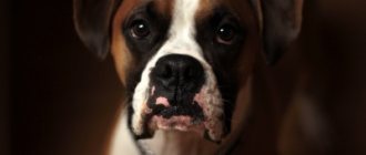
These hairs appear in a dog only at the 4-6th month of life and can be either very delicate or quite hard. With this disease, several hairs most often grow from one point. This disease is most often recorded in English and American cocker spaniels, boxers, Tibetan terriers, collies, and Pekingese.
Clinical picture. During a clinical examination of a dog, a veterinarian notes profuse lacrimation, constant blinking, blepharospasm, irritating hairs have contact with the cornea of the eye. If a dog has curled eyelashes, keratitis is diagnosed.
The diagnosis of the disease is made based on the above symptoms.
Differential diagnosis. Distichnasis is differentiated from trichiasis, entropion and eversion of the eyelids, allergic conjunctivitis, and keratoconjunctivitis sicca.
Treatment. It is carried out in veterinary clinics by electrolysis under an operating microscope. Excision of the third eyelid.
Trichiasis
Trichiasis is a condition when hair from a dog's eyelids or muzzle gets into the eye, coming into contact with the conjunctiva and cornea. Trichiasis can be primary or secondary. Primary occurs in dogs with medial inversion of the eyelids and a large nasolabial fold. Trichiasis occurs in the following dog breeds: Pekingese, Pugs, English Bulldogs, English Cocker Spaniels, Chow Chows, Shar-Peis.
Clinical picture. During a clinical examination of a dog, a veterinarian notes lacrimation, hairs in contact with the cornea cause blinking in dogs, constant discharge from the eyes, symptoms of keratoconjunctivitis, inflammation of the skin in the area of the nasolabial fold.
The diagnosis is made based on the detection of hair in contact with the cornea, provided there is no other eye pathology.
Differential diagnosis. Trichiasis is differentiated from keratoconjunctivitis sicca, entropion and eversion of the eyelids, dystrichiasis, and ectopic eyelashes.
Treatment. Treatment of the disease is surgical. Temporary improvement can be achieved by trimming the hair that gets into the eye.
Treatment of pigmentary keratitis in pugs:
We diagnose corneal melanosis at the earliest stages and carry out comprehensive work with melanosis of any degree.
- First of all, the root cause is treated: immunomodulators for pannus (often occurs in shepherd dogs and their mixed breeds).
- blepharoplasty for eyelid pathologies (relevant for pugs and Pekingese).
- plastic surgery of the nasal fold (relevant for Pekingese).
- replacement of tears during the dry process (relevant for Shih Tzu, pugs, Pekingese, English bulldogs, spaniels and other breeds predisposed to dry eye syndrome). When the root cause of keratitis is eliminated, the pigment growth process slows down or stops altogether, but the existing pigment can remain on the cornea in the same volume.
If you find signs of corneal melanosis in your pet, you should not delay a visit to a veterinary ophthalmologist, because any disease is better treated in the early stages.
Your Pet's ophthalmologist, microsurgeon Yastrebov O.V.
Join us on social networks
Entropion of the eyelids
Entropion is a pathology of the eye in which part of the organ turns inward towards the eyeball. A dog's eyelid inversion can be either upper or lower, one-sided or two-sided.
Unilateral inversion of the eyelid margin is most often the result of heredity and appears in a dog in the first year of life. Congenital entropion occurs in puppies after the eyes open in some breeds with excessively folded skin on the head (chow chow, shar pei).
In this disease, the eyelashes, hair and skin of the eyelid rub against the surface of the cornea, causing inflammation and irritation.
Clinical picture. During a clinical examination, the veterinarian notes the leakage of liquid secretion from the eye, the dog has photophobia (to an electric light bulb, the sun), the dog rubs its eyes with its paw, blinking, and there may be an eye tic.
Treatment. Treatment of entropion of the eyelids is surgical.
How to choose a healthy blue-eyed pet
It is almost impossible to determine whether a puppy will be healthy if its eyes are blue. If in doubt, you can opt for a husky. Sometimes, blue color can be inherited as an independent gene, which will not affect health.
Blue eye color is a beautiful feature that attracts many. Not only huskies become owners of this eye shade. Many other breeds can also boast such representatives. It is worth understanding that dogs with ordinary iris shades are no less loyal and friendly!
Eversion of the eyelids
With eversion of the eyelids, the edge of the eyelid turns outward, while the mucous membrane (conjunctiva) of the eyelid is exposed.
This pathology occurs in dogs with too large palpebral fissure and excess, easily removable skin in the head area.
Cause. Mechanical eversion of the eyelids in a dog occurs as a result of pathological changes in the eyelid itself, as well as tissue scarring after injuries or surgery.
Paralytic ectropion occurs in dogs as a result of facial paralysis.
Clinical picture. During a clinical examination, the veterinarian notes incomplete closure of the eyelids, discharge from the eyes, and inflammation of the conjunctiva.
Treatment. Treatment for this pathology should be aimed at eliminating the cause that caused and maintains the ectropion of the eyelids (removal of a neoplasm, conjunctivitis, facial paralysis, surgical removal).
Blepharitis
Blepharitis is inflammation of the eyelids.
Cause. Unilateral blepharitis in a dog occurs due to injury and local infection. Bilateral blepharitis occurs as a result of allergies, including food allergies in animals, staphylococcal infections in dogs, demodicosis (treatment and prevention of demodicosis in dogs), mycoses and systemic diseases.
Clinical picture. During a clinical examination, the veterinarian notes redness, swelling, itching, scaling, loss of eyelashes and hair, erosion and ulcers in the eyelid area of a sick dog.
Treatment. In the case where the cause of blepharitis is an allergy, dog owners should exclude its contact with the allergen and use antihypertensive drugs (diazolin, suprastin, diphenhydramine, tavegil) in treatment. For staphylococcal infections - antibiotics. For demodicosis, anti-mite drugs.
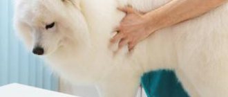
Diagnostics
When diagnosing your pet’s condition, the doctor begins with an external assessment of the condition and a conversation with you. In other words, from the medical history, you will greatly help your pet if you can give the veterinarian as much information as possible.
- What does the dog eat?
- How often does he walk, is there any contact with other animals?
- When did the first signs of the disease appear?
- Was the animal sick before?
And most importantly, never hide from your doctor if you have already tried to treat the disease with something; these details are very important for making the correct diagnosis, and therefore for effective treatment.
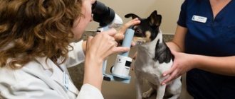
Testing the dog's vision and a thorough examination of the condition of the eye must be carried out under very good lighting.
A keratoscopy is performed to determine the condition of the cornea, and an ophthalmoscope is performed to study the permeability of light. If necessary, a swab is taken from the cornea to detect infections or fungi.
Exophthalmos (protrusion of the eyeball)
Exophthalmos in dogs can be species-specific and is typical for dogs of brachycephalic breeds, with a normal eyeball size, a flat orbit and an overly large palpebral fissure.
Acquired exophthalmos - in which a normal-sized eyeball moves forward due to space-demanding processes in the orbit or its immediate surroundings, or due to an increase in the size of the eyeball as a result of glaucoma in a dog.
Clinical picture. During a clinical examination, the veterinarian notes that the dog has strabismus, an abnormally wide palpebral fissure with protrusion of the eyeball; in some dogs, prolapse of the third eyelid is possible.
Treatment is only surgical.
Convergent strabismus
Convergent strabismus is a noticeable visual deviation from the normal position and joint movement of both eyes of a dog.
Moreover, with paralytic strabismus, the dog's squinting eye does not repeat the movement of the fixed eye.
Cause. Traumatic eye injuries, hypertrophic processes in the orbit (tumors), damage to the central nervous system.
One of the reasons may be congenital underdevelopment of the periorbital muscles, congenital hydrocephalus.
Treatment. Treatment of convergent strabismus involves treating the underlying disease that led to the strabismus.
Corneal melanosis.
Corneal melanosis, which is also called pigmentary keratitis (pigmentous keratitis), is one of the common ophthalmological pathologies of brachycephalics. The essence of the pathology is that with chronic irritation of the cornea (keratitis), melanin cells are introduced into its epithelium and the surface layers of the corneal stroma, migrating from the limbus zone. It is worth noting that pigmentary keratitis is a very general diagnosis. The thing is that the cause of this condition is chronic inflammation of the cornea (keratitis). And the main goals of treatment will be to eliminate the causes that led to keratitis.
Allergic conjunctivitis
Allergic conjunctivitis in dogs occurs as a result of contact with the mucous membrane of the eye of one or another allergen (contact allergy). The allergen can be pollen from flowering plants, dust, etc.
Allergic conjunctivitis in dogs In recent years, allergies to certain food products (food allergy) have often been recorded.
Clinical picture. During a clinical examination, a veterinarian notes in such a dog redness of the mucous membrane of the eyes, mucous discharge from the palpebral fissure. As a result of itching, the dog rubs its paw on the affected eye.
Treatment. If contact dermatitis occurs, it is necessary to rinse the eye affected by inflammation with saline solution or chamomile decoction.
In case of food allergies, it is necessary to exclude the allergic product from the dog’s diet and transfer the dog to a hypoallergenic diet (buckwheat, rice, beef).
The sick dog is prescribed antihistamines (cetirizine, diazolin, suprastin, diphenhydramine, tavegil), and Diamond Eyes eye drops are instilled into the conjunctival sac.
Purulent conjunctivitis
Purulent conjunctivitis in a dog develops due to the entry of various pathogenic microorganisms into the conjunctiva. Purulent conjunctivitis is one of the symptoms of carnivore plague......
Clinical picture. During a clinical examination, a veterinarian notes reddening of the conjunctiva, its swelling, and purulent discharge from the eye of a sick dog.
Treatment. With this form of conjunctivitis, a sick dog is treated with eye drops and ointments that contain antibiotics. Tetracycline eye ointment and Ciprovet drops are widely used. Before applying eye drops and eye ointment, it is necessary to clean the affected eyes of exudate.
Progressive retinal atrophy
Progressive retinal atrophy is an inherited disease characterized by multiple retinal abnormalities that causes blindness in dogs. The first sign is often blurred vision at night, but dogs often behave normally until their vision is almost completely lost. There is currently no cure for the disease. . The level of vision damage varies between breeds and with different progression of the disease, but most dogs eventually become completely blind. Sick dogs should be protected from mating or sterilized, since their offspring will certainly inherit the defect - puppies will be born either predisposed to the disease or carriers.
Follicular conjunctivitis
This form of conjunctivitis is most typical of chronic conjunctivitis and often develops in a dog when toxic substances get into the eye.
Clinical picture. During a clinical examination, a veterinarian reveals many bubbles with transparent contents on the mucous membrane of the conjunctiva. Mucous discharge comes from the palpebral fissure. The conjunctiva itself is crimson in color, and the dog's inflamed eye is squinted.
Treatment. When treating this form of conjunctivitis, eye ointments containing an antibiotic are used. In severe cases of the disease, specialists are forced to resort to excision of the conjunctiva and subsequent symptomatic treatment.
Causes
There are several explanations for these brown spots, although the spot is usually caused by pigmentary keratitis or melanoma of the eye.
- Pigmentary keratitis is a group of melanocytes, which are the pigmented cells that give the spot its color in the eye. They are harmless, but the cause of pigmentary keratitis may need to be addressed. Further pigmentation can be stopped by prescribing appropriate treatment appropriate to its cause.
- Ocular melanoma is a tumor in the eye that can be malignant (meaning it can spread and get bigger) or benign (meaning it just gets bigger). Malignant tumors are dangerous and require urgent medical attention. Benign tumors are usually harmless, but they can grow to a size that puts pressure on local organs or structures. In this case, medical attention will also be required.
It is recommended that you visit your veterinarian or veterinary ophthalmologist as there is a possibility that the brown spot could be a serious problem such as melanoma. The sooner your dog receives treatment, the easier it is to treat it.
© shutterstock
Disease of the lacrimal apparatus
Keratoconjunctivitis sicca - this disease is characterized by a very small amount of the tear film of the eye as a result of insufficient or absent tear production. This disease is observed in West Highland white terriers and is inherited by their offspring. Keratoconjunctivitis sicca in dogs occurs due to disorders of sex hormones, canine distemper, trauma to the frontal part of the skull, neuropathy of the facial nerve, congenital hypoplasia of the lacrimal glands, and from the use of certain medications.
Clinical picture. Veterinary specialists, when conducting a clinical examination of a sick dog, note frequent blinking, dry crusts at the edges of the eye, itching, the presence of mucopurulent discharge from the eyes, viscous mucus is found in the conjunctival sac, follicular conjunctivitis. Later, as the disease progresses, symptoms of ulceration and unevenness of the corneal surface appear, and swelling of the conjunctiva develops. If there are dry crusts in the area of the nostrils on the affected side, we can talk about the presence of damage to the facial nerve in the sick dog.
Treatment. Treatment for this form of keratoconjunctivitis should be aimed at eliminating the underlying cause of the disease. The area of the conjunctiva and cornea is washed generously every two hours with saline before each application of the drug. The inner corners of the eyes of a sick dog are washed with a solution of chamomile or chlorhexidine, since the lacrimal sac in a sick dog is a reservoir for various microorganisms.
During treatment, eye ointment with an antibiotic is used.
Possible Causes of Cloudy Eyes in Dogs
There are structures in the eyes that should normally be completely transparent - these are the cornea, lenses and intraocular fluid. If any of these structures changes its color, the integrity of the tissues is disrupted, or pigments are deposited in them, then the transparency of the eye changes and we may see clouding.
The cornea is the outer transparent layer of the eye that transmits light to the posterior segment of the eye. It is transparent because it has no vessels, nerves, or pigments. If the cornea is injured or its cells stop renewing, it becomes cloudy and the eye becomes bluish. Turbidity can be local (a white spot on the eye) or total, when the eye is completely blue.
The lens is a dense lens that lies immediately behind the pupil. It is held by thin muscles, due to which, when the muscles contract or relax, its curvature changes. As a result of this process, the eye can see objects at different distances. Since light also passes through the lens to the back of the eye, it is normally transparent. If a pathological protein begins to form inside the lens, the integrity of its capsule is disrupted, or it itself changes its position and breaks away from the muscles, the eye becomes cloudy.
Intraocular fluid is a very important component for the normal functioning of the eye. It regulates intraocular pressure, nourishes all internal structures of the eye - the lens, cornea, trabecular apparatus, vitreous body. With the development of inflammation and extensive bleeding, the fluid changes color and the dog’s eyes become cloudy.
Keratitis
Keratitis is an inflammation of the cornea. Causes may include injury, a foreign body in the eye, viral diseases, or a bacterial infection. The main symptom of this disease in dogs is clouding of the cornea. It develops as a result of swelling, and this happens very quickly, literally in a few hours. Keratitis is often underestimated, although it is a rather serious disease that can lead to blindness. In addition to clouding, the eye turns red, tears come out profusely, and the dog scratches and squints at it.
Corneal ulcers and erosions
If a dog's eye suddenly becomes cloudy, the cause is most likely corneal erosion. Erosion is a superficial injury that affects only the upper epithelium. When an ulcer occurs, the deep layers of the cornea - the stroma - are damaged. The damage can be accidental, such as from playing with another dog, fighting with a cat, or inevitable, such as if the dog has abnormal eyelashes, entropion of the eyelid, eye growths, or lack of tears. In addition to turbidity, symptoms such as blepharospasm (squinting of the eye), lacrimation, and redness of the conjunctiva appear at the site of injury. The dog experiences severe pain from corneal ulcers, so it often becomes depressed and has a decreased appetite. Dogs with bulging eyes – Pekingese, pugs, and bulldogs – are most susceptible to corneal trauma. This is due to the fact that they rarely blink due to the structure of the eyelids and eyes.
Uveitis
Uveitis is an inflammation of the choroid of the eye. The choroid lines the inside of the eye, like a web, and, in fact, forms blood vessels. Uveitis can occur as a result of external trauma - a blow to the eye, injury to the cornea, or internal diseases - viral and bacterial, as well as parasites and neoplasms. The symptoms of uveitis are not always the same; it all depends on which parts of the eye are affected by the inflammatory process. The eye may become red and there will be profuse lacrimation. The dog will be afraid of light, and the cornea will become cloudy due to swelling. The eye usually hurts a lot, which causes miosis of the pupil. This is a condition in which the pupil contracts, becomes a small dot, and no longer dilates. The eye becomes cloudy not only due to swelling of the cornea, but also due to the turbidity of the intraocular fluid. And it can become cloudy as a result of the release of inflammatory cells from the vessels or pus in the anterior chamber of the eye. Intraocular pressure decreases, and the eye visually becomes smaller than a healthy one.
Hurry up, choose a box and find out what gift awaits you
Discount on pet insurance
Promo code copied to clipboard
Panophthalmitis
Develops against the background of advanced uveitis or lack of treatment for injuries. Panophthalmitis is characterized by purulent inflammation of all membranes of the eye. In addition to visible symptoms - cloudiness, redness, increase in size, discharge of pus or blood in the anterior chamber of the eye, the dog's temperature rises, there is no appetite, and activity decreases. Over time, the eye dies and vision loss occurs. Unfortunately, even active treatment may not produce results and the affected eye is removed.
Glaucoma
Glaucoma is a complex disease that results in increased intraocular pressure. It occurs with acute pain and a bluish film on the eyes. The eye increases in size and protrudes from the socket. Cloudy eyes in a dog indicate critical blood pressure levels and that the condition requires urgent medical intervention. The disease is hereditary or is a complication of systemic diseases such as diabetes, hypertension, advanced uveitis. The most susceptible breeds are Beagles, Huskies, Beagles, Spaniels, Dalmatians, Samoyeds and Labradors.
Corneal dystrophy
A hereditary disease in which a dog develops a white spot on one eye or symmetrically on both. At the site of clouding in the cornea, metabolic processes are disrupted and it dies. Usually the dog has no other complaints - it is active, there is no discharge from the eyes and no pain. However, if the damage gets worse, the cornea may tear.
Cataract
The disease develops due to the deposition of protein inside the lens, so it partially or completely turns white. It looks like the dog has cloudy pupils. The clouding process is slow and painless, the lens increases in size and swells. Protein can be deposited as a result of genetic predisposition, uncontrolled diabetes, or injury. Cataracts are inherited. Predisposed breeds are Jack Russell Terrier, Cocker Spaniel, Poodle, Retriever, Schnauzer.
Lens luxation
This pathology in dogs is associated with clouding and displacement of the lens from its normal position. The lens breaks away from the ligaments that hold it and moves into the anterior or posterior chamber of the eye. Due to the displacement, the nutrition of the lens is disrupted and it becomes cloudy. Chinese crested dogs, Jack Russell terriers, Tibetan terriers, and wire fox terriers have a breed predisposition to lens displacement.
Pannus
This is superficial keratitis of the shepherd, caused by the sloughing of the cornea's own cells. It manifests itself as clouding and disruption of the contour of the cornea. Usually this is a bilateral, symmetrical lesion. If left untreated, the cornea turns black and the dog goes blind. The provoking factor of the disease is ultraviolet radiation, so the exacerbation of the disease usually occurs in spring and summer. The disease mainly manifests itself in shepherd dogs - German, Belgian, Australian and mixed breeds.
Uveodermal syndrome
A rather rare syndrome in which granulomatous uveitis and depigmenting dermatitis develop simultaneously. The dog develops a veil over its eyes, uveitis develops, and the nasal planum, paw pads, eyelids, lips, and fur on the face begin to become inflamed and turn white. Skin lesions, as is correct, appear later than the eyes. It is found in dogs of the Aquito breed, Alaskan Malamute, Australian Shepherd, Basset Hound, Chow Chow, Dachshund, German Shepherd, Irish Setter, Old English Sheepdog, Samoyed, Sheltie, Shiba Inu, Siberian Husky, St. Bernard. The disease leads to blindness and requires aggressive complex treatment from dermatology and ophthalmology.
Diseases of the cornea.
Keratitis is a disease of the cornea of the eye. The most common types of keratitis in dogs are:
- Purulent superficial keratitis.
- Vascular keratitis.
- Purulent deep keratitis.
The causes of keratitis in dogs are varied:
- Mechanical injuries.
- Burn damage to the ocular surface.
- Hypovitaminosis state.
- Infectious diseases (canine distemper, parvovirus enteritis in dogs, infectious hepatitis in dogs).
- Invasive eye diseases (dirofilariasis).
- Diseases of the endocrine system (diabetes mellitus).
- Weakening of the immune system.
- Genetic predisposition.
- Allergic reactions.
Clinical picture. During a clinical examination of a sick dog, a veterinarian notes in a sick animal:
- Profuse lacrimation from the affected eye.
- Cloudiness of the eye cornea.
- Photophobia.
- Swelling.
- The sclera and conjunctiva are hyperemic.
- Purulent discharge comes from the eye.
- Gray, yellow and white spots appear in the cornea of the eye.
- Redness of the white of the eyes and mucous membranes.
- The ocular membrane is rough.
- The dog blinks frequently.
- Dark smudges appear in the inner corner of the diseased eye.
- The dog becomes nervous, restless or lethargic and depressed, trying to hide from the light, constantly rubbing its eyes with its paws.
If keratitis in a dog is not treated in a timely manner. As the disease begins to progress, inflamed blood vessels grow into the eye cornea, causing it to become lumpy and thickened.
Consequences of keratitis. Keratitis for a dog is fraught with the development of complications such as the development of glaucoma, cataracts, and corneal perforation. Partial or complete loss of vision.
Treatment of keratitis in a dog depends on the cause of the keratitis, as well as on the factors that provoked its development.
Based on this, the clinic’s veterinary specialist prescribes appropriate treatment for the dog. At the same time, in all forms of keratitis, the sick dog’s lacrimal sacs are washed daily with solutions of furatsilin, rivanol, boric acid, which have an antiseptic effect.
Treatment of each type of keratitis is strictly individual. For superficial keratitis, the dog is prescribed chloramphenicol drops or sodium sulfacide, injections of novocaine and hydrocortisone.
For purulent forms of keratitis, the sick dog is treated with antibiotics. Oletterin or erythromycin ointment is applied to the affected eye.
For allergic keratitis, treatment begins with eliminating the effect of the allergen on the body and prescribing a special hypoallergic diet. Antihistamines are used.
In other forms of keratitis, the sick dog is given a course of antibiotic therapy, using broad-spectrum antibiotics, corticosteroids, antiviral drugs, vitamins, eye drops and antiseptic solutions to wash the sore eye.
With advanced keratitis, it is necessary to resort to tissue therapy. To resolve scars on the eye cornea, lidase and yellow mercury ointment are used. Sometimes in a clinical setting it is necessary to resort to surgical treatment by performing superficial keratectomy.
Dog owners should know. That the treatment of keratitis in a dog is long and takes 1-2 months.
Operation
For cataracts and in the later stages of glaucoma, dogs are indicated for surgical treatment. These diseases respond poorly to conservative therapy. The following types of surgical interventions are performed:
- Lens replacement. This operation is performed for cataracts. The affected lens is removed from the animal and replaced with an artificial one. In this case, the eye tissue has to be cut.
- Phacoemulsification. This is a less traumatic method of cataract surgery. All manipulations are carried out through a small puncture. The affected lens tissue is sucked out and a rolled-up artificial lens is inserted in its place.
- Removal of the eye. This operation is performed in severe cases of glaucoma, if there is a risk of malignant tumors.
Dog owners respond positively to the treatment of cataracts using phacoemulsification. Animals tolerate this operation well. After replacing the clouded lens, pets regain lost vision. However, veterinarians emphasize that the question of the need for such intervention can only be resolved on the basis of a comprehensive examination. If the animal is diagnosed with infections, heart disease or retinal damage, then the lens cannot be replaced.
In case of degenerative and dystrophic processes in the cornea, as well as in the later stages of the cataract, surgical correction is performed. The affected tissue is removed surgically. However, after such an intervention, scars remain, which negatively affects the quality of vision. A radical treatment method is a corneal transplant. The changed tissues are removed from the dog and replaced with donor biomaterial or an artificial graft. This operation is very expensive and not available to all animal owners.
Lens luxation
Lens luxation (luxation) - the corresponding part of the eye is displaced from the hyaloid fossa. Lens luxation in a dog can be partial or complete.
Cause. Lens luxation in a dog can be due to genetic predisposition, glaucoma, cataracts, and as a result of severe injuries and infectious diseases suffered by the dog. Lens luxation occurs in dogs as a result of rupture of the ligaments of the lens and the ciliary muscle. Terriers are more susceptible to this disease.
Symptoms. During a clinical examination of a dog with a similar pathology, a veterinarian notes a deformation of the pupil, its displacement away from the center or it is swollen, and the shape of the eyeball itself may change. There is a disruption in the movement of fluid in the ocular body.
Treatment. Treatment of lens luxation is carried out in a veterinary clinic through surgical correction. After removal of the lens, an intraocular lens implant is placed. In especially valuable dogs, it is possible to implant the entire eyeball.
Briefly about the main thing
- A cataract (leukoma) is a scar formation on the cornea, manifested by clouding of the eye. It is easy to confuse leukoma with other pathologies due to its similar external manifestation.
- There are several causes of the disease - injuries, other diseases and improper treatment.
- Diagnostics can only be carried out in a specially equipped clinic.
- Treatment should be prescribed by a specialist. At home, it is safe to use only tetracycline ointment and chloramphenicol eye drops.
- Prevention involves monitoring the pet’s condition and periodically caring for its eyes.
Eyeball luxation
When the eyeball is dislocated, dog owners note that the eyeball comes out of the orbit behind the eyelid completely or partially.
This pathology is most often found in Pekingese, Japanese hips and similar breeds of dogs.
Cause. Dislocation of the eyeball in a dog most often occurs due to mechanical damage to the bones of the head and temples, high muscle tension in dogs with a shallow depth of the bone orbit.
Clinical picture. During a clinical examination, the veterinary specialist of the clinic notes a strong extension of the eyeball beyond its natural boundaries, the conjunctiva is swollen, often dries out, and externally takes the form of a hanging cushion.
Treatment. The treatment of this pathology is surgical.
Lens diseases
Cataract is a disease of the lens accompanied by partial or complete opacity of the lens and its capsule.
Cataracts in dogs can be primary. In which a veterinarian, during a clinical examination, notes isolated damage to the eye area or systemic diseases in the animal.
In Boston Terriers, West Highland White Terriers, and Miniature Schnauzers, cataracts may have a hereditary form.
Primary juvenile cataract is considered the most common form of cataract in all breeds of dogs and mixed breeds. It is usually registered in dogs under 6 years of age.
Secondary or sequential cataracts in dogs are non-inherited cataracts.
Congenital cataracts usually occur in dogs together with other congenital eye changes.
Acquired - occurs in dogs with retinal diseases, eye abnormalities in collies, injuries, diabetes.
Dystrophic and degenerative changes
Corneal dystrophy is a hereditary disease. With this pathology, the division of epithelial and endothelial cells is disrupted in the animal. Sagging and a cloudy blue film appear on the cornea. Dystrophic changes are not accompanied by painful sensations, the disease progresses very slowly.
As an animal ages, salts and cholesterol are deposited in the cornea. This leads to tissue degeneration. Swelling and a gray-blue spot forms. First, it is localized in the lateral part, and then spreads to the entire cornea. At the initial stage, the pet does not experience any unpleasant sensations. But in the future, degeneration is complicated by ulcerative keratitis, which is accompanied by severe pain.