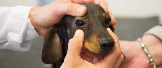
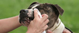
The eyes of animals and humans are a real indicator of their health. If they are cloudy and stop shining, then something is clearly wrong with your pet. Moreover, if you notice a cloudy eye in a dog, immediately take your pet to the veterinarian, as this phenomenon can be caused by many dangerous pathologies. Many of them are potentially fraught with the loss of not only vision, but even the eye itself.
Let's consider several diseases that occur most often in veterinary practice and cause clouding of the eyes of animals.
Nuclear sclerosis and cataracts
It should be noted that nuclear sclerosis itself is not a “classical” pathology, since in any dog with age, the center of the lens begins to sclerotize (that is, become harder). Only the severity of this process and the degree of its severity varies from animal to animal. The most characteristic sign of this phenomenon is the appearance of a blue-gray spot in the center of the eye lens.
As a rule, degenerative changes are recorded in pets over the age of six years. More often the process affects both eyes. The only good news is that the animal does not feel any pain. In addition, the increase in changes occurs very gradually - the animal has time to adapt to the changed vision, it worries less, does not experience such severe stress as it would experience if it suddenly went blind.
But all this is true specifically for nuclear sclerosis. Cataracts are much worse, they develop much faster, and depend less on age. It happens that this pathology is found in very young animals, almost puppies. Today it is known for sure that the disease is hereditary, and at least 80 breeds .
Miniature schnauzers, cocker spaniels, and golden retrievers are thought to be most susceptible. In animals of these breeds, the incidence of cataracts is maximum. The cause of the pathology has not been fully studied. Scientists suggest that the matter is due to some dystrophic, pathological processes, due to which the process of nutrition of the lens is disrupted .
Causes of the pathological process
If your pet has clouding or one eye is affected, you should understand the reasons for this condition. Any of them can lead to disability.
Physiology
Age, injury, and improper care are the most common factors causing vision problems in dogs.
Age
Dogs over 8 years of age may develop various diseases:
- erosion or ulcer of the eye;
- corneal degeneration;
- clouding of the lens (cataract).
The disease develops mainly in older animals. Medicines do not completely eliminate the pathology, but only restore blood circulation inside the eyeball, slowing down the course of the disease.
Injuries
Untreated damage to the retina or deeper layers of the eye leads to the appearance of a cataract. The spot forms at the site of punctures, ulcers, burns, and is most often observed on the cornea.
If treatment is started on time, the pathology can be easily cured. But when a cataract appears on the lens, serious therapy is required, including surgery.
Irregular and improper care
Some dog breeds especially need careful eye care. In lapdogs, pugs, boxers, and bulldogs, there is no tear duct, and fluid does not accumulate at the inner corner of the eyes, but flows out through the lower eyelid.
These dogs need to wash their eyes regularly, otherwise the accumulated secretions will lead to suppuration and clouding of the cornea..
Conjunctivitis
Theoretically, these serious diseases are rare. This is the name for inflammation of the conjunctival cavity. Roughly speaking, with this pathology the mucous membranes of the eyeball become inflamed. In severe cases, a large volume of exudate accumulates directly on the surface of the eye, which leads to clouding. In addition, the eyes of a sick animal become very red, blood vessels appear on them, the dog resembles an albino rabbit.
The predisposing factors of the disease are very diverse. Very often, its appearance is caused by foreign bodies, dust, plant pollen , etc. Also, conjunctivitis is a classic manifestation of many infectious pathologies . In many cases, inflammation of the conjunctiva is caused by allergic reactions. This is especially true for dogs living in urban areas.
Treatment in many cases is quite simple. The eyes are washed with antiseptic solutions , ointments with antibiotics are placed in the conjunctival cavity, and antihistamines are prescribed in case of an allergic nature of the disease.
Piroplasmosis
Clouding of the cornea in dogs with piroplasmosis appears in the first days of the disease. The causative agent of this pathology is the simplest microorganisms - Babesia. They enter the dog's bloodstream when bitten by ixodid ticks. Babesia causes massive destruction of red blood cells, enlargement of the liver and spleen.
With piroplasmosis, the dog's eyes acquire a yellowish tint. The mucous membranes of the mouth are painted the same color. Such symptoms are a sign of hemolytic jaundice, which develops against the background of the death of red blood cells and an increase in bilirubin levels.
Piroplasmosis is also accompanied by the following manifestations:
- a sharp increase in temperature (up to +41 degrees and above);
- severe weakness;
- staining urine red or brown;
- vomiting;
- difficulty breathing.
This parasitic pathology is extremely dangerous. Without treatment, it can lead to the death of the pet from intoxication in the first days of the disease.
Corneal dystrophy
This is a hereditarily transmitted disease of a dystrophic-degenerative nature . Its only “positive” feature is that the pathology does not cause pain and suffering to the sick animal. There are three types of corneal dystrophy, classified depending on the location of the pathological process: epithelial corneal dystrophy, which disrupts the formation of the epithelial layer; stromal corneal dystrophy, in which the surface of the eye acquires a clearly visible bluish tint; endothelial corneal dystrophy, in some cases contributing to the formation of cellular “nodules” directly on the surface of the cornea.
Stromal corneal dystrophy usually does not require complex treatment. Endothelial dystrophy is more severe; a cloudy film appears on the dog’s eye, due to which the animal’s vision drops almost to zero. The epithelial variety is something “in between”. How can this pathology be cured?
Alas, a specific and effective treatment for corneal dystrophy has not been developed to this day. If the area of the defects becomes very large and they significantly impair vision, surgical correction . The problem is that the resulting scars also do not contribute to good vision. In the rarest, experimental cases, a cornea transplant is performed... But here, too, everything is not very good. Firstly, the cost of the operation is very high, and secondly, the results so far are not very encouraging.
Symptoms
At the first signs of pathology, the dog begins to squint. After some time, the surface of the eye becomes cloudy, acquires a whitish or slightly blue tint, and becomes matte.
With any pathology of the lens, the cornea remains transparent, and a cloudy spot can be observed in the area of the pupil.
After injury, turbidity is observed in the anterior chamber of the apple. This is a consequence of the accumulation of pus or blood discharge.
Inflammation of the choroid, uveitis
A rather severe pathology, fraught with loss of vision. In addition, the process is quite painful and causes the animal a lot of suffering. The eyeball contains a huge number of blood vessels, so with any infectious disease there is a considerable risk of developing eye complications . This disease is extremely rare; it is almost always preceded by an inflammatory process somewhere in the body.
Since the pathology is quite painful, a sick animal will constantly touch its eyes with its paws and desperately rub them . The surface of the eye turns red, becomes cloudy, profuse lacrimation appears, and purulent exudate may be released. The animal’s behavior changes - it becomes apathetic, tries to spend more time in dark, remote places. Photophobia develops - the dog cannot look at the light, covers its eyes with its paws, and squints tightly.
Treatment options will depend on the underlying cause of the disease. As a rule, antimicrobial and anti-inflammatory drugs, drops and ointments injected into the conjunctival cavity are prescribed. In extremely rare cases, when the process is very advanced, surgical removal of the eye may be recommended, as the disease can contribute to the development of glaucoma.
If you notice your dog has cloudy eyes, take your pet to the vet immediately. Of course, some diseases that cause this effect are not particularly dangerous to the dog’s health, but in other cases your pet may well be left without eyes
Diagnostics
If you notice that your dog's eyes have become cloudy, you should immediately contact a veterinary ophthalmologist. A specialist will examine and examine the eye to make a diagnosis and prescribe treatment.
The ophthalmological examination includes:
- Eye examination and reflex testing.
Corneal reflexes are checked - if the cornea is weakly sensitive, this may indicate the development of inflammation (uveitis, panophthalmitis, keratitis) and pupillary reflexes - impaired pupil contraction may indicate the development of inflammation, increased intraocular pressure or acute pain.
- Corneal staining.
If the cornea is not damaged, special ophthalmic dyes are applied to the eyes. When you blink, the dye is washed out, and if there are ulcers or erosions on the cornea, the dye colors them brightly. This way the doctor can assess the depth and volume of the lesion.
- Measuring intraocular pressure.
Using a special veterinary device - a tonovet, an ophthalmologist can measure intraocular pressure, which will make it possible to make diagnoses such as glaucoma - when determining high pressure, or uveitis - when determining low pressure.
- Ophthalmoscopy.
This is a study of the back shell of the eye - the retina, using a special apparatus. With it you can examine the optic nerve head and evaluate the vessels that supply the eye. The study allows you to evaluate the visual function of the eye and the consequences of diseases such as glaucoma, uveitis, and uveodermal syndrome.
- Ultrasound of the eye.
The study will assess the size and position of the lens in cataracts and luxation.
- Genetic tests
are needed for certain breeds of dogs to carry genes for diseases such as pannus, uveodermal syndrome, lens luxation, and cataracts.
Treatment
It should be understood that at home it is impossible to understand what is happening to your pet’s eyes, so you should always contact a veterinary clinic.
Treatment of cataract (leukoma) is primarily aimed at eliminating the cause that caused the problem. It is not always possible to get rid of the stain itself, but the main task - restoring vision - is quite feasible.
Since it is very difficult to differentiate various eye pathologies, manifested by clouding and the formation of white spots on the cornea, from each other, you should not count on any eye drops to help.
Preparations containing hormones should never be used without a reasonable prescription from a veterinarian. You risk aggravating the dog’s condition, including loss of vision!
If you cannot show your dog to a specialist in the near future, but you see that the animal needs help, at home you can use:
- Tetracycline ointment.
- Levomycetin eye drops.
By using these drugs you will not harm your pet in any way and will be able to help in some way, even without knowing a reliable diagnosis.
For more reliable treatment, we recommend finding a veterinary clinic that has a veterinary ophthalmologist. Since eye pathologies can be very serious and it is not always possible to get by with therapeutic treatment.
In many cases, surgery is required, and it will be better if this is done by a highly specialized specialist.
Photo: libreshot.com
Physiology of the lens
The lens in the eye of an adult is a biconvex lens of natural origin. Thanks to this biological formation, which is transparent and has no blood vessels, light rays are focused on the retina of the eye, which provides visual perception of the surrounding world. The lens is located in the front of the eye between the vitreous body and the iris.
Age-related clouding of the lens of the eye significantly limits the light flow, making the visibility of objects around blurry and their contours blurred. Pronounced “fog” on a biological lens seriously reduces visual acuity. There is only one correct way out of the situation - contacting an ophthalmologist. Appointments at Dr. Trubilin’s clinic are made daily through the website, by phone and in the WhatsApp mobile application.


