Dermatitis in dogs is a very common allergic disease that can be caused by a wide variety of factors.
- Pododermatitis
The most common types of dermatitis in dogs
Since there are many causes of dermatitis and their combinations, the disease can be of different types. Each of them is characterized by its own distinctive symptoms and treatment approaches. The most common types of dermatitis in dogs are briefly described below.
Pyotraumatic dermatitis in a dog
Pyotraumatic
With pyotraumatic dermatitis in dogs, the infection penetrates from the surface into the deeper layers of the skin, leaving behind plaques and a thickened stratum corneum. The deeper it penetrates, the more papules (pimples) and pustules (pustules) will appear on your pet’s skin.
Treatment consists of using antibacterial drugs.
Allergic
Allergic dermatitis in dogs
Allergic dermatitis is considered the most common in dogs, since any object can be an allergen. Experts note that in most cases, skin allergies are provoked by external parasites, food and cosmetic care products. Symptoms include: severe itching, redness of the skin, scratching, rashes, swelling.
The basis of treatment is elimination of the allergen and symptomatic therapy.
Atopic
Atopic dermatitis
Atopic dermatitis is one of the varieties of the previous form. The clinical picture is the same: dry epidermis, rashes, very severe itching. Additional characteristic signs include the development of otitis media and pododermatitis. The pathology develops mainly in dogs under 5 years of age who have similar heredity. It is noted that atopic dermatitis “loves” such dog breeds as: dachshunds, sharpeis, bulldogs, setters, Dalmatians and a number of others. At the same time, anything can become an allergen.
Treatment consists of identifying and eliminating the allergen, carrying out symptomatic therapy, and preventive measures to prevent otitis media.
Flea
Flea dermatitis in dogs
The salivary secretions of fleas contain more than a dozen allergenic components that lead to flea dermatitis in dogs. The severity of symptoms depends on the individual susceptibility of the animal. Signs of pathology can be: alternating wet areas and areas covered with crusts. In the chronic course of the disease, the pet exhibits excessive skin pigmentation on the abdomen and hair loss. It is characteristic that the dog’s ears, muzzle and paws are almost not affected.
Treatment consists of getting rid of fleas and symptomatic therapy. Prevention is of great importance for recovery - the use of a parasite collar, disinsection of the environment in which the dog lives.
Interdigital dermatitis in a dog
Pododermatitis
Interdigital dermatitis in dogs, or pododermatitis, develops between the animal's toes, mainly on the front legs. If the lesions are single, it can be assumed that the disease is provoked by injury. With extensive damage to the limbs, the cause may be insects, allergies, or infections. Symptoms: bleeding nodules, boils, pain leading to lameness.
Treatment is symptomatic.
Seborrheic
Seborrheic dermatitis in dogs
Seborrheic dermatitis is caused by genetics. With this disease, the process of keratinization of the skin accelerates, which is visually manifested in numerous scales, oily epidermis, but dry hair. Your pet may have brittle nails; The inflammatory process on the skin is clearly visible, accompanied by severe itching. In the future, the disease is complicated by secondary infection with all the ensuing consequences.
Treatment is carried out in two directions: local therapy and a systemic approach.
Acral
Acral dermatitis in dogs
Symptoms of acral dermatitis are dense, ulcerated areas of skin that occur as a result of the dog licking the area excessively. Pathological behavior of an animal can be caused by allergies, parasites, infection and other factors that must be detected and eliminated before treating the skin.
Treatment consists of the initial elimination of the provoking factor and a therapeutic effect on the damaged epidermis. Limiting your pet's access to the itchy area is of great importance. In addition, antidepressant and psychotropic drugs are used in therapy.
conclusions
Itching in dogs occurs for a number of reasons, including the following:
- parasitic skin diseases;
- allergies;
- infections that cause/increase itching;
- rare skin diseases.
When looking for causes of dermatitis in dogs with pruritus, approaches include taking a history, clinical examination, routine dermatological tests and, if necessary, a step-by-step algorithm for diagnosing allergies.
An adverse food reaction includes two possible conditions: food intolerance and food allergy. Elimination diets are used to diagnose these two conditions. In order to confirm and control an undesirable food reaction, specialized diets are used.
If atopic dermatitis is diagnosed, additional tests may be performed to search for significant allergens for the purpose of desensitization therapy (ASIT). Along with this, it is important to use complex treatment with systemic drugs and topical agents.
Bibliography:
- Gnirs K., Prelaud P. Cutaneous manifestations of neurological diseases: review of neuro-pathophysiology and diseases causing pruritus. Vet Dermatol 16:137–146, 2005.
- Ikoma A., Steinhoff M., Stander S., et al. The neurobiology of itch. Nat Rev 535–547, 2006.
- Patel KN, Dong X. An itch to be scratched. Neuron 68:334–339, 2010.
- Vogt BA Pain and emotion interactions in subregions of the cingulate gyrus. Nat Rev Neurosci. 2005; 6:533–544.
- Kwan CL, Crawley AP, Mikulis DJ, Davis KD An fMRI study of the anterior cingulate cortex and surrounding medial wall activations evoked by noxious cutaneous heat and cold stimuli. Pain. 2000; 85:359–374.
- Moulton EA, Keaser ML, Gullapalli RP, Greenspan JD Regional intensive and temporal patterns of functional MRI activation distinguishing noxious and innocuous contact heat. J Neurophysiol. 2005; 93:2183–2193.
- Darsow U., Drzezga A., Frisch M., Munz F., et al. Processing of histamine-induced itch in the human cerebral cortex: a correlation analysis with dermal reactions. J Invest Dermatol. 2000; 115:1029–1033.
- Drzezga A., Darsow U., Treede R., Siebner H., et al. Central activation by histamine-induced itch: analogies to pain processing: a correlational analysis of O-15 H(2)O positron emission tomography studies. Pain. 2001; 92:295–305.
- Rosa AC, Fantozzi R. The role of histamine in neurogenic inflammation Br J Pharmacol. 2013 Sep; 170(1); 38-45.
- Andrew D., Craig AD Spinothalamic lamina 1 neurons selectively sensitive to histamine: a central neural pathway for itch. Nat Neurosci. 2001; 4:72–77.
- Sun YG, Chen ZF A gastrin-releasing peptide receptor mediates the itch sensation in the spinal cord. Nature. 2007; 448:700–703.
- Ikoma A. Analysis of the mechanism for the development of allergic skin inflammation and the application for is treatment: mechanisms and management of itch in atopic dermatitis. J Pharmacol Sci 110:265–269, 2009.
- Garibyan L., Rheingold CG, Lerner EA Understanding the pathophysiology of itch. Dermatol Ther. 2013 Mar-Apr; 26(2):84-91.
- Bell JK, McQueen DS, Rees JL Involvement of histamine H4 and H1 receptors in scratching induced by histamine receptor agonists in Balb C mice. Br J Pharmacol. 2004 May;142(2):374-80.
- Ewa Teresiak-Mikołajczak, Magdalena Czarnecka-Operacz, Dorota Jenerowicz, Wojciech Silny. Neurogenic markers of the inflammatory process in atopic dermatitis: relation to the severity and pruritus. Postepy Dermatol Alergol. Oct 2013; 30(5): 286–292.
- O'Connor TM, O'Connell J, O'Brien DI, Goode T, et al. The role of substance P in inflammatory disease. J Cell Physiol. 2004 Nov; 201(2):167-80.
- Fallahzadeh MK, Roozbeh J., Geramizadeh B., Namazi MR Interleukin-2 serum levels are elevated in patients with uremic pruritus: a novel finding with practical implications. Nephrol Dial Transplant. Oct 2011; 26(10):3338-44.
- Grimstad O., Sawanobori Y., Vestergaard C., Bilsborough J., et al. Anti-interleukin-31- antibodies ameliorate scratching behavior in NC/Nga mice: a model of atopic dermatitis. Jan;18(1):35-43.
- Patrick Hensel, Domenico Santoro, Claude Favrot, Peter Hill and Craig Griffin. Canine atopic dermatitis: detailed guidelines for diagnosis and allergen identification.
- Frane Banovic. The biology and contribution of Staphylococcus pseudintermedius in canine atopic dermatitis". Dubrovnik, 2018
- Guidance for the rational use of antimicrobials. CEVA, 2016
- H. A. Jackson at al. Evaluation of the clinical and allergen specific serum immunoglobulin E responses to oral challenge with cornstarch, corn, soy and a soy hydrolysate diet in dogs with spontaneous food allergy. Veterinary Dermatology 2003, 14, 181–187
- Anja Zimmer Jennifer Bexley Richard EW Halliwell Ralf S. Muller. Food allergen-specific serum IgG and IgE before and after elimination diets in allergic dogs. Veterinary Immunology and Immunopathology, 144(2011) 442-447
- Jeffers JG, Shanley KJ, Meyer EK. J. Diagnostic testing of dogs for food hypersensitivity Am Vet Med Assoc. 1991 Jan 15; 198(2):245-50.
- Mueller, Tsohalis. Evaluation of serum allergen-specific IgE for the diagnosis of food adverse reactions in the dog. Article first published online: 5 JAN 2002 DOI: 10.1046/j.1365-3164.1998.
- Oliver et al. Treatment of canine atopic dermatitis: 2015 updated guidelines from the International Committee on Allergic Diseases of Animals (ICADA) BMC Veterinary Research (2015)
- Olivry. Treatment of canine atopic dermatitis: time to revise our strategy? Vet Dermatol 2019; 30:87–90
- Forsythe P. and Jackson HA (2020) New therapies for atopic dermatitis. In Practice 42, 82–90
About the correct selection of shampoo for seborrhea
It is important to know how the individual components of the shampoo work and whether they have any synergistic effects, since the overall effectiveness of the drug depends on this. When choosing a detergent that can be used to treat seborrhea, we focus on the following factors:
- Moderate activity of the sebaceous glands, the pet's coat looks almost normal.
- Noticeable secretion of sebum and visible oiliness of the coat.
- There is a clearly visible layer of grease and scabs on the skin, and the fur is very greasy.
- The animal emits an unpleasant odor, the fur is matted, greasy and oily.
It is very important for the owner to inform the veterinarian exactly what stage of seborrhea is observed in his dog. In moderate cases, mild shampoos with a low content of antibacterial and antifungal agents, with a pronounced moisturizing effect, are needed
They are good because you can bathe a sick dog every day. Such products almost do not wash away the natural protective layer of the skin, which speeds up and facilitates the healing process.
The prognosis for seborrhea depends on the severity of the disease, the general condition of the dog after completion of treatment, and other factors. If the root cause of the disease has been accurately identified and eliminated, the chances of a relapse will be minimal. If you or your veterinarian have doubts about the completeness of the cure, you will need to show your pet to a specialist at least once a quarter. Some diagnostic tests may be necessary (especially if seborrhea was caused by hormonal imbalances).
Recommendations
You should not self-medicate, because there can be many reasons for the appearance of dermatitis. First of all, you need to visit a veterinarian. He will not only prescribe medications that can be used at home, but will also give recommendations on the pet’s diet.
Feeding
It is necessary to exclude dry food, canned food, carbohydrates, and all grains. Do not give bread, potatoes, red meat.
To determine what food a dog is allergic to, it must be kept on a strict diet for at least 2 months.
If after this time there is no improvement, then the problem is not in the pet’s nutrition.
And if improvements in the condition are noticeable, then it is best to transfer the pet to a specially developed food for animals suffering from allergies.
Care
If a dog has purulent dermatitis, then it should not be bathed, but if seborrhea is diagnosed, bathing is necessary. At the same time, you need to bathe your dog in disinfectant solutions.
Use shampoos containing antimicrobial agents that protect the skin from drying out.
Good antipruritic results can be achieved by using antipruritic shampoos and antihistamines. Sometimes, in addition to urea and glycerin, hormonal and antiallergic agents are added to the composition of such shampoos. It is important to rinse the shampoo thoroughly.
For the best effect, such shampoo should be on the animal’s skin for at least 10 minutes. At first, the detergent is used 2 times a week, with improvement, the number of procedures is reduced to 1 time a week or even 1 time every 2 weeks.
Main characteristics and types of disease
Dermatitis in dogs is a special type of allergic reaction of the body to the penetration of a foreign body or substance into it. The inflammatory process affects not only the epidermis, but also penetrates into the deeper layers of the skin. The disease is characterized by redness of the skin, the formation of ulcers, wounds, and erosions.
With dermatitis, areas of the dog's skin become inflamed, erosions, ulcers and wounds form.
The intensity of manifestations depends on the type of dermatitis:
- Allergies are the most common. It is a reaction to food products, especially often a reaction to dry food of a low price category, such as Darling. Contact with various chemicals can also cause an allergic reaction. Allergies develop from flea or tick bites.
- Atopic is a type of allergic dermatitis that occurs against the background of autoimmune diseases. predisposition to it is laid down at the genetic level.
- Mechanical – the most easily curable, develops after injury or scratching of any area of the skin. Most often, mechanical dermatitis develops after wearing a low-quality collar or due to synthetic bedding.
- Contact – skin reaction to a chemical or physical irritant. Inflammation develops from constant touching of an object or when a chemical comes into contact with the skin. Contact dermatitis is caused by dog grooming products that are used incorrectly.
- Medicinal - develops when a dog is treated with any drug, the individual components of which are intolerable to the animal.
- Sunny – localized on the dog’s face. This type of dermatitis mainly affects dogs with lightly pigmented skin.
- Parasitic – the cause of dermatitis is fleas, nematodes or worms.
- Infectious and fungal - are complications after skin injuries against the background of weakened immunity. Most often, inflammation is caused by staphylococcus, which lives on the skin of any dog. If a fungal infection is added to it, quite severe inflammation develops.
Short description
Atopic dermatitis or atopy is a chronic allergic skin disease. Most often it is hereditary. However, recent studies prove that it is the environment that becomes the trigger for this disease.
Atopy is characterized by an inadequate response of the dog's body to environmental allergens. Once in an animal, a foreign protein triggers the production of antibodies or immunoglobulin E. Excessive concentration of protein compounds in the blood leads to skin itching and other associated diseases. As a rule, it has a chronic form and cannot be completely cured.
Drug treatment
The doctor’s task is to normalize the state of the dog’s immune system and cure skin lesions, preventing secondary infection.
Medications are quite often prescribed for flea dermatitis, both for indoor and outdoor dogs.
Along with insecticidal treatment and strengthening the dog’s immunity, the doctor may prescribe:
- A glucocorticosteroid, usually prednisolone at a dosage of 0.5-1 mg per kilogram of weight. Orally. Duration - from seven to ten days. Then the dosage is gradually reduced. The duration of taking the medicine depends on how successfully the owner fights fleas. The faster you can get rid of insects, the shorter the course of prednisone will be.
- In some cases, when oral administration of prednisolone is not possible according to indications, glucocorticosteroids are prescribed in the form of ointments for external use.
- For advanced forms and secondary infection of the skin, antibacterial ointments are used.
Attention: the veterinarian must take into account the individual characteristics of each animal: age, weight, general physical condition, the presence of other disorders and diseases. Therefore, it is unacceptable for dog breeders, based on personal experience, to independently prescribe treatment for their own or other people’s pets!
Causes and development of the disease
There are many reasons for the development of acral dermatitis. They can be divided into three main groups:
- Itching.
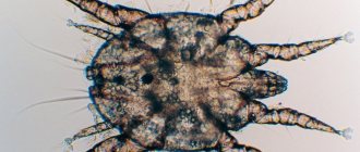
Itching can initially be caused by diseases such as allergies (reaction to insect bites, atopic dermatitis, food allergies), skin infections (bacterial, fungal, leishmaniasis), folliculitis, furunculosis, skin parasites (demodicosis (photo 1), dirofilariasis, sarcoptic mange, pelodera ).
Photo 1. Demodectic mange in a dog - Pain.
The causes of pain are varied - foreign objects, granulomas (nodules) from compression, orthopedic problems, neoplasia (tumors), trauma, furunculosis (inflammation of the hair follicle). - Stress.
Stressful situations, anxiety, fear and boredom lead to the habit of licking the paw, which results in the development of acral dermatitis. According to various sources, psychogenic disorders are observed in 70% of cases. For this group of root causes, dogs that experience a lack of social communication, have a sedentary lifestyle, live in a limited space (enclosure, cage), and are predisposed to anxious behavior are more often suitable.
The so-called “vicious circle” leads to the development of acral dermatitis. Whatever the root cause, the first action that triggers the onset of the disease is licking. Continuous licking leads to hair loss and skin damage. Due to this, the nerve endings are exposed, and this, in turn, stimulates even more licking. The dog experiences itching in this area of the skin and begins to lick its paw to relieve it. Paradoxically, a vicious circle of actions occurs, each time worsening skin conditions and increasing the severity of itching.
That is why it is very difficult to determine the true root cause of the onset of the disease. And an important place in this issue is occupied by studying the life history (history) of the animal and determining the underlying disease, which is the main factor in successful treatment.
Treatment of dermatitis
When visiting a doctor, first of all, you must undergo all the necessary diagnostic measures. The success of therapy depends on the correctness of the diagnosis. To provide an accurate diagnosis, the following are prescribed:
- examination of skin scrapings under a microscope and bacteriological culture;
- determination of the specific sensitivity of bacterial pathogenic microorganisms to antimicrobial drugs;
- leukocyte formula studies;
- examination of stool, urine and blood.
After establishing the cause, the doctor selects the appropriate treatment regimen, its duration and the number of drugs. Two types of treatment are used - medication and physiotherapy.
To facilitate access to the affected areas, the hair is cut off and dead tissue, purulent exudate and crusts are removed using a special swab soaked in antiseptic. The damaged area is dusted with a special disinfectant powder. To isolate the wound, bandages are used, often impregnated with antifungal, anti-inflammatory and antibacterial ointments. This allows the animal to speed up the healing process.
Drug treatment for dermatitis in dogs includes:
- Use of antibiotics for a course of 5 days to 2 weeks. This is an important point to prevent the occurrence of secondary infection. Antimicrobial agents of the penicillin and cephalosporin group are widely used. A treatment course is carried out under the strict supervision of the attending physician, with the use of ointments for dogs based on antibiotics, which promote rapid healing of wound surfaces.
- Skin lesions caused by the action of fungal microorganisms require the use of fungiostatic drugs. In addition, the pet’s skin must be washed using special shampoos that destroy the fungus - Nizoral, Clotrimazole, Ketoconazole.
- To eliminate itching and discomfort in allergic dermatitis, systemic antihistamines are used. The drugs that have proven themselves to be excellent are Suprastin, Diphenhydramine, Fenistil.
- Drug therapy includes increasing the body's defenses by using systemic immunostimulating drugs that increase the activity of T cells. Gamavit, Gamapren, Glycopin are actively used in veterinary medicine.
- The destruction of parasites is carried out by using acaricidal and insecticidal preparations of systemic importance.
Dermatitis of various etiologies can be effectively treated using physiotherapeutic procedures. Irradiation is carried out with ultraviolet lamps and lamps with infrared light. The use of physiotherapy in combination with medicinal methods can significantly increase the rate of restoration of epithelial cells, disinfecting the surface and reducing the amount of purulent exudate released.
Main types of dermatitis
Dermatitis is classified depending on the factor that caused its development:
- traumatic - occurs at the site of bruises, injuries, open wounds and scratching of itchy skin by the animal itself;
- allergic - its causes may be mites, poor-quality food, poplar fluff or plant pollen;
- contact – develops after direct contact of an animal with an allergen;
- atopic – caused by a combination of factors, including genetic predisposition;
- flea infestation – occurs after an animal comes into contact with parasites.
Dermatitis in dogs, the symptoms of which allow the disease to be classified, can be successfully treated in most cases. At the end of therapy, the skin is completely cleansed, the animal’s well-being improves, and the risk of relapse is reduced.
Allergic dermatitis: why is it dangerous for animals?
Allergic dermatitis is considered the primary source of some types of the disease.
Its main subtypes are contact, atopic and flea; the differences lie solely in the mechanism of development of the immune system's response to an irritant. In each of these cases, a specific allergen is present, although other factors can also cause the disease. The main places of localization are the ears and the folds of the paws. This type of dermatitis is very easy to confuse its symptoms with other types. Experts believe that it is also transmitted genetically.
Allergic dermatitis in dogs can develop slowly as the allergen is exposed. The danger of the disease lies in the risk of developing angioedema (in advanced cases) after bites from poisonous snakes, ticks or other insects. Rapid swelling of the animal's larynx or muzzle area can be fatal, so contact a veterinarian immediately.
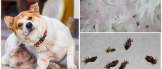
Flea dermatitis: how to protect your animal from infection
Fleas settle on the dog and parasitize it for a long time.
They become the cause of the development of such an unpleasant disease as flea dermatitis. Insects bite through the dog's skin, and in this case their saliva becomes the allergen. The main places where insects are localized have high humidity (sacrum, groin, behind the ears, under the tail, stomach). The disease itself and the presence of fleas are two different things. If the dog is not prone to allergic reactions, a huge number of adults and their larvae may be hiding in its fur, but no symptoms of dermatitis are observed.
Getting rid of fleas does not guarantee that the insects will not attack the animal again. Eggs, larvae and pupae can survive on surrounding household objects, and after some time migrate again to the dog. After its treatment, it is necessary to carry out disinfestation of the apartment.
Flea dermatitis in dogs is a disease that can affect even a clean, well-groomed animal with a flea collar on its neck. As a preventative measure, you should inspect the animal’s fur daily in good lighting. Having noticed the first traces of new “settlers”, they immediately take measures to get rid of them.
Atopic dermatitis: how to recognize it by the first signs
When an animal becomes restless, begins to itch regularly, and loses the desire to move and play, it is worth carefully examining its skin.
If redness, baldness and hyperpigmentation are observed around the eyes, lips, groin and near the anus (up to acanthosis nigricans - darkening of areas of the epidermis in the folds, their thickening), this may indicate a disease such as atopic dermatitis in dogs. Animals aged from 6 months to 6 years are at risk. Among the provoking factors are:
- heredity;
- food allergens of protein origin;
- epithelial cells (both other animals and humans);
- fungal microspores;
- plant pollen during the flowering period;
- dust mites (accumulate in upholstered furniture, blankets and even in the air).
It is impossible to completely cure atopic dermatitis; sometimes it takes the entire life of a pet to fight the symptoms. The acute form of the disease is treated with medication, and after that it is necessary to take preventive measures.
Which breeds are more susceptible
Atopic dermatitis affects young dogs between 1 and 5 years of age, but can be diagnosed earlier. The first symptoms of dermatitis appear at six months of age, when the allergen enters the body and causes the immune system to produce neutralizing antibodies, then the pathology recurs throughout life.
The risk group includes representatives of such breeds as bulldogs (French, American), boxers, cocker spaniels, pugs, sharpeis, beagles, poodles, golden retrievers, Labradors, German shepherds, chow-chows, setters (English, Irish).
However, this does not mean that other breeds are not susceptible to this disease.
Causes of dermatitis – types of skin lesions
Skin inflammation can develop in a variety of pathologies. In some cases, this is an independent disease, manifested by a rash, erosions, ulcers, and purulent processes. But more often dermatitis is a symptom of various pathologies:
- fungal infections - microsporia, trichophytosis;
- parasitic diseases – ticks, fleas;
- pyoderma caused by various bacteria;
- allergic reaction.
Bruise, skin dissection, trauma and other mechanical damage lead to inflammatory processes. Injuries can be caused by other dogs (fights, fights between pack members), animals. Pets often injure themselves - scratching due to flea or tick infestation. In guard dogs, inflammation of the neck is common due to friction from the collar. Pododermatitis in dogs develops due to primary damage to the skin - resistance decreases, the area becomes susceptible to infections.
Another form of contact dermatitis involves inflammation due to exposure to a physical or chemical nature. Thus, in pets with thermal burns, skin lesions appear, but more often such a defect occurs in stray and guard dogs in the winter. Chemical irritation occurs when treating a pet for fleas or bathing it with different shampoos. Dermatitis of the interdigital fissure is often observed in winter due to the treatment of roads with ice reagents.
A common type of chemical dermatitis is inflammation after the thoughtless use of medications. This form of pathology is observed in dogs with individual sensitivity, as well as during self-treatment of pets. The disease appears both as a result of severe irritation (application of crystalline substances, tincture of iodine, a number of ointments to the skin) and in the form of an allergic form. Many antibiotics, vaccines, and steroid drugs cause hypersensitivity.
Inflammation of the dog's skin may be due to genetic characteristics. Atopic dermatitis in a dog develops when a number of immunoglobulins are formed after allergic substances enter the body:
- feed proteins;
- pollen;
- dust;
- vaccines, serums, biological products;
- animal epithelium.
Special cells of the animal's body perceive antigens and transmit them to lymphocytes, forming susceptible cell clones. An allergic immune reaction does not form on the first occasion of contact, but upon repeated exposure to the antigen. The reaction can develop in two ways - slowly (over several months with prolonged contact with the allergen), or rapidly - immediately after exposure to the substance on the body.
Dogs with white fur and fair skin may experience solar dermatitis. Under the influence of ultraviolet rays, sunburn appears, leading to disruption of keratinization - improper growth of the horny epithelium. The disease is caused by excessive exposure to hairless or light-colored skin.
Elimination diet
The PRO PLAN® VETERINARY DIETS HA HYPOALLERGENIC diet can be used as an exclusion or diagnostic diet and is suitable for dogs of any age. The only source of protein in this diet is hydrolyzed soy protein, and carbohydrates are corn starch, which is practically devoid of protein (content less than 0.5%). Or, for the same purpose, you can use PRO PLAN® VETERINARY DIETS HA wet diet for puppies and adult dogs.
Also, the PRO PLAN® VETERINARY DIETS DRM DERMATOSIS diet has proven itself as a product for diagnosing adverse food reactions in dogs. It contains such rare sources of protein as rapeseed, peas, and herring. Corn starch is used as a source of carbohydrates. This composition allows you to easily include this food in the diet of dogs for the purpose of an elimination diet after a preliminary history aimed at assessing the list of products previously used for feeding.
The diet lasts for eight weeks. In addition to the proposed diet, the dog should not consume any other food products, such as table food, dog treats, medications in gelatin capsules, etc. The diet must be strict. After eight weeks, the patient’s condition is assessed: the presence/absence of clinical symptoms of itching and dermatitis. If there is no clinical improvement, it is concluded that the patient is suffering from atopic dermatitis. If the animal shows clinical improvement, a provocative test (reversal diet) is performed.
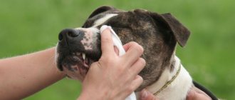
Diagnosis and treatment of dermatitis
Treatment for dermatitis depends on the causes of its occurrence. The greatest difficulty is in diagnosing atopic (allergic) dermatitis, since it requires identifying the specific allergen.
At the appointment, the veterinarian examines the dog’s skin, determines the timing of the appearance of rashes, and conducts research.
The medical history is compiled based on information:
- First appearance of the rash. The time of appearance of skin irritations is an important diagnostic factor, since atopic dermatitis manifests itself in the spring and autumn, while food allergies occur year-round.
- Heredity.
- Which areas of the body are most irritated?
- Color and nature of the rash.
- Under what conditions did dermatitis develop?
- Laboratory tests are used:
- Culture the smear for the fungus.
- General and biochemical blood test.
- Blood serum analysis for hormones.
In the treatment of dermatitis, local remedies, oral medications and physiotherapeutic methods are used. To relieve irritation, bandages with antiseptic drugs are used, which regenerate tissue, relieve inflammation, and destroy fungal infections. For these purposes, applications of ozokerite and paraffin are used. Painful sensations are relieved by novocaine blockade.
If the dog experiences severe pain, it is given novocaine blockades.
The course of treatment for dermatitis includes the following:
- Antibiotics – for the development of a secondary infection or infectious dermatitis. Drugs from the group of penicillins, cephalosporins, and carbapenems are prescribed. The dosage and duration are prescribed by the doctor. To relieve inflammation, ointments containing antibiotics are simultaneously prescribed.
- Fungal dermatitis requires the use of drugs with fungicides: Fungin, Zoomikol. Additionally, shampoos Clotrimazole, Ketonazole, Nizoral are used.
- Itching is relieved with the help of drugs Allervet, Diphenhydramine, Tavegil, Fenistil.
- In case of intoxication, Furasemide is prescribed.
- Together with medications, drugs are prescribed to simulate immunity: Hemobalance, Gamavit, Gamapren, Glycopin, ImmunolVet. To quickly restore the animal’s body and increase resistance, injections of vitamins A, group B, E, PP are prescribed.
- For parasitic dermatitis, it is necessary to treat the dog's skin with insecticidal and acaricidal preparations: Praktik, Sanofly, Certificate, Spot-on, Scalibor.
- The wound surface is irradiated with ultraviolet or infrared lamps. The procedure accelerates skin healing, relieves inflammation, and has an antibacterial effect.
- For atopic dermatitis, an anti-allergenic diet is prescribed.
Folk remedies for dermatitis
A compress of raw potatoes will help relieve inflammation.
Drug treatment of the disease can be combined with folk recipes used to treat wounds and relieve redness:
- Potato compress. Grated raw potatoes are placed on gauze and applied to the affected area.
- Herbal ointment. Chamomile, fireweed tea, a tablespoon, two glasses of hay dust are poured with water and boiled for 5 minutes in a water bath. Strain the broth, add 1 tablespoon of butter and cook until a homogeneous mass is formed. The mixture is mixed with glycerin in equal proportions. The ointment is applied to the affected areas 4 times a day for 30 days.
- Pear lotions. A glass of crushed pear leaves is boiled in half a liter of water for 5 minutes, infused for 12 hours. Used in the form of lotions.
Prevention
Preventive measures include:
- Timely treatment of irritations on the animal’s skin.
- Haircut in summer.
- Washing with special shampoos.
- Regular treatment against parasites.
- Regular wet cleaning of the apartment.
- Balanced diet.
- Regularly inspect the animal for irritation.
Skin diseases in dogs are common. This is mainly a reaction to parasites or an allergy. Young animals are more susceptible to dermatitis. To make their life easier, you need to conduct regular examinations and follow all the veterinarian’s recommendations.
Symptoms of dermatitis
Common signs of the disease that appear in most cases include:
- hypermia (redness) of the affected area;
- intense itching;
- impaired hair growth, local or complete baldness;
- insomnia;
- severe swelling of the skin;
- loss of appetite or its complete absence;
- bleeding from small vessels, resulting in the formation of dried crusts;
- the formation of erosions on the surface of the epidermis and secondary wounds due to scratching the itchy area.
The disease cannot be left to chance; there is a possibility of infection, which will complicate the overall picture and may worsen the prognosis. When the first individual symptoms appear, you should contact your veterinarian.
Providing first aid to a dog
First aid for a dog with dermatitis consists of actions aimed at alleviating the animal’s general condition and reducing pain symptoms . The affected areas are treated with antiseptic drugs.
It is also recommended to remove fur if possible. Self-medication is not allowed, since any type of dermatitis can be a secondary disease. Further treatment is carried out according to the scheme prescribed by the veterinarian after examining the pet.
Fighting the disease at home
Traditional medicine is used as alternative treatment methods. However, this method does not exclude the professional prescription of a specialist. Rather, this is regarded only as additional measures to alleviate the condition of a mustachioed patient, but certainly not as a panacea for the disease.
- Soap solution. One of the most popular techniques to relieve itching and remove skin rashes. Essential oils of cedar, tea tree and lavender are mixed in equal proportions with soap foam. The resulting composition is used to treat the animal’s fur before and after bathing. The owner will have to be patient, because the results of such treatment will not appear soon.
- Lotions. Infusions of chamomile, fireweed, celandine, burdock, and plantain will help reduce unpleasant symptoms. Usually a compress is applied to the affected area or the surface of the skin is wiped with a cotton swab soaked in the broth.
- Medicinal ointments. You can also make your own special paste. Take one tablespoon of all the above plants and mix with 400 ml of hay dust brew. The resulting mixture is poured with boiling water and steamed in a “bath” for about 5-7 minutes. Then filter the cake and add about fifteen grams of butter to the liquid and keep it on the fire until the mixture thickens. Then add another 15 grams of glycerin and apply to the damaged areas for a month four times a day.
- Chlorhexidine. Based on this antiseptic, various sprays and shampoos are produced that relieve inflammation, prevent infection and have an antimicrobial effect. It is allowed to use a regular 1-2% pharmaceutical solution. It is enough to gently wipe the inflamed area with a moistened cotton pad.
- Hydrogen peroxide. Popular antimicrobial agent. It is better to buy a 3% solution.
- Nutrition. During the acute stage and until stable remission occurs, the animal can be given hypoallergenic food. But it is better to exclude beef, wheat and fermented milk, which provoke allergies. Suitable natural foods would be: pumpkin, turkey, potatoes.
- Vitamins. Be sure to support the dog’s immunity, which is weakened by the fight against the unfortunate allergen. Here you will still have to consult a doctor, since prescribing supplements to your pet at your own discretion is highly not recommended.
In conclusion, I would like to remind you: if you develop hypersensitivity to flea saliva, it will be impossible to completely get rid of this type of dermatitis. The problem will be relevant until the onset of the first cold weather. Then the temperature outside will become unsuitable for fleas and the insects will go into hibernation.
During the warm season, it is recommended to constantly treat dogs prone to this infection. Try to avoid areas with tall, dry grass, where these obnoxious insects like to settle.
And finally, I would like to note that properly functioning anal glands in a pet directly affect the barrier function of the skin
It is important to regularly maintain and keep the area clean. If it is difficult to carry out this procedure on your own, seek the help of specialists.
If these glands work well, it will be easier for your pet to tolerate flea dermatitis.
Be attentive to your four-legged friends. Do not neglect taking care of their health and bring your animal to the veterinary clinic for examination on time.
At home
Many owners are thinking about how to help their pet, what additional measures to take and at least alleviate the dog’s condition a little. Of course, alternative medicine helps speed up recovery a little, but cannot replace basic therapy. In any case, the main appointments should be made by a veterinarian, not the owner.
- Chamomile. Decoctions are made from the pharmaceutical composition. The leaves of this plant have been famous since ancient times for their regenerating, antimicrobial and strengthening properties.
- Echinacea. Compresses with this tincture help relieve itching. It is believed that ordinary raw potatoes cope well with this task.
- Comfrey. Tincture of root and leaves will help heal wounds.
- Calendula. The flowers are used to fight fungal infections.
- Aloe. Our grandmothers knew about the beneficial properties of this succulent: it heals, stimulates the immune system, protects against free radicals.
- Curcumin. Root extract has antibacterial, immunomodulatory and anti-inflammatory effects.
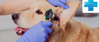
Diagnostics
The primary diagnosis of interdigital dermatitis in a dog is made by a veterinarian after examining the animal. But to choose a treatment method, it is necessary to determine the cause of its development. Therefore, the doctor carefully examines the medical history (the animal’s diet, the presence of concomitant diseases, heredity, etc.). Then the dog is prescribed a series of laboratory tests: a test of blood, feces, urine, smears, scrapings or punctures taken from the affected area. This makes it possible to identify possible pathogens of the disease (infectious agents, fungi or subcutaneous parasites).
To clarify the diagnosis, it may also be necessary to conduct hardware studies (tomography, radiography) or consult specialists in related professions - an orthopedist, oncologist, allergist.
It is important! To ensure high-quality diagnostics, the skin on the dog’s paws should not be treated with anything for 2-3 days before the veterinarian’s appointment. Otherwise, the conclusions drawn during the examination of the animal may turn out to be unreliable.
Causes of the disease
As a rule, the principles of nosology are taken as the basis for the classification of the nature of the disease: according to this doctrine, various disorders of the functioning of the body are combined according to the characteristics of kinship. In this vein, experts rightly classify dermatitis as a skin disease. Dermatitis can be infectious, inflammatory or hereditary in origin. Dermatitis can occur in all breeds of dogs, both adults and puppies. Stressful situations - unfavorable living conditions, change of owner, place of residence - can also lead to the appearance of dermatitis. Often the course of the disease is determined by several concomitant factors that must be taken into account when treating your pet. The lesion causes severe discomfort and causes changes in the dog’s behavior, leading to various complications. Identifying dermatitis in the early stages of its development will allow you to choose a treatment that will help suppress the developing disease in the shortest possible time. Treatment of dermatitis in dogs with modern methods of disease treatment and diagnosis in most cases has a positive prognosis. But this forecast will be justified if you consult a veterinarian in a timely manner.
Hurry up, choose a box and find out what gift awaits you
Discount on pet insurance
Promo code copied to clipboard
Symptoms
First of all, you need to suspect this disease in your pet if it itches all the time.
Flea dermatitis has the following characteristic symptoms:
- Itching. The main sign that worries the dog. In mild cases, the dog itches all the time. The symptoms are much worse and more complex if the disease is severe. In this case, the animal can tear the skin until it bleeds, bite its paws - it does everything to get rid of the annoying itching.
- Redness and swelling of the skin. The skin at the site of the lesion will be hyperemic and swollen. These are signs of an inflammatory process.
- Hair loss. In areas where fleas live, hair may fall out. In some areas it is completely absent.
- Secondary symptoms that are associated with long-term indirect effects on the nervous system. The dog becomes irritable, aggressive, restless, and sleeps poorly.
Local decrease in immunity (on the skin) and constant damage to the skin (scratching, scrapes, open wounds) create the preconditions for the attachment of bacterial flora. Bacterial skin lesions are most often characterized by purulent inflammation. In this case, symptoms of general intoxication (fever, lethargy) may appear.
Predisposition
Recent research has shown that many dogs with atopic dermatitis have abnormalities in the stratum corneum of the skin. It has been proven that in some cases, metabolic failure leads to the development of the disease. The main cause of this unpleasant disease in dogs was thoughtless selection. As a result of interspecific crossings, some breeds turned out to be more prone to this disease than others:
- American Staffordshire Terrier;
- English bulldog;
- Boston Terrier;
- boxer;
- bull terrier;
- West Highland White Terrier;
- golden retriever;
- Dalmatian;
- Jack Russell Terrier;
- Cairn Terrier;
- Cavalier King Charles Spaniel;
- Labrador Retriever;
- Lhasa Apso;
- pug;
- German Shepherd;
- setter;
- French Bulldog;
- fox terrier;
- chow-chow;
- schnauzer;
- Scottish Terrier;
- Shar Pei;
- Shih Tzu.
As you can see, the list is very long. For individuals who have been diagnosed with the disease, subsequent sterilization is recommended to prevent the spread of atopy within the breed.
Differential diagnosis
Thus, the specialist will always focus on those symptoms that can indicate the exact cause of what is happening to the animal. Must be taken into account: age, breed, gender
The owner is required to have as complete a medical history as possible: when, how and after what alarming signs began to appear in his pet. Various allergies (atopic dermatitis) are a more likely underlying cause if the pet is between one and five years of age, while neoplasia (especially cutaneous lymphoma) is more likely if seborrhea begins in older animals, or even middle-aged pets.
The intensity of itching plays an important diagnostic role. If it is minimal, then the animal may have some problems with the endocrine glands. Demodicosis or sebaceous adenitis is also possible. If the itching is significant, it is necessary to first consider the possibility of allergies and ectoparasites. It should be taken into account that many infections, malassezia or pyoderma also cause severe “pruritus”. Thus, the absence of itching is the first “bell” that may indicate seborrhea.
It is necessary to identify the presence/absence of polyuria, polydipsia or polyphagia (increased urination, thirst and appetite). These signs do not indicate seborrhea, but serious kidney pathologies. It is also useful to take into account the presence/absence of a dog’s pregnancy or sexual heat, that is, seasonality is important. The veterinarian should know what food and in what quantities your pet consumes, whether you are giving him corticosteroids, antibiotics, antifungals, antihistamines, etc.
In addition, it is important to take into account environmental factors (is there a possibility of the dog coming into contact with household chemicals, for example). Such a scrupulous approach to diagnosis is due, as we have repeatedly repeated, to the presence of many diseases, the clinical manifestations of which are very similar to seborrhea
Mechanisms of itching formation
Previously, there was an idea that itching and pain (as sensations) were close to each other, since it was assumed that their formation required the same factors, mechanisms and ways of their development. (1–3) Subsequently, certain differences were identified. In particular, results from positron emission tomography of the brain indicate that the perception of the sensation of pain is processed in the primary and secondary somatosensory cortices of the brain. The perception of a sensation such as itching is processed in the prefrontal cortex, premotor areas, primary somatosensory cortex and anterior cingulate cortex. (4–8)
However, it should be noted that the areas of the brain that perceive sensitive information about itch and pain largely overlap each other (that is, they are identical to each other). Of course, there is still no complete clarity on this issue.
Equally important is understanding which mediators can cause itching, pain and inflammation. It is believed that the predominant mediators of itching are histamine, interleukin-31 and substance P, and the predominant mediators of pain are interleukin-6/-8, tumor necrosis factor, hydrogen ions, calcitonin gene-related peptide, adenosine triphosphate. Mediators that may be involved in the development of both itching and pain include endothelin, tryptase, nerve growth factor, and acetylcholine.
Along with this, there are other mechanisms that create itching. A certain role in the development of this process is assigned to gastrin-releasing peptide, neurotrophins, kallikreins, proteases, leukotrienes, prostaglandins and other inflammatory mediators that can activate nerve fibers and sensitize them. In addition to IL-31, a number of other interleukins (such as IL-2,4,13) also affect the development of itching. (9–18)