Dogs are active, lively animals that love to explore the world around them. Therefore, it is not surprising that in the process of learning about the surrounding space they can become injured. The ears of a furry friend are rightfully considered one of the most vulnerable places to injury, since their tissue is very delicate. A dog's ear hematoma refers to a type of injury that is caused by hemorrhage under the pet's skin and is accompanied by rupture of small and medium-sized vessels. As a result, a characteristic cavity appears in the pet’s ear, where tissues are displaced under the influence of blood pressure. The article will discuss the main causes, characteristic symptoms and methods of treating such an injury.
Causes
Otohematoma in a dog is another name for a bruise in the ear of a four-legged friend, which is accepted in veterinary medicine. The main reasons leading to hematoma include:
- Bruises and abrasions. Most often, ear hematoma occurs for this reason in the youngest individuals. A puppy can get injured while walking or even at home, for example, by simply falling off the sofa. An adult can damage its ear in a fight with another dog.
- Entry of various foreign objects into the auricle.
- Vitamin deficiencies and intense head shaking.
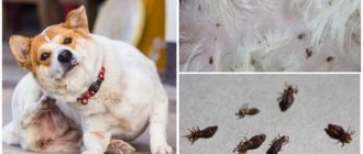
- Combing. If inflammatory skin processes occur on the outside of the ear (arising from chronic otitis, allergies or parasite bites), then the dog can injure itself with its paws when trying to relieve the itching.
Ear structure
To understand how difficult it is to diagnose otohematoma, you should first understand the structure of the organ, which consists of the following components:
- outer ear;
- middle ear;
- inner ear;
- bone labyrinth;
- membranous labyrinth.
The structure of a dog's ear.
The outer part consists of the concha, the motor mechanism and the external auditory canal. The middle part includes the tympanic membrane, the tympanic membrane cavity, the chain of auditory ossicles, and the auditory tube. In the internal cavity there are receptors that are responsible for the level of hearing.
Bone labyrinth
The bony labyrinth is a system of hollow compartments that are located on the temporal part of the skull and serve to accommodate membranes.
- As for the membranous labyrinth, it is a whole system of adjacent hollow compartments consisting of connecting membranes. These compartments are filled with endolymph and in structure resemble an oval bladder containing three semicircular canals and the membranous canal.
- And also the named bladder serves as a container for the endolymphatic duct.
- The walls of the bladder consist of villous epithelium.
The bony labyrinth in a dog's ear contains membranes.
Symptoms of the disease
A hematoma on the ear of a furry friend most often does not have rich symptoms, so the owner needs to be twice as careful to detect it. The most striking features include:
- The dog has an increase in body temperature.
- Otohematoma appears on the pet's skin in the form of a hemisphere, which in its consistency is soft to the touch.
- If the owner touches the hematoma, or the animal itself scratches the damaged ear, this is expressed in a strong painful sensation at the site of dislocation. The pet may whine and try to prevent the owner from touching the area where the ears are damaged.
- The lesion acquires a characteristic reddish tint, which veterinarians call hyperemia.
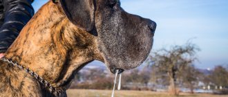
The changes also affect the usual behavior of the tailed pet. He constantly tilts his head in the direction where it hurts. The ears may become itchy. In general, with otohematoma, animals are characterized by restlessness and restlessness. At the first signs, you need to carefully examine your pet, or even better, take him to an appointment with a specialist. Next, we’ll look at how auricular hematoma in dogs is diagnosed and treated.
Pathogenesis of the disease
After the vessel ruptures, blood flows out and permeates the surrounding tissue. The pressure of the flowing blood pushes and separates the tissues, which leads to the formation of a pathological cavity (hematoma). The process of bleeding stops when the pressure inside the vessel is not balanced by the resistance of the walls of the created cavity. Therefore, hematomas with arterial injuries are always larger than with veins. In addition, the size of the hematoma is determined by blood pressure, the type and extensibility of the skin, and blood clotting.
When hematomas form, a process of self-blocking of hemorrhage occurs in parallel. First, the ends of the damaged vessels curl up and are drawn into the surrounding tissue. At the same time, the lumen of the vessels narrows. Thanks to this, a strong blood clot forms within 2-3 days.
The process of self-blocking hemorrhage does not occur if there is only a crack or puncture near the surface of the artery. In this case, a parietal pulsating hematoma is formed, and then a false aneurysm is created from it.
In the created cavity, the blood coagulates, a small thrombus resolves on its own, while in case of a large hematoma it remains for life. In this case, the blood clot is replaced by connective tissue, then a capsule is formed, in which calcium salts are later deposited, resulting in a cyst. When microorganisms penetrate, an abscess or phlegmon occurs.
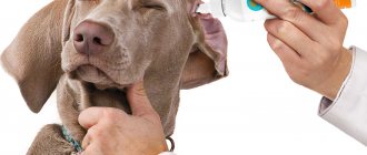
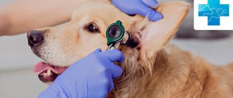
Diagnostics
In a veterinary hospital, the first thing the doctor will do is palpate the area of the ear affected by the hematoma. Further procedures will include a medical history, blood and urine tests, and possibly an ultrasound and x-ray. This is done in order to exclude internal causes of hemorrhage. Often, diagnostics can establish that the true cause of otohematoma is not ordinary canine trauma, but a serious disease of the liver or some other vital organ. Further treatment of the hematoma depends on the degree of mechanical damage and whether a blood clot has formed in the ear.
Postoperative nutrition for the animal
The attending veterinarian will provide specific recommendations on what the dog can do after surgery. This directly depends on the type of surgery and anesthesia.
The dog breeder wonders what to feed the dog after surgery? We offer the following:
- Feeding should be done little by little so as not to burden the body, since a lot of energy is spent on digesting food.
- After surgery, the dog does not eat or drink for several hours. This is especially true for operations performed on the peritoneum.
- There is no need to worry about your dog not going to the toilet after surgery. This is natural, because she doesn’t eat anything. And to avoid constipation, you need to stick to a diet. It is better to give dietary food, which is sold in special canned food. Hard food softens in warm water. This type of diet is observed for about 30 days. It is better to return to your normal eating rhythm gradually. To do this, the usual food is gradually mixed into the diet.
- In the postoperative period, it is preferable to give the dog broth, cottage cheese, kefir and porridge.
- There must be fresh drinking water near the dog.
- The owner must inform the treating veterinarian about negative reactions to food in the form of vomiting, diarrhea, constipation.
Treatment options
Owners should be aware that treatment at home is excluded. The maximum that the owner is allowed to do is relieve the pet’s pain. To do this, you need to treat the lesion with an antiseptic, and then apply a cold compress there. With some skill and skill, you can apply a pressure bandage. Such manipulation is carried out according to the following instructions:
- The dog's ear is pressed quite tightly (but without fanaticism) to the surface of the head.
- The animal’s head is bandaged in the form of a “kerchief”.
- Make sure that the bandage is firmly in place, as your pet will probably try to pull it off.
- If it is clear that the dog is in great pain, then it is permissible to inject him with a dose of novocaine, based on the weight of the animal.
After primary care has been provided, the pet should be taken to the doctor. After diagnosis, there may be two options for the development of events. Here they are:
- Conservative treatment, which involves the veterinarian removing the contents of the hematoma using an ordinary syringe. That is, no incisions are made, no stitches are placed. The doctor simply treats the affected area with an antiseptic, and then inserts a needle into the inflamed area, pumping out the liquid. After this, an antibiotic is injected into the wound. It is important to understand that this procedure must be completed 5 to 7 times until the signs of hematoma completely disappear, this will reduce the risk of relapse to zero. The advantages of this treatment include the fact that the dog does not experience severe stress, and deformation of the cartilage tissue of the pet’s ear is impossible. However, there are disadvantages - complete recovery with such treatment does not always occur.
- Surgical treatment of otohematoma in dogs involves anesthesia and subsequent surgery. In this case, the fur at the site of injury is completely removed, and the hematoma itself is opened. It is washed and stitched so that the ear tissue and cartilage do not grow together. Surgical intervention is mandatory if it is determined that a blood clot has formed in the otohematoma. This method will almost certainly completely cure the pet, but it may cause deformation of the cartilage, making the dog look less attractive. But this is not so important if the owner wants his pet to be completely healthy.
If the hematoma is not treated, then the animal may develop necrosis of the ear, which in itself is very dangerous, as well as self-healing by resorption, because the auricle will still be hopelessly deformed. Therefore, it is better to carry out therapy under the supervision of a veterinarian.
First aid
If the hemorrhage does not stop for a long time, then the dog’s body temperature may rise significantly (to critical levels), and its general condition worsens. In this case, you should not hesitate and it is better to provide emergency assistance yourself. It is recommended to do the following:
- Apply an ice or cold compress to the area of injury: ice placed in a bag, a cloth moistened with cold water, or a heating pad filled with it. Such an event will help stop bleeding and reduce pain, but it should be used correctly. To prevent ice from damaging your skin, you should first wrap it in cloth. The compress can be kept on one area for no longer than 15 minutes, which will eliminate the possibility of frostbite to the tissue. After 2-3 hours, it is recommended to repeat the procedure.
- The damaged area can be treated with iodine solution - you can apply a mesh, which has an anti-inflammatory effect.
- A tight bandage is applied to the hematoma. It is best to use bandages for this. But if the dressing is done in the field, then you can take any sterile material for these purposes, a clean handkerchief or napkin will do.
If the subcutaneous hemorrhage reaches a large size, then a paraffin compress can be applied. In addition, exposure to heat helps speed up the resorption process: warming up under a special lamp, using warming ointments. But thermal procedures should be carried out after the swelling subsides, no earlier than a day after the hematoma appears.
Watch a video about ear hematoma in dogs:
Prevention
An owner who wants his four-legged friend not to be treated for such an unpleasant phenomenon as a hematoma must carefully monitor his pet. Regular examination of your pet’s ears and washing them, as well as accurate knowledge of allergens that are dangerous to your pet and the causes of otitis in dogs will help to avoid its occurrence. In addition, you need to avoid getting injured as a result of fighting and playing with other animals. It is important to understand that ignoring the appearance of a hematoma is dangerous for the life of your beloved pet.
What is an ear hematoma in dogs?
This pathology refers to the accumulation of lymph and blood located between the cartilage and the skin of the auricle. Normally it shouldn't be there. A hematoma usually appears in a pet due to vascular trauma. In veterinary medicine, the following types of such formations are distinguished:
- venous;
- arterial;
- hybrid;
- pulsating.
Based on localization, diffuse hematomas and neoplasms with clear boundaries are distinguished. Such small pathologies are not life-threatening for the pet and can be easily eliminated. Large injuries may require emergency surgical treatment.
How does otohematoma occur?
Direct trauma to our dog's ear, scratching the ears, or repeated shaking of the head can cause the blood vessels in the ear to rupture. This causes blood to drain and pool under the skin of the ear (between the skin and cartilage), causing an oscillating bulge at the top that will vary in size depending on the amount of blood.
If we allow time to pass without contacting a veterinarian, further trauma to the area (such as scratching or shaking) may cause new bleeding to begin again and increase the size and severity of the process.
Otitis
Otitis is an inflammation of the ear. Otitis can be external, middle and internal. The causes of this disease in dogs are hypothermia, injury or bruise in the head, the presence of worms, decreased immunity, a poorly balanced diet, and allergies. The causative agents of otitis are bacteria and fungi (staphylococci and streptococci). They can be constantly present in the body, but with strong immunity they do not multiply particularly. Only a malfunction in the body leads to the rapid growth of pathogenic microflora.
Symptoms:
— The dog may scratch its ear for a long time and often shake its head, tilting it to the affected side;
— When examining the ear, swelling, purulent discharge with an unpleasant odor and redness are visible;
— The dog reacts painfully to touching its ears;
— The ear is hot, and the dog loses its appetite and looks tired.
Treatment:
1. Anti-inflammatory, analgesic and anti-itch drops (Framycetin sulfate (Sofradex), Otipax, and others).
2. If the dog has scratched the ear a lot and crusts have already formed on it, then they can be carefully removed with a swab containing hydrogen peroxide, and the wounds can be treated with a solution of brilliant green.
3. If the ear produces “squelching” sounds due to accumulated pus, it can be carefully cleaned with boric alcohol and sprinkled with powder from one streptocide tablet.
4. In addition to drops, intramuscular antibiotic injections may be required. The diet changes (often a poorly balanced “menu” is a predisposing factor in the development of otitis media), and vitamin therapy begins.
============================================================================================================================================================================================
Post-operative instructions
A flawless surgical operation can turn into a failure if we do not strictly adhere to the subsequent care of the dog.
In addition to following your veterinarian's directions for administering medications and cleaning the incision, the main thing is to avoid scratching or hitting the wound to avoid further bleeding and to ensure the ear heals properly. This requires our dog to wear an Elizabethan collar until the stitches are removed (10-15 days after the intervention).
Anti-inflammatory medications (corticosteroids) help control any pain and itching that may result from the procedure, so your dog will be calm about it; thus, we must prevent him from playing roughly or hitting the furniture in the house if he is not used to handling the Elizabethan collar.