- Anatomy of the spinal column
- Symptoms
- Forms of the disease
- Intervertebral disc disease type I
- Intervertebral disc disease type II
- Intervertebral disc disease type III
- Diagnostics
- Treatment
- Prevention
Discopathy in dogs is an outdated and generalized name for a group of diseases that affect the intervertebral discs in dogs, causing their destruction and, as a result, damage to the spinal cord.
In everyday life and in the literature, the name “disc herniation” is also often found; in the modern classification of neurological diseases, this term is also not recommended for use, because it incorrectly describes the essence and pathophysiology of the process.
Most leading practicing veterinary neurologists consider it most correct to use the term, which is a tracing-paper from the English language - intervertebral disc disease - IVDD.
Understanding the essence of the disease is impossible without understanding the anatomy of the dog’s spinal column and spinal cord.
Anatomy of the spinal column
The spinal column consists of individual vertebrae, which have a complex shape and consist of a body, an arch and processes.
There are five sections in the spine of dogs: cervical ( C
- vertebrae cervicales), thoracic (
T
- vertebrae thoracicae), lumbar (
L
- vertebrae lumbales), sacral (
S
-vertebrae sacrales) and caudal (
Cd
- vertebra caudales).
A dog, like any mammal, always has 7 cervical vertebrae. There are usually 13 thoracic vertebrae, but there may be one more or less (12 or 14). There are usually 7 lumbar vertebrae, but there may be 6 or 8; sacral vertebrae 3 or 4, caudal vertebrae usually from 15 to 25.
Intervertebral discs are cartilaginous pads located between the vertebral bodies throughout the entire spinal column, except from the atlanto-axial joint (between the first and second cervical vertebrae) and the sacral region. There are also intervertebral discs between the caudal vertebrae, but discopathy in this section does not lead to problems with the animal’s health, so it is not an independent medical problem. The main function of intervertebral discs is to act as an elastic shock-absorbing element, absorbing shocks and loads.
The thickness of the intervertebral discs varies from 1.5 to 4 mm. The discs in the thoracic spine are thinner than those in the cervical and lumbar spine.
Anatomically, the disc consists of a gel-like central (pulpous) nucleus and a fibrous (connective tissue) ring. The disc is very strong, as strong as bone.
The nucleus pulposus is a vestige of the notochord. In the intervertebral disc, it is not located in the center, but is slightly shifted upward, towards the spinal canal in which the spinal cord is located.
The spinal cord is located inside a bony canal formed by the processes of the vertebrae and is covered with protective meningeal membranes. Motor and sensory nerve fibers emerge as part of the large spinal nerves in the space between two adjacent vertebrae on each side.
Any compression of the meningeal membranes or roots of the spinal nerves causes severe pain, and compression of the spinal cord itself causes damage to first the superficial (sensitive) and then deeper (motor) nerve fibers, which determines the symptoms of BMPD.
Causes
There are several causes of discopathy in dachshunds, these are:
- Genetic predisposition. The reason is a mutation of certain genes, as a result of which the growth process of the vertebrae changes and the spinal canal narrows. In this case, the disease develops in dogs at a young age.
- Special body structure (elongated spine and short legs).
- Age-related changes. Most often, dogs that have reached 4-9 years of age suffer from this disease, even if the pet was absolutely healthy before that.
- Sedentary lifestyle. It was noticed that in dogs constantly engaged in burrow hunting, discopathy occurs much less frequently than in dachshunds leading a “domestic” lifestyle.
- Obesity. Body weight puts pressure on the vertebrae, causing them to experience additional stress and wear out faster.
- Excessive physical activity often contributes to rapid wear of the spinal discs.
Symptoms of discopathy in dogs
The symptoms that occur with BMPD are associated with compression (squeezing) of the spinal cord by the altered intervertebral disc, which moves upward towards the spinal canal.
The symptoms and degree of neurological deficit directly depend on how severely and “deeply” the spinal cord is affected.
1. The first degree of neurological deficit is manifested in the fact that the dog experiences pain, the gait becomes stiff, the dog refuses typical motor reactions - jumping, running, and does not want to climb stairs. There may be a slight proprioceptive deficit, i.e. decrease in fine skin sensitivity (as a rule, this is difficult to notice with the naked eye and is determined using special tests at an appointment with a veterinarian).
2. The second degree of neurological deficit is manifested by a more pronounced decrease in proprioception (it is noticeable that the dog slightly drags its pelvic limbs) despite the fact that independent movement is possible. There may be no signs of pain.
3. Third degree of neurological deficit: the pelvic (or both pelvic and thoracic, if compression occurred in the cervical region) limbs are paralyzed, but the dog consciously controls urination. Deep pain sensitivity is preserved - the dog responds to a strong painful stimulus (strong compression of a finger of a paralyzed limb with a clamp) by trying to turn around, bite, licking its lips or any other “head” reaction. (NOT withdrawal of the limb - this is a flexion reflex that is not relevant to the assessment of deep pain sensitivity).
4. Fourth degree of neurological deficit: paresis (paralysis) with preservation of deep pain sensitivity, but without urinary control. The bladder can empty itself after fullness, but the dog cannot consciously control the bladder sphincter.
5. Fifth degree of neurological deficit: paralysis accompanied by a lack of deep pain sensitivity (first 24 hours).
6. Sixth degree of neurological deficit: paralysis, accompanied by a lack of deep pain sensitivity, if more than 24 hours have passed since spinalization (the onset of paralysis).
Why does the disease appear?
Discospondylitis occurs when bacteria or fungi reach the intervertebral discs.
They can get there in several ways:
- hematogenous (through blood);
- direct pollution;
- together with a foreign body.
Bacteria or fungi enter the body through a wound or infection. And blood is an excellent vehicle that can deliver them to any organ. As it passes through the space of the intervertebral disc, it can leave bacteria there. This path is considered the most common.
Important! Caution should be exercised when transporting a patient with potential spinal pathologies or injuries.
The second method is associated with a deep bite or injury in the spine area. As a result of interaction with teeth or the environment, the wound becomes contaminated with bacteria. If this happens directly next to the disk, then microorganisms have a chance to enter it immediately. Infection can also occur due to infection during surgery.
A migrating foreign body will seem like the strangest way to transmit bacteria to most dog owners. But sometimes microorganisms, as well as grass seeds, get inside through breathing, ingestion or through a wound. If the item contained bacteria or was contaminated with fungi, it can lead to discospondylitis.
The main microorganisms that cause the disease:
- the bacterium Staphylococcus pseudintermedius or Staphylococcus aureus;
- various species of Streptococcus and Escherichia coli;
- Aspergillus fungi;
- bacterium Brucella canis.
A pet can also become infected through a transfusion of body fluids or through transplacental transmission from mother to puppy.
Intervertebral disc disease type I
This type of disease is characterized by rupture of the fibrous ring of the intervertebral disc and the release of altered contents of the nucleus pulposus into the spinal canal. This type of development of the disease is characteristic of dogs of chondrodystrophic breeds, which have a predisposition to early “aging”, dehydration and calcification of the nucleus pulposus of the intervertebral disc.
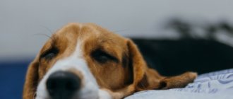
Chondrodystrophic breeds include:
- dachshunds of all varieties
- beagles
- bulldogs (French and English)
- pugs
- welsh corgi
- basset hounds
- Pekingese
- spaniels
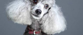
- poodles
- Lhasa Apso
- shih tzu
The disease manifests itself in young and middle-aged animals, usually acutely - symptoms develop within 1-5 days.
The spinal cord can be affected both directly at the level of the spinal disc in which the nucleus pulposus ruptured, and above the bodies of adjacent vertebrae, because The nucleus pulposus “leaks out” and is randomly distributed throughout the spinal canal.
What are the most common health problems?
There can be many causes of illness in a French bulldog, especially if its health is not constantly monitored. Monitor your pet more carefully and do not let even the most minor health problems, as you think, take their course.
Dermatitis
French Bulldogs are known allergy sufferers. Almost anything can cause an allergic reaction in them, since the breed has a low immune threshold and is prone to a strong reaction of the body to an irritant.
The main symptoms of allergies can be quite varied, but the main one among them is atopic dermatitis , which manifests itself in almost all cases.
Dermatitis is an inflammatory skin reaction to an allergen. In dogs, it manifests itself in the form of rashes, inflammation in certain areas of the skin and purulent blisters on the least protected skin (for example, the testicles, skin on the stomach and between the toes).
Most often, dermatitis is visible on the dog, but it is even more likely to show it with constant scratching and restlessness. Due to severe itching, the bulldog may scratch himself in an attempt to itch, which only worsens his situation with pain and the risk of introducing an infection into the blood.
There are less common allergy symptoms - eating disorders. A French Bulldog may refuse to eat, may vomit and have diarrhea. In any case, it is necessary to pay attention to such a reaction of the body, whatever the reason.
Possible causes of dermatitis:
- the dog is allergic to food - in this case, review its diet, eliminating foods one by one to identify the allergen;
- drug rejection. If your pet has recently undergone any treatment, then the cause of the allergic reaction may be due to the medications;
- The dog accidentally swallowed substances that were dangerous to itself. These are household chemicals, hard-to-digest products, junk food not intended for dogs and full of preservatives and dyes;
- The bulldog was bitten by contagious parasites. Dog parasites often carry diseases. Carefully inspect your dog for bites and apply preventive insect repellent ointment;
- congenital allergy. Like people, French bulldogs are often born with allergies to certain environmental factors - dust, fluff and pollen of certain plants, etc.
NOTE!
The only way to cure dermatitis in a dog is to eliminate the allergen, first identifying it quickly. If the allergy is seasonal and the allergen cannot be removed, buy your dog special medications that alleviate the symptoms. Veterinary ointments will help with itchy wounds and skin areas.
Musculoskeletal disorders
The body constitution of French bulldogs implies the development of such problems in any case, especially if the dog is aged - then the chances double or triple.
How quickly your French Bulldog begins to have difficulty walking is up to you.
The dog needs:
- moderate physical activity;
- a complete diet with phosphorus and calcium;
- maintaining normal body weight.
Arthritis and osteochondrosis are the most common diseases of this type in bulldogs. Low withers and widely spaced paws create ideal conditions for the development of problems with the musculoskeletal system.
If you do everything right, your dog will encounter such problems in old age. We can only help by preventing further complications and critical periods.
Prevention includes improving the diet, removing any stress from the dog, and taking bone-strengthening medications.
In irreversible cases (disability or borderline disability), the dog can be given a prosthetic wheelchair to facilitate movement.
The prosthetic wheelchair is the most popular because it is designed for the back legs, which are usually the first to fail in French bulldogs. Unfortunately, not every clinic can make such a device for your pet.
Hind leg failure
The French Bulldog seems to become significantly lower in the spine from the withers to the tail. This structure is very inconvenient in the long term and causes a lot of problems for the dog in old age or even adulthood.
Due to a low seating position, pressure on the spine increases, spinal disc dystrophy and various forms of arthritis develop.
If at first only the vertebral discs are affected, then over time they are completely erased and the animal loses the ability to control its hind legs. The effect is similar to a spinal injury and is also irreversible.
Arthritis, if it does not deprive the dog of the ability to lean on its hind legs, makes it unbearable due to pain in the bones. The bulldog stops moving of its own accord, fearing pain. The best solution in this situation is prosthetics in combination with analgesics and dietary nutrition to avoid obesity.
Heart diseases
In small breed dogs, heart problems almost always turn out to be problems with the mitral valve .
Mitral valve insufficiency is very easy to determine - the dog gets tired very quickly, becomes out of breath, and may even faint. When you try to feel the pulse, you will find a serious arrhythmia.
To diagnose the problem, you can contact a modern veterinary clinic, where your pet will have a chest x-ray, cardiogram and ultrasound cardiography. This is enough to accurately understand the cause of the deficiency.
In such cases, veterinarians advise eliminating nitrates from the animal’s diet as much as possible, and also prescribing heart-strengthening medications - medications keep the weak heart muscle in working condition.
Red and watery eyes
If your French Bulldog's eyes are red or watery, this could indicate an infection, recent eye injury, or a progressive vision problem in the dog.
When infected, the dog will try to scratch the eye with its paw, will become restless and whine, and blink frequently. In such cases, the eye should be carefully rinsed; if this does not help, take it to the veterinarian. It is possible that a foreign object has entered the eye.
IMPORTANT!
The infection can be very dangerous - an inflammatory process begins in the eye, which, if neglected, can deprive the animal of its vision.
If the dog does not itch, then injury may be suspected. Look into the eye and look for a bruise there - if there is one, then the bulldog most likely hit his eye on something.
Vision problems are not as easy to detect as previous health problems. Usually, the owner visually notices eye diseases (cataracts, retinal detachment) too late, already in the presence of “sores” and cloudy membranes of the eye. Tearing may be a symptom.
Scabies
The first thing to do if your pet itches intensely and incessantly is to check the house for parasites. Fleas, lice, bedbugs - all these blood-sucking insects transmit infection to pets. Short-haired, squat and not very fast, the French Bulldog will be an easy target for insects.
Carry out pest control in the house, and thoroughly rid the animal of pests using tablets, gels, special collars and a good bath with special shampoo. If after this the scabies do not stop, then the cause is dermatitis.
Dermatitis as an allergic reaction is treated by eliminating the irritant. Simply remove the allergen factor and over time the dog’s symptoms will disappear.
There is also a worse option - your pet could become infected with real scabies (an infectious dermatological disease) or shingles from another animal or simply on the street.
In this case, the bulldog must be urgently taken to the veterinarian for treatment. If there are other animals in the house, they will have to be isolated.
Severe hair loss
If your French bulldog loses hair outside of periods of natural, seasonal shedding (spring and autumn), or moments of age-related changes (puppies change color quickly and with hair loss, old dogs - simply due to deterioration in the quality of wool with age), then there are reasons for enough worry.
A dog may shed when it does not receive enough nutrients for the body to function properly. When there is a calorie or nutrient deficiency, preserving the coat is not a priority, so it actively falls out, being replaced very slowly with a new one or not being replaced at all.
It could also be due to an allergy you already know.
NOTE!
If your French Bulldog gets everything he needs from food, but still sheds a lot, then it may be due to intestinal helminth parasites, due to which the animal does not receive the nutrients fully.
There may be a genetic predisposition to shedding - this is similar to the human tendency to go bald.
Diarrhea
Diarrhea is an unnatural condition of stool for almost any creature. This indicates an intestinal disorder, harms the dog in every possible way, causes severe gas production and irritates the animal’s anus.
Diarrhea can be either a one-time incident - the dog ate something wrong or picked up garbage on the street, or chronic - the result of some disease or long-term poisoning, allergies.
In the first case, you shouldn't worry too much. Put the dog on a daily diet (do not give the product that allegedly poisoned the animal) and reduce the amount of food for the next two days.
Chronic diarrhea should not be treated so lightly - it is better to take your dog to the veterinarian and find out what is so upsetting its intestines.
Snore
Due to the flattened shape of their muzzle, French bulldogs (and indeed all bulldogs), as well as sometimes Pekingese, pugs and boxers, snore. This is the norm for this breed and there is no need to worry about this - these are the features of the respiratory system of these dogs.
Another thing is when snoring is interrupted by whistling, and the dog itself is obviously having a hard time and restlessness in its sleep. It is also worth paying attention to the fact that your pet snores if, while awake, the dog also chokes and wheezes.
All of these symptoms may indicate an abnormally shaped nasal septum or brachycephalic syndrome.
Considering the problems of such dogs, veterinary clinics have long been performing nose correction operations on them. After such an operation, the dog can breathe almost fully, and its condition improves significantly.
Intervertebral disc disease type II
Type 2 intervertebral disc disease is characterized by protrusion of the fibrous ring into the spinal canal (“bulging disc”), sometimes with extrusion of parts of the nucleus pulposus through the cracks of the ring. Its development is caused by age-related degeneration of the intervertebral disc. Basically, this type is characteristic of non-chondrodystrophic breeds. Manifests chronically with a gradual increase in neurological symptoms from grade 1-2.
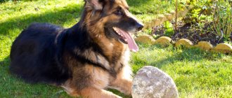
Although BMPD type 2 can occur in dogs of any breed, the most predisposed are:
- german shepherds
- labradors
- dobermans
- Rottweilers.
The disease usually manifests itself in older animals. The spinal cord is compressed directly at the level above the intervertebral disc. Very often, protrusions (disc depressions) are multiple.
Intervertebral disc disease type III
This type of disease is quite rare in both chondrodystrophic and non-chondrodystrophic breeds of dogs, mainly in young animals.
As a rule, symptoms arise abruptly after a significant load, the fibrous ring ruptures, and the nucleus pulposus or part of it is “shot” into the spinal canal at high speed, which leads to severe compression, swelling or rupture of the spinal cord, causing paralysis to occur acutely ( neurological deficit grade 4-5). This type has a poor prognosis.
Diagnostics
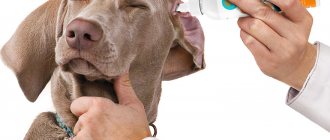
A preliminary diagnosis is made based on a neurological examination and medical history. A veterinary neurologist determines the degree of neurological deficit and the level of damage - i.e. answers the question of conduction at the level of which segments of the spinal cord is impaired and to what extent.
Further diagnosis consists of neuroimaging methods: this is MRI of the corresponding part of the spinal cord or, if this study is not available, myelography.
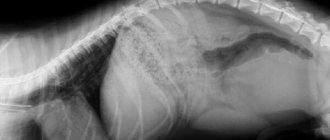
Myelography is the injection of a radiopaque substance under the dura mater of the spinal cord. After this, the contours of the spinal cord become visible on a regular x-ray (without contrast, both the spinal cord and intervertebral discs are transparent to x-rays), due to which you can see areas of deformation, compression or destruction (myelomalacia) of the spinal cord.
Thanks to these studies, you can find out exactly which disc is damaged and determine the location of surgical intervention if necessary.
Treatment
Treatment depends on the degree of neurological deficit.
If we are talking about the initial stages, when there is only pain and impaired movement, conservative (non-surgical) methods are used - painkillers and anti-inflammatory drugs are prescribed and strict rest (limitation of movements) with the help of cellular maintenance for a period of at least 3 weeks. Exact localization of the lesion (data from MRI, CT or myelography) is not required to prescribe conservative therapy.
Often such therapy is enough to prevent further development of the disease and relieve the dog of pain for a period of several months to several years, and with some luck - forever. However, you need to understand that the disappearance of symptoms does not mean that the dog has recovered - if the intervertebral discs are affected by degenerative changes, their “recovery” will no longer occur. If, after a period of cellular dormancy, the dog experiences a relapse, diagnostics are performed to localize the lesion (MRI) and surgical treatment.
If the degree of deficiency is higher than the second (the animal is paralyzed), then, as a rule, symptomatic treatment is not enough; it is necessary to eliminate the cause of compression (compression) of the spinal cord as soon as possible in order to prevent damage to the deep structures of the spinal cord and irreversible changes in it.
The operation performed to provide decompression is called laminectomy and consists of partial removal of the vertebral arch above the site of displacement of the intervertebral disc and extraction of the disc substance from the spinal canal.
Removing the vertebral arch ensures that spinal cord compression does not develop again in the same location, but does not preclude re-displacement of the disc in another (adjacent) intervertebral space - this is uncommon, but occurs in dogs with multiple degenerative changes in the discs.
The earlier the operation is performed, the better the prognosis for surgical treatment. There are isolated cases of recovery of animals operated on with degree 6 neurological deficit, but in general the prognosis at this stage is poor.
Physiotherapeutic rehabilitation methods for spinal patients play an important role in the recovery of animals after surgery.
In addition, if laminectomy is unsuccessful or performed too late, with the help of intensive physiotherapy it is possible to develop the so-called spinal (reflex) walking in a hopelessly paralyzed dog.
Appearance of the sphenoid vertebra
The French Bulldog often suffers from such a pathology as the formation of an irregular vertebra (wedge, trapezium). Wedge-shaped vertebrae (sometimes 2-3 or more) are a defect that is not considered a disease. This is typical mainly for this breed.
A trapezoid or wedge-shaped vertebra compresses neighboring vertebrae, then gradually moves to the side and begins to put pressure on the spinal cord. Also, when jumping or suddenly changing body position, it may dislocate or a hernia may appear.
Veterinarians believe that wedge-shaped vertebrae cause pain, weakness in the limbs and are the cause of herniated discs, spinal curvature, lordosis and kyphosis. Veterinarians have expressed the opinion that high jumping is contraindicated for the French bulldog.
Correct diagnosis is important as sphenoid vertebrae is often confused with disc disease. And if in the latter case drug therapy is possible, then with a changed vertebra it is not effective.
surgery is shown. However, in this case, success depends on the area affected. If 5-9 vertebrae are damaged, surgery is not performed due to difficult access.
Prevention
Unfortunately, it is impossible to prevent the development of intervertebral disc diseases with absolute reliability.
In a global sense, the main method of preventing degenerative diseases of the intervertebral discs is breeding hygiene - the exclusion from breeding of dogs that have had this pathology and their immediate relatives.
For a particular dog, especially if we are talking about a chondrodystrophic breed, the main and accessible prevention of discopathy in dogs is maintaining good physical shape (developed muscle corset, lack of excess weight).
Author: Kurganskaya (Shuraleva) Natalia Ivanovna
(c) Veterinary center for the treatment and rehabilitation of animals “Zoostatus”. Varshavskoe highway 125 building 1. tel. 8