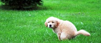
Ophthalmological diseases of dogs are primary (main congenital, acquired diseases) and secondary, which arise against the background of various systemic, functional pathologies and diseases. In addition, eye diseases in animals are classified into congenital and acquired.
Eye diseases in dogs, based on etiology (causes), can be:
- Infectious. Development is promoted by various viruses, bacteria, pathogenic protozoa, and fungi.
- Non-infectious. Among the main reasons are mechanical damage, trauma, bruises of the eyelids, foreign objects in the eye, congenital anomalies, abnormal eyelash growth, and neoplasms.
- Pedigree. Eye diseases typical for certain breeds of dogs. For example, brachycephalics are diagnosed with eversion, entropion of the eyelids, and prolapse of the third eyelid.
Eye diseases in dogs can be caused by allergies, chemical and thermal burns, inflammation of the sebaceous and meibolian glands, parasitic and invasive diseases, inflammatory processes, and hypovitaminosis.
As you can see, eye diseases in dogs have very different etiologies and genesis, as well as characteristic symptoms. Some diseases are typical for puppies and young pets, while others most often appear in animals as they age.
Content
1. What is the causative agent of the disease 2. Where and how a dog can become infected with distemper 3. Incubation period 4. How the disease manifests itself in practice 5. Variants of the course of the disease 6. Is it possible to cure a dog 7. What conditions must be created for the animal 8. Video: 9. Let's sum it up
Distemper in dogs is a dangerous viral disease that manifests itself in its most acute form in young dogs, puppies. It is almost impossible to cure an animal infected with paramyxovirus; after contracting the disease, the dog itself becomes a carrier of distemper and can, upon contact, transmit the pathogen to another dog. At an early stage, the symptoms are almost invisible, but if the animal is affected by the virus, treatment in the vast majority of cases will not lead to anything good.
That is why one of the most important tasks of a dog owner is strict control of the animal’s condition, knowledge and ability to recognize the very first signs of possible penetration of the virus into the pet’s body. A caring owner knows how to protect the health and life of his dog and knows how to do it. Well, the information presented below will help you understand the symptoms, causes of the disease, methods of transmission, diagnosis, self-control, treatment procedures and other important issues of caring for your dog!
Adenoma of the third century
Adenoma of the third eyelid , or correctly said – prolapse (prolapse) of the lacrimal gland of the third eyelid.
Usually observed in young dogs aged from 6 months to 2 years, most often in brachiocephalic breeds (Shih Tzu, cocker spaniels, pugs, bulldogs, etc.), beagles, etc. This occurs due to genetically determined weakness of the ligaments that fix the gland to the edge of the eye socket. Symptoms:
— An oval red formation appears in the inner corner of the eye, protruding from under the edge of the third eyelid. It is most often observed in one eye, but can also be bilateral.
- There may be moderate lacrimation and squinting of the eyes (blepharospasm).
Treatment:
1. The gland must be returned to its place surgically.
============================================================================================================================================================================================
What is the causative agent of the disease
Paramyxovirus (contained in the body's RNA) is the main and only causative agent of the fatal disease. A characteristic feature of the pathogen is increased resistance to external influences, preservation of all harmful parameters in almost any environmental conditions. The only effective influence that destroys the virus completely is boiling water! The first rule: when caring for a sick animal, it is necessary to regularly and thoroughly disinfect it, cleaning dishes, bowls, toys and other objects surrounding the pet. Sterility and constant disinfection increases the likelihood of getting rid of the disease.
The viral disease is extremely dangerous and has amazing persistence. After the dog has fully recovered, the dangerous elements of the disease carrier remain in the body, manifesting itself in all sorts of secretions - saliva, feces, urine. All these feces pose the highest danger to healthy individuals.
The canine distemper virus is characterized by:
- The ability to reproduce very quickly.
- The strongest pathogenic effect on the dog’s body.
- Lethal effects on various internal organs and systems of the animal.
- Preservation of activity in the secretions of a sick dog.
As soon as the virus enters the dog’s body, it immediately becomes infectious and is a dangerous carrier of infection (with all the ensuing consequences).
Dry eye syndrome
Dry eye syndrome , or keratoconjunctivitis sicca, is an inflammation of the conjunctiva and cornea caused by insufficient tear production and, over time, leading to decreased vision.
Symptoms:
— Redness of the eye, squinting (blepharospasm)
- Mucous or mucopurulent discharge from the eyes
— Changes in the cornea: normally the cornea is transparent and shiny, with dry keratoconjunctivitis it becomes dull, with visible opacities, brown pigmentation, can grow with blood vessels, and corneal ulcers often appear.
Often dry keratoconjunctivitis is complicated by a secondary infection and is mistaken for chronic conjunctivitis.
Treatment:
1. The eyes must be cleared of secretions; to do this, they are washed with a 0.05% chlorhexidine solution.
2. Locally, for secondary bacterial infections, drops and ointments with broad-spectrum antibiotics (cyclosporine) are used.
3. Preparations that moisturize the surface of the eye must be used.
============================================================================================================================================================================================
Where and how can a dog become infected with distemper?
Unfortunately, a carnivore can become infected with the virus in a variety of ways. The virus enters the body directly through the respiratory system or digestive system. Once viruses enter the body of a carnivore, they find an ideal environment for rapid spread. The circulatory system contributes to this, delivering the infection to all major organs, to its most remote places and tissues.
An already infected carnivore begins to spread the disease itself. Experts recommend paying close attention to the following places, which are the most likely sources of infection:
- A carnivorous dog already infected with the virus.
- Sick wild animals (foxes, wolves, ferrets, etc.). One of the reasons why the closest and tireless attention should be paid to hunting dogs.
- Places of permanent or most frequent habitat of animals - where they eat food, eating utensils, sleeping places, toys.
- Any type of premises allocated for a pet: dog enclosures, storage rooms in apartments, etc.
- The virus often settles and can accumulate on the outer clothing of a person in contact with an infected animal.
As previously noted, the virus retains its danger and aggressiveness for quite a long time; it can only be overcome by the most thorough sterilization, the use of a set of measures to maintain cleanliness, disinfection, cleaning and scalding with boiling water of all objects and things that come into contact with an infected carnivorous individual.
Trichiasis
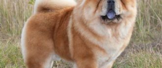
Trichiasis is a condition when hair from a dog's eyelids or muzzle gets into the eye, coming into contact with the conjunctiva and cornea. Trichiasis can be primary or secondary. Primary occurs in dogs with medial inversion of the eyelids and a large nasolabial fold. Trichiasis occurs in the following dog breeds: Pekingese, Pugs, English Bulldogs, English Cocker Spaniels, Chow Chows, Shar-Peis.
Clinical picture. During a clinical examination of a dog, a veterinarian notes lacrimation, hairs in contact with the cornea cause blinking in dogs, constant discharge from the eyes, symptoms of keratoconjunctivitis, inflammation of the skin in the area of the nasolabial fold.
The diagnosis is made based on the detection of hair in contact with the cornea, provided there is no other eye pathology.
Differential diagnosis. Trichiasis is differentiated from keratoconjunctivitis sicca, entropion and eversion of the eyelids, dystrichiasis, and ectopic eyelashes.
Treatment. Treatment of the disease is surgical. Temporary improvement can be achieved by trimming the hair that gets into the eye.
Incubation period
After direct infection of the animal has occurred, during the first few days (everything depends on the degree of immunity), a secretive period of development and progression of the disease continues. There are practically no external signs, but the rapid spread of the harmful virus continues in the body, which, together with the blood, is transferred to all the most distant organs and systems of the body.
On average, the duration of the incubation period ranges from 5 to 7 days, but there have been cases of an increase to 2-3 weeks or a sharp, almost immediate manifestation already on the second day after the deadly infection penetrates the dog’s body.
Follow us
Find out more about your animal's personality on our social networks
The disease is not seasonal. At least, veterinarians do not have any accurate data. Moreover, the virus shows a high level of resistance even in conditions of extremely low temperatures and retains its activity and aggressiveness at – 24 – 26 °C. However, every owner should know that, according to statistics, the highest percentage of infection occurs in spring and autumn.
At an early stage after distemper penetrates inside, this lesion does not manifest itself in any way on the dog. She looks absolutely healthy, remains mobile, active, eats well, however, she becomes an increased source of danger to other carnivores. The canine distemper virus can continue to spread even after the dog has recovered, so it must be isolated from the outside world for this period of time.
Who is at high risk?
First of all, these are carnivores with weakened immune systems, who have become ill or have suffered some kind of disease and have not yet fully recovered. Stray, homeless animals are a separate topic for discussion; they are not only dangerous, but, to the extent possible, they should be isolated from contact with other animals and people until the end of quarantine. A viral disease will more quickly affect those dogs that do not receive a balanced diet. But puppies, whose age ranges from 2 to 6 months, up to 1 year, who suckle a mother who previously suffered from this disease and was successfully cured, develop stable immunity; they are not afraid of distemper.
What external factors determine whether an animal is infected?
A viral infection at the very beginning after damage to the body practically does not manifest itself in any way. There are no symptoms or they are almost invisible. And only an attentive and caring owner is able to determine, by the most insignificant indirect signs, the possible damage to the body of a carnivorous animal by this dangerous virus. You should remember the first signs of a possible dog infection with distemper. This will allow you to promptly and promptly respond to the disease at its earliest stage, take all necessary preventive measures, undergo examination and begin treatment. The owner of a four-legged pet must be extremely attentive to the following deviations in the behavior and appearance of a carnivore from the norm:
- The animal begins to show a certain lethargy and laziness.
- A depressed state is recorded.
- Appetite decreases, and in some cases, vomiting complications are recorded.
- The mucous membrane around the eyeball becomes red.
- In the area of the eyes and nose, the formation of mucous secretions that are more abundant than in traditional standard conditions occurs.
- The fur becomes somewhat disheveled (this symptom is observed in the vast majority of carnivores).
- A dog infected with the distemper virus begins to show fear of bright light and, at the first opportunity, tries to go into the shade, huddle in a dark corner, away from the sun's rays.
It should be understood that all of the above signs have varying degrees of manifestation in a particular individual. It all depends on the breed, age, and also immunity. In the first couple of days, the dog’s body temperature rises sharply, can reach 40 ° C and last for 2 - 4 days. Cases have been recorded when adult individuals coped with the disease on their own and in this case the main signs of plague disappeared. Weak individuals suffering from various types of diseases, carnivores with a disrupted nutritional system, young dogs are categories of increased danger. All of them become seriously ill after being infected with the virus, and their condition deteriorates significantly quite quickly.
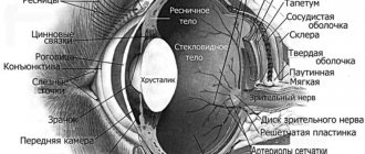
Diseases and problems of the choroid and cornea
The choroid and cornea of the eye may suffer mainly from the progression of inflammatory processes. Failure to promptly contact a veterinary ophthalmologist can lead to complete blindness of the dog. Moreover, this can happen in a short period of time, since such pathologies have intensive development dynamics.
Ulcerative keratitis
In the eyes of an animal, ulcerative keratitis develops as a result of sun or thermal burns, when exposed to mechanical forces during impacts, or when foreign objects enter the eye. In addition, ulcerative keratitis is a secondary disease against the background of allergic abnormalities, vitamin deficiency, bacterial and viral infections. Another reason for this pathology is endocrine diseases (for example, diabetes mellitus).
With such a lesion, tearing develops. In this case, the dog rubs its eyes with its paws, which indicates itching, discomfort and the presence of foreign bodies on the cornea. The eye may be very sore. Blue eye syndrome also occurs when the pigmentation of the pupil changes under the influence of pathological factors.
In these circumstances, veterinarians prescribe drug therapy with antimicrobial, antihistamine, painkillers, as well as external agents to localize the inflammatory process.
Uveitis
Uveitis is an inflammatory ophthalmological disease. It is accompanied by damage to the choroid of the eye and disruption of the blood supply to its tissues.
Signs of intense inflammation of the irises are changes in their color, fear of bright light, half-closed red eyelids, and decreased visual acuity. Uveitis occurs due to injuries to the head and eye area, viral and bacterial infections.
If a dog's eye is inflamed in the area of the iris, anti-inflammatory drugs are mainly used to treat uveitis, as well as drugs to reduce pain.
How does the disease manifest itself in practice?
Depending on the pronounced clinical manifestations, experts divide plague into 4 main subcategories. As a rule, they do not occur in isolation, but complement one another in the process of further progression of the disease and damage to the body of the carnivorous individual.
The classification provides the following options:
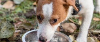
| Pneumonic plague | It actively develops and progresses when the virus accumulates in the lungs and respiratory tract. The symptoms here are fever, cough. The pet categorically refuses to take any food, while drinking a lot and often of water. Further progression of the disease includes diarrhea, gag reflexes, and copious discharge from the mucous membrane. |
| Intestinal type | This form of the virus primarily manifests itself in the form of vomiting and diarrhea (the color of liquid stool has a yellowish tint), which has a pungent and extremely unpleasant odor. A careful examination of the surface of the tongue can reveal a fairly thick layer of white plaque. The enamel of the teeth is also damaged, on the surface of which clearly visible dark spots appear. The animal becomes lethargic, inactive, weakened, eats practically nothing and may even lose consciousness from time to time. |
| Skin | The cutaneous form of plague is considered the mildest type of viral disease. It is here that both the pet and its owner have an excellent chance of a speedy recovery without any pathologies. The virus manifests itself as a slight redness that appears on different parts of the body, primarily on the pads of the paws, in the nose and on the surface of the ears. A little later, these rashes turn into small ulcers. The danger of the disease lies in the high probability of secondary bacteria entering the wounds, after which inflammatory processes are activated and this causes the animal some pain and increased discomfort. If treatment is not started in time, the situation may worsen, become irreversible and lead to the death of the pet. |
Distichnaz
With this disease, single or multiple hairs appear in a row on the free edge of the eyelid, which should be hairless.
These hairs appear in a dog only at the 4-6th month of life and can be either very delicate or quite hard. With this disease, several hairs most often grow from one point. This disease is most often recorded in English and American cocker spaniels, boxers, Tibetan terriers, collies, and Pekingese.
Clinical picture. During a clinical examination of a dog, a veterinarian notes profuse lacrimation, constant blinking, blepharospasm, irritating hairs have contact with the cornea of the eye. If a dog has curled eyelashes, keratitis is diagnosed.
The diagnosis of the disease is made based on the above symptoms.
Differential diagnosis. Distichnasis is differentiated from trichiasis, entropion and eversion of the eyelids, allergic conjunctivitis, and keratoconjunctivitis sicca.
Treatment. It is carried out in veterinary clinics by electrolysis under an operating microscope. Excision of the third eyelid.
Variants of the course of the disease
Canine distemper can develop at lightning speed or manifest itself as a chronic disease. The most common variants of the course of the disease:
- Fulminant. In this case, there are no characteristic obvious clinical signs, and death occurs, as a rule, within 1 day.
- An extremely dangerous form of distemper is determined by a sharp jump in temperature and the simultaneous refusal of the animal to eat any food. A coma gradually sets in and the dog dies within 2-3 days.
- All obvious and most common variants of the manifestation of plague are recorded precisely in the acute form of the disease. These include lethargy, fever, depression, decreased reflex reaction, gastrointestinal disorders, photophobia.
- When the disease is chronic, the virus persists in the animal’s body for many days, up to several months.
The distemper virus poses the highest degree of danger to a carnivore when it affects the central nervous system. With this variant of the disease, the animal dies in more than 90% of cases.
Exophthalmos (protrusion of the eyeball)
Exophthalmos in dogs can be species-specific and is typical for dogs of brachycephalic breeds, with a normal eyeball size, a flat orbit and an overly large palpebral fissure.
Acquired exophthalmos - in which a normal-sized eyeball moves forward due to space-demanding processes in the orbit or its immediate surroundings, or due to an increase in the size of the eyeball as a result of glaucoma in a dog.
Clinical picture. During a clinical examination, the veterinarian notes that the dog has strabismus, an abnormally wide palpebral fissure with protrusion of the eyeball; in some dogs, prolapse of the third eyelid is possible.
Treatment is only surgical.
Is it possible to cure a dog
The only right decision at the slightest suspicion of an animal being infected with distemper is to contact a veterinarian. Only an experienced, qualified specialist is able to accurately diagnose, assess symptoms and develop the best option for treatment and recovery procedures. Every owner of a four-legged friend should know that a complete, specific medicine that can specifically cope with the distemper viral infection has not yet been developed. Therefore, when organizing and implementing medical procedures, veterinarians prescribe complex therapy. Specific treatment methods depend on the diagnosis performed, the depth and type of damage to the pet by the distemper virus, and the presence of certain severe symptoms. One of the areas actively used in treatment by veterinarians is the prescription of drugs that strengthen and enhance the animal’s immunity.
As a rule, the following types of solutions are prescribed to a carnivore as intramuscular injections:
- Hexamine (40 percent)
- Glucose (40%)
- Calcium gluconate
- Vitamins (ascorbic acid)
In some cases, the veterinarian will recommend the use of the drug Mydocalm as a means to reduce muscle tone, and if increased excitability is detected in the dog, phenobarbital is prescribed.
What is categorically not recommended is carrying out treatment on your own, without the help of a qualified specialist. Only a complete diagnosis, as well as recommendations from a veterinarian, will allow you to most accurately determine the type and degree of complexity of distemper damage to the body and organize effective treatment.
Cataract
Cataract is clouding of the lens, leading to a decrease in visual acuity, up to its complete loss. The clouding may be partial or complete. The release of protein from the lens (phacolysis) leads to chronic phacolytic uveitis (endophthalmitis), a severe intraocular inflammation. The disease can be congenital or hereditary and can occur at any age.
Symptoms:
— Decreased or loss of vision in the dog, blurred eyes.
Treatment:
Currently, no conservative (therapeutic) methods for treating cataracts have been developed. To restore visual function and treat secondary complications, surgical treatment is effective, during which the affected lens is removed and, if indicated, an artificial lens is implanted.
============================================================================================================================================================================================
What conditions must be created for the animal?
For a speedy recovery of the dog and a complete cure for distemper, it is necessary not only to give the correct drugs and medicines, but also to create external conditions that will contribute to recovery.
Here are the main ones:
- Prepare the room, pantry, disinfect and sterilize. Ensure a stable microclimate, no drafts.
- Curtain the windows, dim the lights, darken the room. At the time of illness, the dog develops photophobia.
- Constantly treat the affected areas of the skin with a special disinfection solution, remove mucous and purulent formations around the eyes and near the nose.
- Review your diet. Give your pet light food. Liquid porridges with soups, give a little boiled, finely chopped meat. Twice a week - milk, cottage cheese.
Follicular conjunctivitis
This form of conjunctivitis is most typical of chronic conjunctivitis and often develops in a dog when toxic substances get into the eye.
Clinical picture. During a clinical examination, a veterinarian reveals many bubbles with transparent contents on the mucous membrane of the conjunctiva. Mucous discharge comes from the palpebral fissure. The conjunctiva itself is crimson in color, and the dog's inflamed eye is squinted.
Treatment. When treating this form of conjunctivitis, eye ointments containing an antibiotic are used. In severe cases of the disease, specialists are forced to resort to excision of the conjunctiva and subsequent symptomatic treatment.
Eyeball luxation
When the eyeball is dislocated, dog owners note that the eyeball comes out of the orbit behind the eyelid completely or partially.
This pathology is most often found in Pekingese, Japanese hips and similar breeds of dogs.
Cause. Dislocation of the eyeball in a dog most often occurs due to mechanical damage to the bones of the head and temples, high muscle tension in dogs with a shallow depth of the bone orbit.
Clinical picture. During a clinical examination, the veterinary specialist of the clinic notes a strong extension of the eyeball beyond its natural boundaries, the conjunctiva is swollen, often dries out, and externally takes the form of a hanging cushion.
Treatment. The treatment of this pathology is surgical.
Uveitis
Inflammation of the mucous membranes and irises, from which no dog is immune. The reasons are different: trauma, internal chronic diseases, infection or virus. Symptoms: inflammation of the eye, change in iris color (cloudness), decreased vision, photophobia (the dog hides in dark places), severe pain and half-closed eyelids. Treatment is aimed not only at eliminating pain and inflammation, but also at preserving vision.
| List of drugs for treatment | ||
| Name of medicine | Price | Release form and application |
| Rimadyl (base – carprofen) | 500-650 rubles per plate of tablets (10 pieces) | The product is used to reduce inflammation and relieve pain; The drug comes out in the form of a syrup, the dose is selected only on the recommendation of a veterinarian |
| Knuckles | 80-100 rubles per ampoule | Anti-inflammatory substance (not steroid), available in the form of an injection solution based on diclofenac |
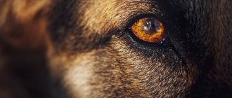
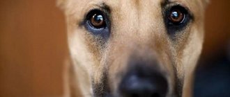
Caring for your pet's vision
A dog’s eyes are, if not the mirror of the soul, then certainly an indicator of its health. Using them, an experienced breeder or veterinarian can tell whether an animal is sick or not. Clean, dry eyes, with a vibrant shine, without swelling or redness - this is a sign of a completely healthy pet.
If everything is fine with the eyes, then in most cases they do not require any care. Sometimes discharge may accumulate in the inner corners of a dog's eyes. If their number is insignificant, then it is enough to remove them with a piece of gauze or a cotton pad. It is not recommended to use cotton wool for these purposes, since its particles can get inside and cause discomfort and even inflammation.
The following liquids can be used for such processing:
- boiled water;
- saline;
- weak solution of furatsilin.
Traditional tea brewing for people is also suitable, but there is one small caveat - it can leave colored areas on dogs with light hair.
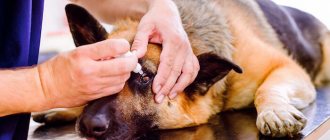
Treatment of the disease
The veterinarian conducts an external examination and a full diagnosis of the dog’s condition, and, depending on the results, prescribes treatment.
If the cause of lacrimation is inflammatory diseases of the eye structures, then anti-inflammatory and antibiotic-containing eye drops are prescribed (the most effective are Bars, Tsiprovet, Diamond Eyes, Tobrex) and ointments (the most popular are tetracycline and chloramphenicol). Treatment can take place at home, but under the supervision of a specialist.
The owner needs to know the technique of instilling eye drops into a dog and applying antibiotic ointment:
- Before instillation, you should rinse your eyes and remove any secretions and crusts that have formed.
- Holding the dog tightly, tilt the head back slightly and carefully drop the medicine into both eyes. Leave in this state for a few seconds so that the medicine washes the mucous membrane.
- It is necessary to place the ointment in the conjunctival sac, carefully pulling back the lower eyelid. Afterwards, you should close your eye for a while, waiting until the medicine is absorbed.
- Bilateral treatment should always be performed, even if one eye is healthy.