A change in the usual color of the skin in certain areas of a pet’s body can be caused by a number of factors, both hereditary and acquired.
Spots on a dog's skin may indicate the development of atopic dermatitis, hormonal disorders, or metabolic problems.
Acanthosis nigricans in dogs
Acanthosis nigricans is considered one of the most serious dermatological diseases in dogs. Its appearance is explained by changes in the superficial layers of the dermis, often as a result of genetic changes. The maximum number of cases of acanthosis nigricans has been recorded in Dachshund dogs, but this does not mean that the disease cannot be diagnosed in dogs of other breeds.
Changes in skin pigmentation
Pigment spots can be of any color (from dark black to reddish). Each type is caused by a specific condition or disease.
Black dots
The appearance of black spots on a dog's skin is most often caused by lentigo. It consists of single spots or group spots of dark black color. Their occurrence is caused by macular melanosis, that is, excessive accumulation of melanin in the body. With canine lentigo, the appearance of flat, hyperpigmented spots of a benign nature is observed. Their localization is the groin area or abdomen.
Features of pathology and description
It is believed that the disease is based on genodermatosis, in which excessive hyperpigmentation occurs. But acanthosis of secondary origin is much more common, but its causes have not yet been established, although the study of this issue continues. The disease is considered a type of anti-inflammatory hyperpigmentation.
Causes of acanthosis nigricans may include:
- Inflammatory reactions in the most delicate groin and axillary areas.
- Endocrine diseases, incl. hypothyroidism, hypersecretion of corticosteroids.
- Obesity, when a dog’s weight is diagnosed above normal and as a critical body mass.
- Nutritional allergies due to improper feeding.
- Infections of the upper layers of the dermis.
Important! Contrary to popular belief among owners, acanthosis nigricans is not contagious. It is not transmitted from animal to animal, much less to humans. Another thing is that a dog with damage to the dermis does not look the best, so at the first signs of acanthosis you need to urgently contact a veterinary clinic.
Diseases caused by pathogenic fungi
Dermatophytoses
Skin diseases resulting from parasitism by pathogenic fungi of the genera Microsporum and Tpichophiton are collectively called dermatophytoses (dermatomycosis). Dermatophytes have the ability to infect healthy hair or skin. The infected part of the hair is very fragile and often breaks off, so the hair in the affected area has the appearance of being cut off (hence the outdated name “ringworm”), up to complete baldness. In addition to the hair shaft, areas of interfollicular keratin can also be affected, which is manifested by peeling of the skin surface. The severity of inflammation varies. Infection with non-host-specific fungal species causes a stronger inflammatory response than with host-adapted species. The hyphae of some (not all) Microsporum species produce a substance called pteridine, which can absorb ultraviolet rays and fluoresce. This property underlies the luminescent diagnosis of microsporia. However, the absence of fluorescence does not exclude dermatophytosis.
It is believed that the cause of dermatophytosis in cats in 95% of cases is Microsporum canis. The remaining 5% of cases are caused mainly by Microsporum gypseum and Trichophiton mentagrophites. Microsporum canis is well adapted to cats as hosts, so the disease is often asymptomatic. It is very difficult to isolate the pathogen from healthy animals that have not previously suffered from microsporia, while the carriage level in cats suffering from dermatophytosis reaches 100%.
Dermatophytosis is more common in kittens and occurs in a more severe form. In adult cats, the disease is more common in individuals with weakened immune systems, pregnant or lactating. Sick cats pose a danger to humans and other animals.
Dermatophytosis is much less common in dogs than in cats. In most cases, Trichophiton mentagrophites, Microsporum canis, Microsporum gypseum are responsible for the occurrence of dermatophytosis in animals of this species. Clinical signs of dermatophytosis are very diverse and are not always limited to the classic picture of the disease. In most animals, the most typical manifestations are erythema, alopecia, scaling and crusting. Lesions are localized on the scalp and neck, less often at the base of the tail and limbs. The spots are small at first, gradually expand and reach a significant size. Hair loses normal pigmentation, becomes brittle and is easily epilated. The skin in the affected area is dense, red-brown or grayish in color. Animals scratch the affected areas, which often contributes to the transfer of the disease from the head to the limbs. As a rule, the itching is mild. The exception is infection caused by Trichophiton mentagrophites. Dermatophytosis can occur in conjunction with other diseases, mainly ectoparasitic: demodicosis (in dogs) and notoedrosis (in cats).
Malassezia dermatitis in dogs
Recently, a number of skin diseases in dogs (atopic dermatoses, otitis externa) have been complicated by yeast fungi of the genus Malassezia, especially Malassezia pachydermatis. Dogs of all breeds are susceptible to Malassezia dermatitis, but basset hounds are particularly susceptible. Skin lesions that are associated with Malassezia pachydermatis may be localized or generalized. Affected areas usually include the external auditory canal, muzzle, ventral neck, axillary cavities, groin area, and interdigital skin folds. Most often, the disease is characterized by erythema, alopecia, and dry or oily seborrhea. In chronic cases, lichenification and hyperpigmentation are observed. The itching varies from mild to extremely severe. Skin lesions are often accompanied by an unpleasant odor, especially in places such as the neck, axillary fossae, and ears.
Acarodermatoses
Tick-borne dermatoses of dogs and cats occupy a significant place among skin diseases in these types of domestic animals. Diseases caused by these arthropods are usually accompanied by severe itching, scratching, baldness, and secondary pyoderma, which not only causes suffering to the sick animal, but also greatly upsets its owners. In addition, sick dogs and cats are a source of disease for other animals and for people. Therefore, the closest attention must be paid to the treatment of these dermatoses. For veterinary dermatological practice, acarodermatoses such as notoedrosis , sarcoptic mange , otodectosis and demodicosis . These diseases affect both dogs and cats, but with varying frequencies. Thus, notoedrosis and otodectosis are more often recorded in cats, sarcoptic mange and demodicosis - in dogs.
Atopic dermatitis
Atopy is a hereditary predisposition to the formation of antibodies against allergens from the environment (pollen, poplar fluff, house dust, etc.). Since atopy is a polyetiological disease with a variety of clinical manifestations, its diagnosis and treatment pose a certain difficulty for veterinarians. In terms of frequency of occurrence among all allergies, atopic dermatitis is second only to allergic dermatitis from flea bites. Often the latter accompanies the former, complicating and confusing the clinical picture of the disease. Food allergy, and in dogs also pyoderma, can also contribute to the complication of the disease. The clinical manifestations and diagnosis of atopic dermatitis in dogs and cats are largely similar, but there are also differences.
In dogs, atopic dermatitis most often occurs between the ages of 1 and 3 years and affects from 3 to 15% of the entire population of animals of this species, regardless of gender. Breeds susceptible to this disease include terriers (WHWT, Scotch, Fox), golden and Labrador retrievers, boxer, cocker spaniels, German shepherd, Shar-Pei, Dalmatian, English bulldog, miniature schnauzer, Irish and English setters. The most typical clinical signs of atopic dermatitis in dogs are pruritus, alopecia, erythema, hyperpigmentation, and lichenification, which are found on the face, feet, chest, ears, abdomen, and tail. Depending on the source of the allergen, atopy can be seasonal or cause trouble for the animal and its owner for most of the year.
Dogs with atopic dermatitis are more likely than others to be affected by a yeast infection (Malassezia), which is facilitated by inflammation and oily seborrhea. In the interdigital spaces, ideal conditions for the proliferation of fungi are created due to increased humidity and relatively higher skin temperature in these areas.
Forms of acanthosis
There are two forms of the disease:
- Primary acanthosis nigricans.
Diagnosed exclusively in dachshunds, gender and age are not traced. When dark spots appear in dogs under one year of age, it can be assumed that acanthosis is transmitted by heredity. Often the manifestation of “elephant skin” is associated with endocrine disorders or kidney diseases.
- Secondary acanthosis nigricans.
It is found not only in dachshunds, but also in dogs of different breeds, incl. mongrel. But the special vulnerability of pets with short, hard, smooth hair has been noted. In this case, the symptoms of skin changes act as a “background sign”; in addition, the symptoms of the main disease appear.
Important! You should not equate various acquired or chronic skin problems in dogs with acanthosis. Pseudoacanthosis often manifests itself with similar external symptoms and they are provoked by a malfunction of the endocrine system, dermatitis or allergic reactions. More often, this is a consequence of improper maintenance and feeding of dogs, giving fatty foods, internal fat and little physical activity.
Diseases manifested by the appearance of spots on the skin
Genetic diseases:
- lentigo;
- epidermal nevus;
- acanthosis nigricans.
Past illnesses:
- acanthokeratoderma;
- atopic dermatitis;
- acute weeping dermatitis.
Acquired deviations:
- a reaction to inflammation caused by bacteria, infections or viruses;
- hormonal disorders;
- drug allergies;
- papillomavirus;
- pigment tumors;
- metabolic failures;
- unbalanced diet or overeating.
Each type of lesion has its own characteristic color of the spots and their location.
Main symptoms of the disease: how to recognize
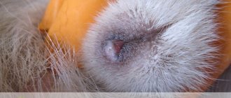
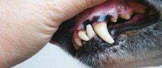
The first thing the owner may notice is the appearance of unevenly dark-colored spots on the pet’s body without signs of inflammation. More often they appear in the armpits and groin, this is especially noticeable in dogs with light pigmentation of the skin and coat.
At first, the dark area will be small and resemble a brownish blot; there may be several such “blots” at the same time. An attempt to wash or scrape does not lead to results, because... the lesion affects the dermis.
What areas of the body are affected:
- Ears, outer and inner sides.
- Folds on the neck and face, especially in dogs whose “folding” is pronounced (Shar Peis).
- Abdomen, groin area.
- Knee and elbow bends.
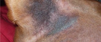
The main symptom of acanthosis nigricans is thickening of the affected part of the dermis with further hyperpigmentation and melanin deposition. Moreover, the skin may acquire a bluish tint. Over time, the skin becomes rougher, and hyperpigmented folds and growths appear.
Gradually, the size of the darkened areas increases; they can merge into one, larger spot, which already affects large areas of the dog’s body. Subsequently, symptoms of seborrhea appear.
Often, acanthosis is accompanied by symptoms of an inflammatory process, especially with constant irritation and infection of the skin. Then the spots along the edges become as if outlined, a red border stands out.
In the secondary form of the disease, alopecia (bald spots) may appear on the depigmented area of the skin, and hair may partially or completely fall out. If the dog touches the spot and shows signs of pain, and this effect develops only in the secondary form, primary acanthosis does not cause concern to the pet.
What breeds have pigmentation?
Any dog can develop dark spots on its skin, especially those that have already reached an advanced age. There are several breeds that exhibit changes in skin color due to abnormal changes caused by hereditary predisposition.
Breeds that are characterized by the appearance of pigmentation:
- French bulldogs suffer from the age-related appearance of the so-called “spotting”, in which several small or one large spot appears on the belly.
- Pugs, miniature schnauzers and sharpeis are prone to developing benign localized spots (lentigines).
- Partial pigmentation in the area of the nose and lips is characteristic of the Akita Inu, Samoyed dog, and Siberian Husky.
- The skin of the Chow Chow and Akita Inu is affected by an acquired autoimmune disease, after which an inflammatory process with purulent discharge may appear.
- Dachshunds suffer from acanthosis nigricans, or elephant skin as it is popularly called. With such hyperpigmentation, rough, modified areas of skin appear. Most often they cover folds in the dog’s neck, groin or armpits.
- Lupus vulgaris occurs in Scottish Sheepdogs.
- Doberman Pinschers, Rottweilers, Siberian Huskies, Alaskan Malamutes and Labrador Retrievers are characterized by depigmentation of the coat, which changes its usual color to a snow-white color.
- Chihuahuas have a color change caused by hormonal changes in the body. In both males and females, spots appear during puberty. They most often occur in the groin area around the genitals.
- The Poodle, Old English Sheepdog, Australian Seal Terrier, Bedlington Terrier and Yorkshire Terrier all have coat colors that change. The cause of the change may be a previous illness or disturbances in the functioning of the sebaceous glands.
In most of the listed breeds, pigmentation occurs due to a mutation of the genotype, less often due to acquired diseases.
The color of an animal depends on a specific combination of genes that make up dozens of different alleles. The main shade of color, as well as the degree of pigmentation, depends on the predominant number of alleles that dominate the genotype.
Additional Information
! Alleles are different forms of a gene and how it interacts with other genes.
Diagnosis of the disease
The dog must be examined in a veterinary clinic. Diagnosis is complex, despite the fact that visual signs speak volumes about this disease. Research is being carried out to exclude similar symptoms of demodicosis and to establish the possible cause of the disease.
What research is being conducted:
- Skin scraping is done followed by microscopy.
- Exclude/confirm endocrine diseases.
- General laboratory tests (blood, urine) are taken.
A skin biopsy is not performed if acanthosis nigricans is suspected.
Determining the causes of black spots
Determining the etiological factor of the disease and correct diagnosis are necessary for the rational organization of treatment. It is necessary to completely collect anamnesis, conduct a clinical examination and prescribe functional diagnostic methods. A big mistake is prescribing symptomatic treatment. In addition, the symptomatic picture is often blurred by the use of antibiotics and other medications. First of all, you should familiarize yourself with the complaint of the owner who has noticed changes in the behavior and appearance of the animal. At the same time, check the general condition of the pet:
- behavior (activity, reaction to stimuli) – apathy is characteristic of hypothyroidism;
- changes in appetite, increased thirst - occurs with improper use of steroid medications, hyperadrenocorticism;
- the presence of signs of digestive disorders occurs during intoxication;
- nervous clinic is also characteristic of hyperadrenocorticism;
- conjunctivitis and other eye diseases due to allergies;
- otitis, diseases of the ear canals due to allergic reactions, metabolic pathologies;
- exhaustion as a consequence of neoplasms, metabolic disorders, chronic diseases.
It is important to find out the nature of your pet's environment. Are there other animals in the house, or can the dog come into contact with stray animals? It is also worth finding out the presence of similar signs in people - fungal diseases and mites are common. It is necessary to clarify the place where the pet is kept (bedding, room).
Then they begin to study the location of the pathological process. It is necessary to clarify the location of the spots and the primary source of their appearance. Perhaps the spots have a seasonal tendency to appear (allergies, flea dermatitis) or the disease progresses. The presence of itching is determined and whether the spots cause anxiety to the dog. It is important to know the previous treatment - the use of special medications, flea and tick treatments .
A skin examination includes examination of all integuments, and not just in areas with pronounced lesions. Mucous membranes are also studied. The nature of the coat is noted - oily skin, hair color and structure, and the presence of bald patches. The skin on the abdomen, neck, perineum, and base of the ears is carefully examined. During the examination, the temperature of the skin, the condition of the surface, and the presence of various types of damage are noted.
Taking an anamnesis and a complete clinical examination allows you to make a preliminary diagnosis and choose a direction for further examination of the dog. A wet paper test should be performed to check for fleas.
If necessary, skin scraping is done in the area of the pathological process. It is carried out with a scalpel at the border of the affected and healthy areas. Pathological material is decolorized with an alkaline solution and examined under a microscope. Cytological examination material is also collected for bacterial and fungal infections.
How to treat disease in dogs
Treatment is mainly carried out when visible complications appear, pathogenic microflora joins the underlying disease, and inflammatory processes develop. Here it is necessary to contact a veterinary clinic and carry out specific therapy.
At the initial stage of the disease, it is appropriate to treat the dog at home using shampoos with an antimicrobial effect. Antihistamines are prescribed if the dog suffers from itching, and glucocorticoids (for renal failure). Sedatives and sedatives are indicated for excessive excitability of the pet.
In the secondary form of acanthosis, treatment is mainly aimed at eliminating the disease that provoked skin changes. If you use only symptomatic remedies and try to remove cosmetic defects without eliminating the cause, there will be no therapeutic result.
Symptoms
The development of pigmentation can be sluggish or rapid. It all depends on the reason that provoked its appearance. There are no precursors of pigmentation, but genetic or acquired factors can be used to judge the likely appearance of spots. The nature of the spots themselves determines the cause of their occurrence.
The following symptoms indicate genetic pigmentation:
- The abdomen, limbs, torso, eyelids or oral mucosa are covered with dark spots.
- The groin and armpits are modified, but without characteristic signs of inflammation.
About acquired pigmentation they say:
- Spots of strange shape that grow in size and thereby change the entire color of the animal.
- Spots appearing on the nose.
- The color of the coat completely changes from light shades to a rusty brown color (most often due to an infectious disease).
- Red spots on the pads of the fingers, paws, ears. They clearly show signs of inflammation or irritation. They may become thicker or itchy, and the skin around them may become bald.
Important!
If you find such spots, they can be extremely dangerous.
What complications may there be?
Despite the fact that with timely treatment, acanthosis nigricans can be cured or its spread can be stopped, if the owner does not contact a veterinary clinic in a timely manner, the pet may develop a number of complications.
What could be:
- Formation of deep ulcers and abscesses. Occurs when the animal is restless, scratching spots and infection gets into the wounds.
- Baldness, roughening of the skin over large areas of the body.
- The appearance of other problems with the dermis, which aggravate the course of the underlying disease.
- Possible development of oncological processes in the dermis, both benign and malignant.
- Blood poisoning (septic phenomena) due to infection in the wounds. In this case, the lack of urgent veterinary care leads to death.
If the dog is constantly worried, the spots cause her discomfort, this affects her quality of life, the appearance of anorexia (refusal to feed), and weight loss. Complete apathy or, conversely, aggression cannot be ruled out.
Treatment method and prognosis
Depending on the results of the examination, the doctor selects the treatment method. These may be antibiotics or immunosuppressants.
If pigmentation is genetically based, treatment is not prescribed for the pet. In such cases, he is excluded from breeding and his spots are monitored. Their control is necessary due to possible thickening and development of seborrhea or another disease. Such development of pigmentation will require taking special medications prescribed by a specialist.
To treat diseases of acquired pigmentation, the doctor prescribes a course of therapy. After taking medications and applying ointments, the main cause of the pet's spotting or change in coat color is eliminated and the animal's condition is normalized.
If suspicious spots appear on a dog’s skin, it is important to show your pet to a doctor: after all, these could be signs of skin cancer or another disorder in the animal’s body.
Malasseziosis
This name hides an interesting pathology of yeast origin. It is interesting because the causative agent of malassezia is not only not dangerous for an animal under normal conditions, but is also a common representative of the symbiotic microflora of the skin. But with a significant deterioration of immunity, yeast can cause illness. As a rule, such an outcome is either a consequence of a long, long and severe pathology of a different origin, or a long, unreasonably prolonged supply of corticosteroids, antifungals and antibiotics.
In addition, the first lesions of Malassezia never appear on the abdomen. They always occur between the toes and on their pads, less often in the genital area of the animal. And only subsequently do the lesions spread to the stomach, and sometimes to other parts of the pet’s body, including the face.
Symptoms of the disease are as follows::
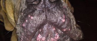
- Since the first foci of the disease appear on the paws, you need to look at them. Loose, whitish-yellow growths appear between the fingers of Malassezia.
- A strong and rather specific odor emanates from the lesions. Some compare it to the aroma of old cheese, others think it is similar to the “amber” from equally old socks... In any case, it is difficult not to notice this sign.
- At first, the fungus does not cause any particular inconvenience to the dog, but soon pain and itching appears. The dog tries to lick these places, rubs them with his paws (which only worsens the condition of the skin and increases pain).
Features of caring for animals with acanthosis
A sick animal requires special care, which consists of measures to prevent secondary infection and boost immunity.
- You should not expose your dog to stress; it is recommended to avoid long trips and changes in place of residence during treatment.
- Work with a specialist to develop a good diet that will take into account the characteristics of your dog, its age, and the presence of chronic diseases.
- Do not exclude physical activity; training should be carried out under the guidance of a specialist. These rules are especially important for individuals who are obese.
- Be sure to give vitamins that stimulate the immune system.
- Keep the dog's bedding and sleeping area clean; it needs to be periodically treated for parasites and washed.
The dog's bedding should be clean.
- After going outside, check the animal's fur to make sure there are no ticks on it. Bites, especially on damaged skin, reduce its defenses.
- Do not allow your pet to stay in the open sun for a long time, as ultraviolet radiation increases the production of melanin several times.
- If a pet with acanthosis has wounds, if possible and necessary, cover them with sterile gauze and promptly treat them.
- Consult your doctor about the possibility of antiparasitic treatment; it will strengthen the body’s protective functions, but only with the right drug and dose.
- If there are large growths and severe thickening of the skin with many folds, you should avoid injuring these places and look through the space between the folds so that dirt does not accumulate in them.
Attention! If the cause of acanthosis was associated with a stressful situation, it is recommended that such individuals take a course of sedatives when changing their usual environment, getting scared, or moving. It is better to determine their dose and type with your veterinarian.
Proper nutrition is the key to a healthy dog
Hyperadrenocorticism
This disease, also known as Cushing's syndrome, is another explanation for why our dog has black skin. In this case, it is the excessive production of glucocorticoids by the adrenal glands located on the kidneys, although this disease can also have an exogenous origin, as it can develop from drugs consisting of glucocorticoids that are administered to the dog as part of long-term treatment.
Endogenous cases are usually associated with the presence of tumors. Excess glucocorticoids cause alopecia in a symmetrical pattern, that is, the same on both sides of the animal. The skin becomes black, the stomach hangs down. The dog is lethargic and loses muscle mass. An increase in water intake and urine output may also be observed. It mostly affects middle-aged and elderly dogs. With the help of analytics, the veterinarian can confirm the diagnosis. Medicines are usually prescribed.