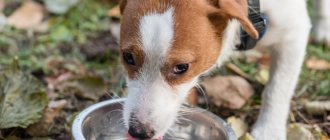
It's no secret that animals feel and see the world differently than people. At the same time, in order to better understand your pet and create more effective training programs, it is important for the owner to imagine how dogs see, how they hear, what they respond best to, etc. After all, for example, what colors dogs recognize determine what shades the shells and retrieval items should be. Given the importance of these issues, we decided to publish a series of articles devoted to the sense organs of animals. In this material, we talk about what kind of vision dogs have, and we will talk about hearing and smell in subsequent publications.
How does a dog's eye see: the structure of the eye, what kind of vision do dogs have?
The structure of a dog's eye
The outer layer of the eye is called the cornea, it protects the organ of vision from external influences. The light flux entering the eye is regulated by the pupil. And the pupil refracts and conducts light.
The dog's eyeball consists of an upper and lower halves. The task of the upper part is to recognize the brightness and contrast of objects: in other words, the upper half is responsible for visual acuity.
In the lower part there is a retina, endowed with a dark pigment that absorbs and extinguishes excess light. Thanks to it, the dog can see in bright light.
Dogs can see somewhat in the dark. Being neither nocturnal nor diurnal animals, they can boast that they navigate in the dark 3 times better than humans.
This is due to the presence of tapetum - a layer with reflective pigment.
Dogs are characterized by mild presbyopia (they are slightly nearsighted and farsighted at the same time). Studies that confirmed this fact showed that their vision differs by approximately half a diopter from the ideal human one, which, of course, is very little.
What do they see and how to find out about it
Cynthia Cook
When you go to the eye doctor, they give you a chart to look at, right? What about your dog? Has she been to an ophthalmologist? Many owners are unaware that some veterinarians specialize in eye diseases. Veterinary ophthalmologists cannot ask their patients about their vision. However, patients have almost the same problems as people - and a little more! How do we know how well our dogs see?
To understand what dogs see, it is important to understand that vision is the result of the interaction of functional elements of the eyes and brain. Visual information is transmitted from the eyes to the optic nerve (Fig. 1).
In dogs, approximately 75% of the optic nerve fibers intersect with the opposite side of the chiasm, located at the base of the brain, near the pituitary gland. From here, the optic nerves go to the centers of the brain that control the pupil and to the corresponding area of the cerebral cortex. Therefore, it is possible for a dog with brain damage to have completely normal eyes and still be blind. It is also important that the brain controls how visual information is interpreted. We have all seen examples of optical illusions, when objects are not what they seem to us. The same thing happens in dogs, and because the way the brain records and processes information is different from ours, their perception in normal and abnormal situations is also different.
The owner is in the best position for timely diagnosis. Astute owners often notice immediate changes in behavior—hesitation, flinching, and for sporting dogs, altered behavior on the track. Differences in behavior between twilight and bright light are usually an early indicator of visual impairment. Walking down stairs with confidence requires good vision. A dog that is hesitant to go downhill but runs up well may be suffering from a visual impairment. But if a dog goes down quickly but struggles to get up, it is more likely to suffer from musculoskeletal disorders.
We believe that vision is necessary for normal orientation. However, a blind dog in a familiar and uncomplicated environment can give the impression of being practically healthy. People often complain to me about cases of sudden blindness. Upon examination, it is clear that blindness occurred a long time ago. When interviewing the owners, it turns out that they have recently moved (or rearranged furniture) and their dog (who has been blind for several months) has shown a “sudden” decrease in vision. Dogs successfully use smell and hearing, sensitive to a degree we cannot imagine, to “represent” their surroundings. Our sporting dogs are much more dependent on vision. They regularly find themselves in new situations and navigate between obstacles at high speed; There is no substitute for vision in performing these tasks.
Vision includes field of view, depth perception (the ability to judge distance), perception of light, motion and color, and acuity. All of these functions must be perceived by the brain to create useful vision. While we can't ask dogs to read an eye chart, by comparing anatomical and behavioral studies we can draw some scientific conclusions about their vision. All of these elements have species-specific variations. We will only talk here about what we know about dog vision.
line of sight
The visual field varies greatly among species and among dogs and among breeds. The placement of the eyes determines the "panorama", or width of the visual field, and the size of the forward field that is visible to both eyes (binocular field). Binocular vision helps in judging distances (depth perception). Humans have a field of view of 200 degrees, of which 140 are binocular. Dogs' eyes are typically positioned closer to the sides, giving them a wider field of vision with less binocular vision.
Most dogs have a field of 240 degrees, of which 30 to 60 degrees is the binocular part. The closest, lower central part of most dogs is covered by the nose. Some breeds may have ear or coat problems. Brachycephalic breeds (pug, Boston terrier) have a shorter nose, but the eyes are more widely spaced; therefore, they have a smaller binocular part of the visual field. The resulting image is also influenced by the height of the dog (eye height).
Depth perception
The ability to judge distances is very important for sporting dogs. They must accurately determine the height and length of the jump, and immediately apply this to stride length, speed and height. To perceive depth, it is necessary to compare the image received in each eye. Therefore, the object must fall into a relatively small binocular part of the dog's field of vision and be in focus in each eye at the same time. The area in which an object is in focus depends on the size of the pupil and the eye's ability to accommodate. Like a camera, a smaller pupil gives greater depth of field. Dogs have a relatively large pupil, which improves their twilight vision acuity through depth perception. Accommodation is the ability of the eye to change focus, basically by changing the shape of the lens (see below).
Light
Dogs have better vision sensitivity in dim light (at dusk) than humans for several reasons. Their eyes are larger and can expand to maximize light capture. It is interesting to note that the size of the eyeball as a whole varies unusually little in adult dogs of all sizes. The eyes of a Chihuahua and a Great Dane are almost the same size.
Dogs also have a special reflective layer behind the retina (tapetum). The color of the tapetum in dogs varies from green-blue to yellow and is what gives the “glowing eyes” in headlights. Although the tapetum greatly influences sensitivity to light, it is also responsible for scattering too much light, which can reduce visual acuity.
The photoreceptor cells in the retina consist of rods and cones. In general, cones are responsible for bright light and color perception; rods are sensitive to low intensity, twilight light. Rods are the predominant type of photoreceptor in dogs, and they are more sensitive to low light than similar cells in humans.
Movement
Dogs are better equipped to react to moving objects, which is to be expected from predators. Owners often tell me that their dogs (with normal vision) don't seem to notice them until they move or speak. Many dogs ignore stationary objects but will reflexively chase them if they move. Their large field of peripheral vision, combined with a rod-rich retina, makes dogs well adapted to tracking movement (prey) in twilight light.
Color
Although there is a general belief that dogs have black and white vision, there is evidence that they can see colors to some extent. Color perception is determined by the presence of cone photoreceptors in the retina. In humans, there is an area in the retina (the "macula") made entirely of cones. This part of the retina is used in almost all activities. Dogs do not have such a spot, but in the center and horizontal of the retina there is a slightly higher concentration of cones. In this "central" region, cones still make up only 10-20% of the photoreceptors. However, dogs are likely to distinguish more shades of gray than humans due to their larger number of rods.
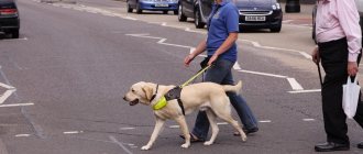
The limited number of cones found in dogs consists of only two types: violet-sensitive (429 nm) and yellow-green (555 nm). So dogs have dichromatic vision (like colorblind people), while humans have trichromatic vision. All colors whose wavelengths range from 500 to 620 nm (visible to humans as green, yellow and red) appear yellow to dogs. Light that looks blue-green to a human probably looks blue-gray to a dog. Behavioral tests on dogs suggest that dogs can distinguish red (which they see as yellow) from blue, but often confuse it with green. So it appears that guide dogs recognize traffic signals based on their location rather than their color. Since red, yellow and green look the same to dogs, it is best to contrast the yellow areas with the blue.
Acuity
Visual acuity is defined as “vision clarity”: the ability to distinguish between two separate objects. It depends on the sensitivity of the eye (the ability to bring precise focus to an object on the retina) and the interpretative ability of the brain (the number of visual receptors that form an image in the brain). Our visual acuity is usually assessed using the Snellen chart (20/x). The top number represents the distance in feet at which a person can distinguish two objects, relative to the bottom number, the distance at which a person with normal visual acuity (20/20) can distinguish two objects. The fraction 20/100 is used to describe a person who can distinguish between two objects at 20 feet that a normal person can see at one hundred feet.
(The Snellen Chart is the most common chart used to test visual acuity. This chart contains rows of capital letters called test types; the size of the letters decreases from line to line from top to bottom. The largest letters are located at the top line of the table; they are of such a size that they can be easily read by a person with normal vision from a distance of 60 meters. A person with normal vision can easily read the lines of letters below from distances of 36, 24, 18, 12, 9, 6 and 5 meters accordingly. The person whose eyesight is being tested sits at a distance of 6 meters from the table and closes one eye, while the other begins to read the letters from this table. If he can only read the lines above the line that a person with normal vision can easily reads from a distance of 12 meters, then his visual acuity is expressed as 6/12. People with normal vision can read one of the lower lines of letters from a distance of 6 meters, i.e. normal visual acuity is considered 6/6; many people can also read a line that a well-sighted person can read from a distance of 5 meters. Smaller versions of this table, the creation of which is based on the same principle that was used in the development of its usual version, can be used to test a person’s near vision. https://dic.academic.ru)
The refractive power of the eye as a whole is a combination of the length of the apple, the curvature of the cornea, and the position and refractive power of the lens. In an ideal situation, the image will go directly to the photoreceptors (rods and cones) of the retina. The refractive power of the eye can be measured by retinoscopy (see below). There are several terms to describe different states of refraction.
EMMETROPIA (commensurate refraction of the eye) - the ability to clearly focus, in a relaxed state, on an object at a distance (more than 6 m).
MYOPIA (myopia) - near objects are in focus, and distant objects are blurry. This is a common result of an eye that is longer (axial length=front to back) than normal.
HYPERMETROPIA (farsightedness) - near objects are blurry; usually caused by the apple being shorter on its axis.
ANIZOMETROPIA (uneven refraction) - the two eyes do not focus equally. This affects binocular vision and depth perception.
ASTIGMATISM - the horizontal and vertical planes of focus are different; not usually found in dogs.
PRESBYOPIA (senile farsightedness) - decreasing ability of the lens to change shape; occurs with age and increasing density of the lens
Refractive studies in dogs have shown that most have near-emmetropic vision. One study of 240 “normal” dogs found that some breeds (German Shepherd and Rottweiler) were more likely to be nearsighted; however, German Shepherd guide dogs were much less susceptible to myopia. This leads to the conclusion that the screening of dogs selected for guide work may have excluded those whose vision was less than satisfactory.
In addition to the ability of the eye's optical mechanisms to send an image directly to the retina, acuity is also determined by the density of photoreceptors and the proportions of cells that are sensitive to small amounts of light (rods) relative to those that are color sensitive and require more light (cones). Continuing the camera analogy, sticks can be compared to high-speed film - in low light the image will be grainy and unclear.
Information from photoreceptors is transmitted through a series of intercellular connections; first into bipolar cells, then into ganglia, the axons of which make up the optic nerve. The number of cells that converge in this chain affects visual acuity. In humans, the rods in the macula are related to the ganglia in a 1:1 ratio, which means that information from each part of the retina has a corresponding connection with the brain. In non-primate mammals, including dogs, many photoreceptors send information to one nerve cell, and then many nerve cells send it to one ganglion. This creates a more blurry image in the brain.
When talking about visual acuity, it is important to understand the role of accommodation, when contraction of the lens changes its refraction to adapt to seeing a close object. The muscles in the ciliary body contract, which makes the lens more convex and thereby improves focus on near objects. Dogs have very poorly developed ciliary body muscles and limited ability to accommodate. In humans, the range of accommodation gradually decreases with age and leads to presbyopia. In children, the range of accommodation is 14 diopters, by the age of 50 it decreases to 2 diopters. In dogs, the weaker muscles of the ciliary body mean that accommodation has much narrower limits - in young dogs the maximum is 2 to 4 diopters.
How vision is clinically assessed
So if a dog can't read a chart, how can we tell how well he can see? We have several ways to evaluate a dog's eyes and vision, including both functional and anatomical assessments.
One of the difficulties in early diagnosis of vision problems is that the dog is able to compensate for visual impairment in one eye. One eye may be completely blind, and the dog's behavior is virtually unchanged except for decreased depth perception and limited peripheral vision on the affected side. For athletic dogs, behavioral changes are more noticeable. On clinical examination, determining the vision in each eye may be difficult. Attempting to physically place the eye patch is so distracting to the dog that it is often impossible to evaluate his behavior. Ophthalmologists use an easy-to-wear, non-opaque contact lens to cover one eye and assess the health of the other.
When light is directed into the eye, the pupil normally constricts, which is called the direct pupillary response to light. Due to the intersection of the optic nerves, both pupils narrow, even if only one eye sees light. The response of the other eye is called the “common pupillary response” and is important in assessing the vision of the eye if the pupil does not respond.
The image reaching the retina must pass through several layers (cornea, anterior chamber, lens, vitreous body), which are normally transparent. Any dullness or distortion of these structures will complicate or distort the image. The transparency of the eye is a unique and main feature of this organ, making it extremely sensitive to abnormalities that may be congenital (possibly genetic) or acquired as a result of inflammation or injury.
There are several specialized surgeries commonly performed by veterinary ophthalmologists. Most can be easily accommodated within a half-hour examination period without discomfort to the dog or the need for anesthesia.
A microscope examination uses a hand-held microscope and a narrow beam of light to identify and locate shadows or imperfections in the ocular environment (cornea, aqueous humor, lens, and vitreous). Indirect ophthalmoscopy uses a light source and a condenser lens to examine the cornea and optic nerve. Opacities within any of the ocular structures can be seen using either of these procedures. The extent to which darkening affects vision can be determined by the degree of damage to the retina.
Measuring intraocular pressure helps determine the presence of glaucoma. A local anesthetic is used and a small pen-like instrument is used to touch the cornea to determine the fluid pressure inside the eye. For dogs, the norm is 10-25 mmHg.
Electroretinography (ERG) is used to measure the electrical signal emitted by the retina in response to light. This test is usually performed in a darkened room under general anesthesia, and is used to determine forms of retinal dystrophy. Some forms of retinal dystrophy cause visual impairment before other symptoms appear. But ERG allows for early diagnosis.
Retinoscopy is a method for assessing the refractive components of the eye and visual acuity itself. This manipulation is painless and can be easily performed without sedatives. The retina is examined through a special instrument and a series of lenses until emmetropia is achieved. A discrepancy of 2 diopters or less does not appear to have much effect on vision. However, if there is a refractive error or difference between the eyes of more than 2 diopters, difficulties in depth perception are likely to occur.
In the next article we will look at cases of visual impairments, including hereditary ones such as cataracts, glaucoma, lens luxation and retinal dystrophy. We will also look at what behaviors indicate vision problems, diagnosis, and treatment.
How do dogs see colors?
How do dogs see colors?
It turned out that if a dog had been in a human eye doctor's office, it would have been diagnosed with color blindness. Research has shown that there is no difference between the colors red and green for dogs (which has led some to wonder how guide dogs navigate traffic lights).
How do dogs see colors?
And this also confirms: it makes no difference for a pet whether you give him an orange or light green toy, he will evaluate it according to completely different criteria. And, of course, this data debunks the myth that wolves are afraid of red flags.
When should you be alarmed?
With the onset of darkness, such dogs quickly “get lost”; they are even capable of colliding with some things and objects.
Don't think this is just a coincidence. It is better to immediately show your pet to a specialist and listen to his advice. However, even complete blindness is not a death sentence for dogs. Animals successfully compensate for the lack of vision with their outstanding hearing and sense of smell. No matter how hard it is to talk about it, a blind dog feels much more confident in this world than a blind person. But both people and dogs who find themselves in such a difficult life situation need additional care and support. Therefore, if possible, do not make any drastic rearrangements of furniture in the house where a “special” pet lives. And don’t forget: the sooner you seek help from a qualified specialist, the more effective the treatment will be.
Original publication: Can Dogs See at Night? Author: Kylie Ora Lobell. Source: cuteness.com Photo: pixabay.com
How do dogs see in the dark?
The reflective layer inside the eyeball is a kind of mirror, reflecting and amplifying light when it hits the retina.
Some veterinarians compare this part to car headlight reflectors. It is due to this layer that pets' eyes glow in the twilight. But its purpose is not only aesthetics, but primarily to amplify the signal: even the slightest illumination is duplicated in the dog’s eye, and it can perfectly distinguish objects in semi-darkness.
Dogs perceive different shades of gray much better than humans, and therefore can navigate well in the dark. The picture they get is probably similar to a night vision camera.
The ability to see in the dark is due to the fact that many wild canines hunt at night.
Useful tips
Let's look at some useful tips for keeping dogs that will help avoid eye problems:
- Walk your pet often to keep it healthy and strong. Sitting on the sofa negatively affects the functioning of the hormonal system, which worsens the well-being and health of the animal. However, avoid conflicts on the street to avoid mechanical eye injury.
- It is recommended to take your dog for a preventive examination 1-2 times a year. If the animal is sick, you must go to the doctor without fail. Some diseases are not dangerous, but they still need to be treated, since they can cause complications in internal organs, including the eyes.
In conclusion, we note that the quality of vision also depends on the breed. Small dogs usually have less acute vision, but age-related problems do not appear until much later. Large breed dogs are more aggressive, so they are more likely to get into conflicts with other animals, which can lead to eye injury.
How do dogs see people and the world around them?
Some researchers have received information that dogs do not concentrate on people's faces, and for them the face is no more expressive than the back of the head.
According to the results of such studies, the image of the owner is vague for pets, and they are guided by sounds and smells. However, many dog breeders will not agree with such conclusions, observing that the dog looks into the owner’s eyes when communicating, and not at the ear or shoe.
Being hunters, dogs perceive 3 times more images per second (as you know, for people this is the notorious 24 frames, and for dogs - over 70). This is also related to the fact that many pets are not interested in television programs: they perceive it as something not only fuzzy, but also terribly slow). Therefore, some enthusiasts began to shoot videos specifically for the entertainment of dogs.
Like many predators, dogs instinctively react to moving objects. Related to this is the recommendation to stop if a dog rushes towards a person: a static target will seem less attractive to it (although, perhaps, the reasons for the extinction of rage are different).
But the unusualness of the perception of dynamic objects does not end there: they see moving objects one and a half times better than lying or standing ones (measurements showed that the range of objects was 900 and 600 meters, respectively).
Does good hearing help compensate for vision?
If a pet goes blind for one reason or another, owners wonder if the quality of his life has decreased to such an extent that he constantly experiences discomfort. Of course, the owner will have to make some adjustments to the pet’s environment: fence off the space, do not walk the dog in unfamiliar places and on a long leash, do not leave unfamiliar objects in the dog’s path, and carefully go down the stairs with the pet. But fortunately, blind dogs can adapt perfectly to life in new conditions, and most strangers will not even notice that these animals are special.
It is believed that dogs can hear frequencies twice as high as those that are accessible to the human ear. Thanks to their acute hearing, dogs can become good hunters and guards: long before the owner hears about the approaching danger, a vigilant pet will warn him. On the other hand, excellent hearing makes dogs vulnerable because they are more sensitive to noise. This is why pets are so afraid of thunderstorms or fireworks: for them these sounds sound much louder than for us.
Photo:
Main differences from human vision
Dogs see close up blurry; for them, anything closer than 30 cm is a blurred spot. Therefore, it is useless to bring an object to your pet’s eyes so that he can see better, since the effect will be exactly the opposite.
Viewing angles when turning the head and eyes in humans and dogs
But dogs are distinguished by a large field of view: in humans it is no more than 200 degrees, and in dogs – up to 250. This is due to the fact that the field of vision of animals seems to be stretched to the sides, while in humans it is presented in the shape of a circle.
But the breed matters: short-faced Molossians do not see as well on the sides as narrow-faced greyhounds. But brachycephals can better see a nearby object.
We recommend reading:
- TOP 8 Best flea and tick drops for dogs
- Rating: Dog Breeds by Intelligence and Trainability Stanley Coren
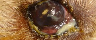
Vision problems
Canines can have many vision problems - let's look at them, and also find out methods of treatment and prevention.
Why do vision problems occur?
After birth, puppies' eyes open at 10-14 days of life, but good vision does not appear immediately. Research shows that puppies begin to perceive the world well at the age of 1-3 months, depending on the breed. Eye problems in young and adult individuals arise for the following reasons:
- Mechanical injuries. Problems with the visual system often arise due to mechanical injuries. Twigs, pebbles, other animals and even favorite toys act as traumatic objects.
- Lack of vitamins. For the normal functioning of the eyes, animals need to absorb vitamins C, E, A and some others from food. If there are not enough of them, the dogs will see worse.
- Eye contamination. During a walk, animals' eyes and fur around the eyes can become clogged with dust and small particles of debris. They cannot get rid of such garbage on their own.
How to warn them
Basic recommendations:
- Feed your animal correctly. The diet should contain not only proteins, fats, carbohydrates, but also vitamins and minerals. Therefore, check the quality of the food or include mineral/vitamin supplements separately in the diet.
- Watch what the animal is playing with. Your favorite toys should not have sharp edges that could cause injury. Never allow fights with other animals.
- Wash your eyes after walks and trim the hair around your eyes. Dust and debris will not accumulate around the eyes, which will help avoid visual problems.
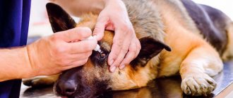
Prevention and treatment
To prevent disease, it is recommended to periodically take the animal to a veterinarian for a general examination. If an animal's vision suddenly deteriorates, then it is imperative to go to the veterinarian. Self-treatment of animals is contraindicated, since traditional methods and pharmaceutical drugs against such diseases are ineffective. And some medications can be harmful, so it's better not to take risks.