The appearance of discharge from the eyes in dogs can be physiological or pathological. The pathological type of discharge from the eyes in pets is diagnosed more often and requires the intervention of qualified doctors. The type of discharge may be watery, mucous, clear, cloudy, brown, green, yellow, and bloody.
The lesion may affect only one visual organ, or may spread to both eyes at the same time. The development of the pathological process in the eye depends on factors and is characterized by a sluggish course or rapid development.
The abundance of discharge from the eyes indicates serious changes in the dog’s body. For owners' information, clear and mucous discharge from the pet's eyes is a normal physiological process. The appearance of brown discharge should also not cause anxiety in the dog owner. The reason for seeking veterinary medical help is the appearance of purulent discharge from the dog’s eyes, sometimes mixed with blood.
At home, it is not possible to independently determine the cause of the development of pathological processes in the eyes of a dog, accompanied by the appearance of specific secretions. Any delay in diagnosis and lack of treatment leads to deterioration of the dog’s vision, and can also cause the animal to completely lose the ability to see.
In advanced cases, the dog's affected eye is surgically removed. Lack of medical care can cause the death of your four-legged friend. It is important to know how to properly care for your dog, what preventive measures need to be followed, and in what cases to seek help from veterinarians in order to prevent the development of serious complications.
How to Treat a Dog's Watery Eyes
By nature, dogs have slight watery eyes, which does not indicate illness. If the discharge is progressive in nature and becomes noticeable on the dog’s face, it becomes necessary to take measures to treat the pet.
In most cases, the cause of tearfulness in a pet's eye is dust or street sand that gets on the surface of the eye. It happens that an animal develops an allergy to certain foods. Everyday factors - gusty winds, the appearance of abundant humidity in the atmosphere - can cause dog tears. Select dogs are considered to have teary eyes as a breed characteristic. In the examples listed above, do not panic; the inconvenience will go away after the pathogens disappear.
If the situation does not change for a long time, on the contrary, deterioration is visible, you should immediately consult a doctor. If you respond at the initial stage, it remains possible to eliminate the ailment at home. The use of tea leaves or chamomile lotions is considered effective and has a mild nature. You will need to use strong tea leaves to get results. Rinsing with freshly brewed tea will require at least 5-8 applications of the drug per day until complete healing occurs.
Increased tearing occurs due to irritation of the surface of the pupil, in case of discomfort or non-standard anatomical location of the eyelid. When a dog has an eye abnormality: an extra row of eyelashes or excessive growth of eyelashes in the corners of the eyes, medical attention will be needed, and in extreme cases, the need to resort to surgical intervention.
Sometimes excessive tears occur due to allergies or a polluted environment. This often happens if the owner or family members smoke. An additional reason is too much hair near the eyes; in such cases, it is recommended to periodically trim the hair.
To avoid unpleasant symptoms, you should take good care of your pet and pay special attention to the dog’s eyes. Once a day, preferably using damp cloths, without adding aromatic additives, carefully clean the formations in the eye area.
Diseases of dogs accompanied by lacrimation
Watery eyes in dogs can be caused by the following eye diseases:
Diseases of the eyelids
Distichnaz
With this disease, single or multiple hairs appear in a row on the free edge of the eyelid, which should be hairless. These hairs appear in a dog only at the 4-6th month of life and can be very delicate or quite hard. With distichnasis, several hairs most often grow from one point. This disease is most often recorded in English and American cocker spaniels, boxers, Tibetan terriers, collies, and Pekingese.
Clinical picture. During a clinical examination of a dog, a veterinarian notes profuse lacrimation, constant blinking, blepharospasm, irritating hairs have contact with the cornea of the eye. If a dog has curled eyelashes, keratitis is diagnosed.
The diagnosis of the disease is made based on the above symptoms. Differential diagnosis. Distichnasis is differentiated from trichiasis, entropion and eversion of the eyelids, allergic conjunctivitis, and keratoconjunctivitis sicca.
Treatment. It is carried out in veterinary clinics by electrolysis under an operating microscope. Excision of the third eyelid.
Trichiasis
Trichiasis is a condition when hair from a dog's eyelids or muzzle gets into the eye, coming into contact with the conjunctiva and cornea. Trichiasis can be primary or secondary. Primary occurs in dogs with medial inversion of the eyelids and a large nasolabial fold.
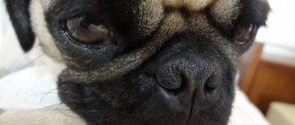
Trichiasis occurs in the following dog breeds: Pekingese, Pugs, English Bulldogs, English Cocker Spaniels, Chow Chows, Shar-Peis.
Clinical picture. During a clinical examination of a dog, a veterinarian notes lacrimation, hairs in contact with the cornea cause blinking in dogs, constant discharge from the eyes, symptoms of keratoconjunctivitis, inflammation of the skin in the area of the nasolabial fold. The diagnosis is made based on the detection of hair in contact with the cornea, provided there is no other eye pathology.
Differential diagnosis. Trichiasis is differentiated from keratoconjunctivitis sicca, entropion and eversion of the eyelids, dystrichiasis, and ectopic eyelashes.
Treatment. Treatment of the disease is surgical. Temporary improvement can be achieved by trimming the hair that gets into the eye.
Entropion of the eyelids
Entropion is a pathology of the eye in which part of the organ turns inward towards the eyeball. A dog's eyelid inversion can be either upper or lower, one-sided or two-sided.
Unilateral inversion of the eyelid margin is most often the result of heredity and appears in a dog in the first year of life.
Congenital entropion occurs in puppies after the eyes open in some breeds with excessively folded skin on the head (chow chow, shar pei).
In this disease, the eyelashes, hair and skin of the eyelid rub against the surface of the cornea, causing inflammation and irritation.
Clinical picture. During a clinical examination, the veterinarian notes the leakage of liquid secretion from the eye, the dog has photophobia (to an electric light bulb, the sun), the dog rubs its eyes with its paw, blinking, and there may be an eye tic.
Treatment. Treatment of entropion of the eyelids is surgical.
Eversion of the eyelids
With eversion of the eyelids, the edge of the eyelid turns outward, while the mucous membrane (conjunctiva) of the eyelid is exposed. This pathology occurs in dogs with too large palpebral fissure and excess, easily removable skin in the head area. Cause. Mechanical eversion of the eyelids in a dog occurs as a result of pathological changes in the eyelid itself, as well as tissue scarring after injuries or surgery. Paralytic ectropion occurs in dogs as a result of facial paralysis.
Clinical picture.
During a clinical examination, the veterinarian notes incomplete closure of the eyelids, discharge from the eyes, and inflammation of the conjunctiva.
Treatment. Treatment for this pathology should be aimed at eliminating the cause that caused and maintains the ectropion of the eyelids (removal of a neoplasm, conjunctivitis, facial paralysis, surgical removal).
Diseases of the conjunctiva
Conjunctivitis
conjunctivitis is the most common disease. Conjunctivitis is accompanied by dysfunction of the conjunctival mucosa and often occurs with infectious diseases. Additionally, the causes of conjunctivitis in dogs can be allergies, clogged tear ducts, viruses, foreign body injuries, irritation of the conjunctiva as a result of eyelid pathology.
Allergic conjunctivitis
Allergic conjunctivitis in dogs occurs as a result of contact with the mucous membrane of the eye of one or another allergen (contact allergy). The allergen can be pollen from flowering plants, dust, etc. Allergic conjunctivitis in dogs In recent years, allergies to certain food products (food allergy) have often been recorded.
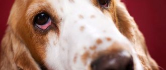
Clinical picture. During a clinical examination, a veterinarian notes in such a dog redness of the mucous membrane of the eyes, mucous discharge from the palpebral fissure. As a result of itching, the dog rubs its paw on the affected eye.
Treatment. If contact dermatitis occurs, it is necessary to rinse the eye affected by inflammation with saline solution or chamomile decoction. In case of food allergies, it is necessary to exclude the allergic product from the dog’s diet and transfer the dog to a hypoallergenic diet (buckwheat, rice, beef). The sick dog is prescribed antihistamines (cetirizine, diazolin, suprastin, diphenhydramine, tavegil), and Diamond Eyes eye drops are instilled into the conjunctival sac.
Purulent conjunctivitis
Purulent conjunctivitis in a dog develops due to the entry of various pathogenic microorganisms into the conjunctiva.
Purulent conjunctivitis is one of the symptoms of canine distemper (canine distemper). Clinical picture. During a clinical examination, a veterinarian notes reddening of the conjunctiva, its swelling, and purulent discharge from the eye of a sick dog.
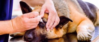
Treatment. With this form of conjunctivitis, a sick dog is treated with eye drops and ointments that contain antibiotics. Tetracycline eye ointment and Ciprovet drops are widely used. Before applying eye drops and eye ointment, it is necessary to clean the affected eyes from exudate.
Follicular conjunctivitis
This form of conjunctivitis is most typical of chronic conjunctivitis and often develops in a dog when toxic substances get into the eye.
Clinical picture. During a clinical examination, a veterinarian reveals many bubbles with transparent contents on the mucous membrane of the conjunctiva. Mucous discharge comes from the palpebral fissure. The conjunctiva itself is crimson in color, and the dog's inflamed eye is squinted.
Treatment. When treating this form of conjunctivitis, eye ointments containing an antibiotic are used. In severe cases of the disease, specialists are forced to resort to excision of the conjunctiva and subsequent symptomatic treatment.
Disease of the lacrimal apparatus
Keratoconjunctivitis sicca
Keratoconjunctivitis sicca is a condition characterized by very little tear film in the eye as a result of insufficient or absent tear production. This disease is observed in West Highland white terriers and is inherited by their offspring.
Keratoconjunctivitis sicca in dogs occurs due to disorders of sex hormones, canine distemper, trauma to the frontal part of the skull, neuropathy of the facial nerve, congenital hypoplasia of the lacrimal glands, and from the use of certain medications.
Clinical picture. Veterinary specialists, when conducting a clinical examination of a sick dog, note frequent blinking, dry crusts at the edges of the eye, itching, the presence of mucopurulent discharge from the eyes, viscous mucus is found in the conjunctival sac,
Follicular
Later, as the disease progresses, symptoms of ulceration and unevenness of the corneal surface appear, and swelling of the conjunctiva develops. If there are dry crusts in the area of the nostrils on the affected side, we can talk about the presence of damage to the facial nerve in the sick dog.
Treatment. Treatment for this form of keratoconjunctivitis should be aimed at eliminating the underlying cause of the disease. The area of the conjunctiva and cornea is washed generously every two hours with saline before each application of the drug. The inner corners of the eyes of a sick dog are washed with a solution of chamomile or chlorhexidine, since the lacrimal sac in a sick dog is a reservoir for various microorganisms. During treatment, eye ointment with an antibiotic is used.
How to treat
When you find a formation in your pet in the eye area, try to find out the cause. In the case of purulent discharge with a clear yellowish or greenish tint, after which wet spots remain in the area of the eye cornea with a complication of the process, measures must be taken immediately. A healthy pet will not experience these symptoms.
If eye rot occurs, carefully examine your pet: there is a possibility that a foreign object has hit the surface. Consider whether the dog has been injured recently. In the latter version, pus is present exclusively in a single eyeball. If the discharge comes from both eyes, the circumstances of the disease are different.
An experienced and specially trained doctor can determine the cause. With different diseases, there is still a possibility that the same symptoms will occur. Untimely services provided will lead to deterioration of the condition until complete loss of vision.
It happens when an eye disease often has a manifestation that is similar for a group of dog breeds. This is caused by the anatomical structure of the eyes, leading to the risk of developing such pathologies. Small breeds of dogs often suffer from the same diseases: pug, terrier or chihuahua.
For treatment you will need to use eye drops. When the pet's condition becomes more complicated and the disease is more dangerous, antibiotics or similar potent drugs are used.
Typology
There are 2 types of eye discharge: purulent and lacrimal. If the latter are not the result of pathologies or serious diseases, then the former are caused by a wide variety of health problems and mechanical injuries to the eyes and mucous membranes.
Most often, discharge from a dog’s eyes is a consequence of the following processes:
- allergies to insects, household chemicals, some food product or an unknown item eaten;
- fungal or bacterial diseases - a whole group of pathogens and microorganisms “work” on this, even leading to an increase in temperature, weight loss and a decrease in activity level;
- viral diseases - have a very powerful effect and do not go away quickly enough (enteritis, hepatitis, plague and rabies);
- injury to the eye from granular substances, chemical compounds or a solid object.
Causes
Most often, the bacterium enters the human body through wounds and microcracks in the skin. The infection, having penetrated through the wound, begins to multiply in the blood, spreading throughout the body and affecting the lungs, heart, brain, liver, kidneys, and joints.
With staphylococcus, diseases can be very different, such as pneumonia, meningitis, osteomyelitis, endocarditis, sepsis and many others.
Staphylococcus infection can occur in the following ways:
● Through contact and household use when using the patient’s personal items;
● Airborne droplets during close contact with an infected person;
● Fecal-oral on dirty fruits, vegetables and other food products, dirty dishes and hands;
● Vertical when a child passes through the birth canal of an infected mother during childbirth.
● Infection often occurs during surgery through medical instruments and during various manipulations.
Types of staphylococcal infections
There are several most common types of staphylococci:
● Hemolytic staphylococcus.
Most often, this infection affects the upper respiratory tract, causing purulent sore throat, pharyngitis, tonsillitis, bronchitis and other inflammatory diseases. These bacteria are very resistant and difficult to treat.
● Golden.
This microorganism is extremely resistant to almost all types of penicillin antibiotics, antiseptics, high temperatures, and active direct sunlight. It causes various skin lesions, such as eczema, abscess, boils, lesions of the gastrointestinal tract, upper respiratory tract, mucous membranes, and in the worst case leads to toxic shock.
● Epidermal.
This microorganism lives on the surface of the skin and mucous membranes of any healthy person and does not cause any harm. But, if this bacterium enters the blood of a person with a weakened immune system, which most often occurs during surgical operations, the use of improperly processed instruments, catheters, blood poisoning occurs, which leads to inflammation of the inner lining of the heart.
● Saprophytic.
Despite the fact that this species is the least dangerous, when infected it leads to general intoxication of the body due to the release of dangerous toxins and enzymes during its life processes. These microorganisms often cause inflammation of the urethra and bladder. This is mainly typical for women due to the anatomical features of the structure of their genitourinary system. If left untreated, cystitis leads to kidney inflammation and problems conceiving a child.
Restoring the sense of smell
If you have a long-term loss of smell, you should immediately go to the clinic for an appointment with an otolaryngologist. If you treat yourself, you can worsen the current condition.
Recovery can be:
- Surgical.
Such intervention may be required in advanced cases (for example, with a tumor or deviated septum). Then the surgeon performs the operation.
- Medicinal.
The doctor prescribes a list of necessary medications, from nasal rinses to antibiotics, antiviral and antifungal agents.
- Physiotherapeutic.
Treatment may require physical therapy. The doctor prescribes heating or inhalation, and in some cases laser treatment is performed.
- People's
No one has abolished traditional medicine, but in no case should you prescribe decoctions for yourself. The doctor may prescribe a decoction for rinsing the nose from chamomile and oak bark or nasal drops based on aloe juice.
- Complex.
Those. This is the application of all types from physical therapy to surgical.
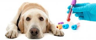
Diagnostics
Only a doctor can diagnose the presence of infection based on the results of a staphylococcus test.
It is worth remembering that testing is recommended only if symptoms of the disease are present. The presence of bacteria in biological material may mean that a person is its carrier, which in itself is the norm.
For analysis, material is taken from the area where the infection is believed to be developing. To detect a pathological process, several tests are carried out to track the dynamics of bacterial growth. If their number increases rapidly, the presence of a staphylococcal infection can be diagnosed. Also, additional analysis will determine the specific type of infection so that the doctor can choose a personal treatment regimen.
Diagnostic techniques
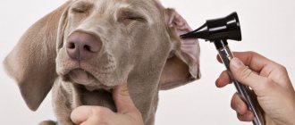
When visiting a veterinary clinic, be sure to remember how and after what your pet developed such discharge in the corners of the eyes.
Also, the specialist will definitely resort to some diagnostic techniques to help accurately determine the type of pathology:
- Medical examination of the eyelids, cornea, conjunctiva, anterior and posterior chambers of the eyeball.
- Schirmer's test. A piece of filter paper is attached to the corner of the eye. After some time, the area soaked with the released tear is measured.
- Application of fluorescent compounds to the surface of the cornea. This method is used to diagnose corneal ulcers and microscopic lesions.
- Tonometry to measure intraocular pressure. Used at the slightest suspicion of glaucoma.
Additional diagnostic tests are required in more complex and non-obvious cases. These may include:
- Full medical examination.
- Cytological examination of scrapings from the surface of the conjunctiva/cornea.
- Taking swabs, biopsy. If your dog's eyes are constantly running, there is no problem taking samples of pathological material! It is only necessary to collect fresh secretions containing a sufficient amount of the pathogen. This will simplify the diagnostic process.
- Growing a pathogen culture on special nutrient media.
- Complete blood count (CBC) and serological tests. These techniques are used to detect systemic infections, parasitic infections, and certain types of cancer.
- Sometimes they resort to fluoroscopy or ultrasound examination of the sinuses of the skull.
- In difficult situations, MRI is strongly recommended.
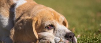
Keratoconjunctivitis sicca
The disease is an inflammation of the conjunctiva and cornea of the eye.
Keratoconjunctivitis sicca occurs when the secretion of tear fluid is impaired. Foreign particles are not washed off from the eye, the cornea is not sufficiently wetted, and the eye easily becomes infected. Purulent conjunctivitis begins. The eyes turn red, become dull and dull, and irritation and itching appear. The dog scratches its eyes, green thick discharge and ulcers are observed. The dog blinks frequently, and vision deterioration is noticeable. If the disease is not treated, the cornea becomes cloudy, becomes rough, grows with blood vessels, and the dog goes blind.
Selected dog breeds are predisposed to the disease. These are cocker spaniels, shih tzus, poodles, pugs, Pekingese, chow chows, chihuahuas and other small dogs whose tear production is impaired due to the special structure of their eyes.
General information
Exudation from the eyes is a common sign of systemic infections in both dogs and other pets. It can begin suddenly, but more often the inflammatory process progresses gradually, which may take several weeks.
Exudate can be:
- Watery;
- Slimy;
- mucopurulent (brownish-yellow);
- ichorous (brownish liquid with a completely unbearable smell of rot).
There is one simple rule - the more exudate is released from the eyes, the more serious the disease that led to this result.
Let's look at some reasons for this condition:
- Obstruction of the lacrimal ducts. This leads to the fact that tears, which are a natural antibacterial agent, do not wash the surface of the eye, as a result of which an inflammatory process develops, complicated by the introduction of pathogenic or conditionally pathogenic microflora.
- Excessive secretion of tear fluid. If the skin around the eyes is constantly wet, it can easily become inflamed with the same result.
- Keratitis (inflammation of the cornea).
- Conjunctivitis.
- Blepharitis (inflammation of the eyelids).
- Congenital and acquired anatomical defects of the eyelids.
- Corneal ulcers.
- Glaucoma.
- Uveitis, that is, inflammation of the choroid of the eyeball.
- Injuries. They lead to the entry of pathogenic and conditionally pathogenic microflora into the tissues of the organ, which contributes to the development of inflammation.
- Keratoconjunctivitis (dry eye syndrome). A very severe pathology that can lead to complete blindness/the need for surgical removal of the eyeball.
- Postoperative complications. This often happens when animal owners do not follow the instructions of the treating veterinarian.
Treatment and care of sick animals
The more accurate the diagnostic results, the more successful the prescribed treatment. The latter depends on the specific root cause of the disease. Thus, for systemic infections, broad-spectrum antibiotics and other antimicrobial agents are prescribed, tetracycline ointment is placed in the conjunctival cavity, etc. Some pathologies (advanced glaucoma, keratoconjunctivitis, corneal ulcers) require highly qualified surgery.
How to care for a sick pet at home? First, you must meticulously follow all your veterinarian's instructions. Secondly, you need to regularly remove the released exudate , not allowing it to dry out and form crusts. The easiest way to do this is to use warm, sterile saline. If possible, it is necessary to place the dog in a portable crate. This will prevent them from scratching their eyes on their own, with subsequent aggravation of the inflammatory process.
Take your dog to the veterinarian at least once a week for additional medical examination. This is extremely important for timely correction of the prescribed treatment. If a specialist regularly sees the dog, he will immediately understand whether the prescribed medications are working, and whether it is time to adjust the dosage and type of medications. Never use human ophthalmic medications. They are designed for human physiology, so introducing them into a dog's eyes can lead to unpredictable results.
Conditions for effective treatment
Effective treatment for oral thrush involves eliminating the underlying cause. It is very important to sanitize the oral cavity: cure teeth destroyed by caries, remove non-viable teeth and roots that can no longer be restored. These are chronic foci of inflammation, so simultaneous sanitation will shorten the treatment time. Tartar and plaque should also be removed. This is especially true for cases of candidal stomatitis associated with injury to the gums by the sharp edges of hard dental deposits.
Patients with removable dentures should be re-trained in hygiene and disinfection of orthopedic structures. If the time to use a prosthesis comes to an end, it is important to replace it in a timely manner. Treatment of candidiasis will be useless if a person uses the denture incorrectly and again creates conditions in the oral cavity for the growth of fungi.
Unsuitable crowns, bridges and other structures must also be replaced. It is also important to eliminate chipped enamel, which becomes a source of injury to the gums, mucous membranes of the cheeks and tongue.
Smokers should reduce their smoking episodes if possible or quit the bad habit. If the disease has developed while taking corticosteroids, it is important to explain the rules of treatment: you should rinse the mouth with plenty of warm water after spraying the drug.
When treating oral candidiasis that has developed during antibacterial therapy, measures should be taken to restore the normal microflora of the intestine and oral cavity. You may need to consult another specialist or therapist: you will need to take probiotics and prebiotics.
For all patients treated for candidiasis, several general recommendations apply:
- maintaining oral hygiene;
- refusal of foods rich in carbohydrates;
- giving up sugary drinks.
It is necessary to exclude from the diet foods that can irritate the mucous membranes: dishes prepared with vinegar, marinades, spicy, peppery foods, smoked foods, sour fruits and berries. You should also not eat confectionery, baked goods made with yeast, or sugar. It is better to give preference to warm dishes. You need to follow this diet for another 1.5-2 months after recovery.
In some cases, it is advisable to use toothpastes with glucose oxidase, lysozyme, and lactoferrin. They help improve the defenses of the oral mucosa and can be part of a comprehensive prevention of inflammation. The choice of toothpaste should be agreed with your doctor; he will recommend the optimal product, and also tell you which brush is suitable.