A dog's eyes, like a person's, are the organ through which they communicate with the outside world. A dog's eye is an organ specially adapted for the animal to perceive light waves. With the help of vision, your dog navigates the world around him, perceives the intensity of light, color, shape of objects, distance to them, as well as the movement of objects in space. With the help of vision, a dog in the wild obtains food for itself, has the ability to move in the direction it needs, and, in the event of an attack, to defend itself
Eyes, like a keen sense of smell and keen hearing, are important for the full life of an animal. Therefore, owners of all diseases that can lead to decreased visual acuity or even blindness of your pet must be noticed and treated in time. How to recognize when a dog’s red whites of the eyes are a pathology that requires immediate treatment, and when is this the norm due to the breed?
By the condition of the eyes you can always determine whether your dog is healthy or not; the eyes are a “mirror” not only of the soul, but also of the health of the animal.
Before talking about whether red eyes in a dog are a particular disease, the dog owner needs to have a general understanding of the structure of the eye.
A dog's eyes are located in the orbits - bony sockets formed by the bones of the skull, where they are held by several muscles that ensure their mobility and orientation in different directions. The dog's eye itself is protected by auxiliary organs - eyelids and glands.
The dog has three eyelids. The upper and lower eyelids are folds of skin, the inner surface of the eyelids is lined with mucous membrane. The outside of the eyelids is bordered by eyelashes, which protect the eyes from dust and other foreign particles. A dog's third eyelid is a simple film in the inner corner of the eye that dog owners usually cannot see. This film covers the eye when it is closed or irritated, as well as during nervous disorders.
The eye in the cornea area comes into contact with the external dry environment, so it needs protection of the lacrimal glands, which produce tear fluid - a secret that moisturizes the surface of the cornea. A dog's tears accumulate in the space between the eyelids and the eye and are then drained through a narrow channel that begins at the inner corner of the eye and opens into the nasal cavity. When there is excessive lacrimation or blockage of the tear duct, tears flow from the eyes and, when oxidized, form red stripes on the fur that look like blood.
The eye consists of two parts. The anterior portion includes the cornea, iris, and lens. They absorb beams of light from the dog, like a camera lens. The cornea and lens are clear and act like optical lenses, and the iris acts as a diaphragm, regulating the amount of light entering the eye through the pupil (the hole in the iris).
The back of the eye consists of the vitreous body, the choroid (choroid) and the retina, which converts optical light signals into nerve impulses that are transmitted to the visual center of the brain.
Thinking of the eye as an analogy to a camera, the back of the eye is like a photographic film on which the dog's brain captures the image.
If there is redness in the eyes, your dog will strive to hide from the light in a secluded place in his kennel, and to find a shaded corner at home to rest. The dog becomes apathetic and there is a decrease in appetite. Sometimes the dog may itch, shake its head, and whine. The dog's owner notes that redness of the sclera of the eye is often accompanied by an increase in body temperature and nasal discharge.
Often, redness of a dog's eyes is accompanied by inflammation of the conjunctiva, and depending on the form of conjunctivitis, mucous, white, greenish, yellow and brown discharge is additionally noted.
Main causes of redness
The sudden appearance of redness is due to pathological or non-hazardous reasons. In the first case, complex treatment is required to normalize the condition, and in the second, it is enough to simply eliminate the irritating factor.
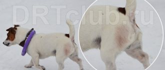
Those at risk include those with long hair, bug-eyed and folded breeds. Due to the peculiarities of their appearance, these animals are more often than others exposed to negative external factors - and suffer from pathologies of the visual organs.
Injury or foreign object
Four-legged pets damage the cornea or deeper layers of the eyeball during excessive scratching, playing and fighting. Also, the integrity of the mucous membrane can be disrupted by a foreign object stuck there: dust, a blade of grass, plant seeds and even your own eyelashes.
Redness can also be caused by a head injury resulting from an accident or a rough blow. In this case, hyperemia is observed on both sides.
Eyeball diseases
If the whites of the dog's eyes are red, it is recommended to check for enophthalmos and exophthalmos. These pathologies are accompanied by retraction or prolapse of the eyeballs. In both cases, the victim requires urgent surgical care.
Other conditions with similar symptoms include keratoconjunctivitis, blocked tear ducts, and glaucoma. They all involve different treatment methods, so don’t rush to solve the problem on your own.
Diseases of the eyelids
This group includes pathologies that change the natural position of the eyelids, inflammatory processes and tumors. These include:
- ectropion and entropion, or eversion and inversion of the eyelid;
- blepharitis - inflammation of one or both corners of the eyelid;
- adenoma of the third century.
Without timely help, these diseases can lead to loss or atrophy of the eyeball, as well as partial or complete loss of vision. Conditions complicated by secondary infections are particularly difficult to tolerate.
Allergy
Hyperemia is a characteristic sign of an allergic reaction. It often occurs when inhaling tobacco smoke, pollen or household chemicals, as well as eating a certain group of foods or medications.
Stress
If a dog has red eyes, then maybe it is just scared. A similar reaction occurs when there is strong anxiety associated with moving, losing an owner, or facing an aggressive opponent. After calming the animal, everything returns to normal, so specific treatment is not usually used in this situation.
Heatstroke
A sudden rush of blood occurs due to overheating in the sun or prolonged exposure to a stuffy room. For this reason, in the warm season it is better to reschedule walks in the early morning or late evening.
Increased load or pressure
Too active loads affect the pressure, causing it to deviate sharply from the norm upward. Also, red eyes in a dog occur due to increased intraocular pressure. It is observed against the background of the development of glaucoma, diabetes mellitus, kidney and heart pathologies.
Infectious diseases
This group includes diseases caused by bacteria, viruses and fungi. These include:
- chlamydia;
- toxoplasmosis;
- leptospirosis;
- plague;
- viral conjunctivitis and keratitis;
- blastomycosis;
- histoplasmosis;
- cryptococcosis.
Some of these infections are dangerous to humans. If infection is suspected, it is recommended to temporarily isolate your pet.
Helminthiasis
Parasitic nematode worms also cause red eyes in dogs. Typically, the lacrimal apparatus harbors thelaziosis pathogens transmitted by infected flies.
Preventive actions
You need to carefully monitor your pet in order to come to his aid in case of illness. Primary vaccination with annual revaccination is the most necessary preventive measure. Once you get your puppy, try to take him to the vet within the first three days.
thorough inspection and constant monitoring
Take your pet's veterinary passport if the breeder gave you one. Be sure to bring a box of stool to test for worm eggs. If you have to wait in line, try to keep your puppy on your lap or in a transport container to prevent contact with other animals.
To avoid accidental infection, do not allow your puppy to play on the floor or sniff foreign objects. A puppy who has not been immunized should not leave the house. A vaccinated pet must undergo a three-week quarantine after vaccination, because it is considered conditionally ill.
Testing stool for worms is a very important preventive measure. A puppy is considered healthy after a fecal test is performed twice. Avoid walking in nature near swampy puddles or slowly flowing small rivers. Such water can become a source of leptospirosis, which is carried by wild ducks and rats.
If you frequent dog shows, try to get your puppy vaccinated against parainfluenza. The virus of this disease is transmitted by airborne droplets and is typical for kennel dogs. Modern means of protection and a thorough examination of the puppy after a walk will help protect against ticks, which can cause fatal diseases. In spring and autumn, ticks are most active and dangerous. There are known cases of tick attacks even near a house on asphalt.
A thorough examination of your dog's skin will reveal the manifestations of various skin diseases that cause itching and irritation of the skin. Use preventatives designed to protect your dog from fleas, which can cause many problems.
In winter, at low sub-zero temperatures, protect your puppy or adult dog from hypothermia. The dog's genitourinary area may be significantly affected. Warm overalls will help protect your dog from this type of disease.
Poor hygienic care of your dog's eyes can cause brown spots on the inner corner of the eye ("poodle eye").
Associated symptoms
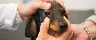
Before going to the veterinary clinic, you should carefully note all accompanying symptoms. With their help, it will be easier for the veterinarian to understand why the dog's eyes turned red.
Depending on the cause, the condition may be accompanied by the following symptoms:
- damage to the skin and mucous membranes, bleeding, as well as severe pain;
- frequent blinking, photophobia and prolapse of the third eyelid;
- an increase in the size of the pupil and loss of its reaction;
- drying and inflammation of the conjunctiva;
- formation of crusts and ulcers;
- local loss of hair and eyelashes;
- excessive tearing and the appearance of purulent discharge;
- loss of coordination and loss of consciousness;
- intense itching, runny nose, sneezing, coughing and shortness of breath;
- fever, nausea, vomiting and diarrhea.
If you notice at least a few of these symptoms, avoid self-medication and seek help. This will help preserve your pet's vision.
Adenoma of the third century
Adenoma of the third eyelid , or correctly said – prolapse (prolapse) of the lacrimal gland of the third eyelid.
Usually observed in young dogs aged from 6 months to 2 years, most often in brachiocephalic breeds (Shih Tzu, cocker spaniels, pugs, bulldogs, etc.), beagles, etc. This occurs due to genetically determined weakness of the ligaments that fix the gland to the edge of the eye socket. Symptoms:
— An oval red formation appears in the inner corner of the eye, protruding from under the edge of the third eyelid. It is most often observed in one eye, but can also be bilateral.
- There may be moderate lacrimation and squinting of the eyes (blepharospasm).
Treatment:
1. The gland must be returned to its place surgically.
============================================================================================================================================================================================
Why do you need diagnostics in a veterinary clinic?
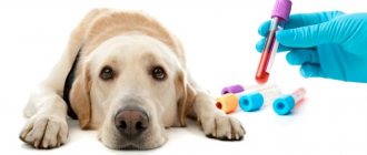
Diagnosis in a veterinary clinic is necessary to exclude diseases with similar symptoms and select effective, and most importantly, safe treatment. A set of laboratory and instrumental studies helps to identify the exact cause of the ailment. These include:
- tests of urine, blood and feces, revealing deviations in baseline indicators and the presence of parasites;
- bacteriological culture, used to determine the exact type of pathogen and drugs to destroy it;
- measurement of intraocular pressure;
- Ultrasound of the abdominal cavity, determining the presence of pathologies of internal organs;
- allergy test;
- X-ray of the chest and skull, which helps detect closed injuries;
- Schirmer test, which allows you to determine the level of tear production.
The list presented can be supplemented or shortened by a veterinarian. After receiving all the results, an individual treatment plan is selected for the animal.
Blepharitis
Blepharitis is inflammation of the eyelids. In dogs, the disease is often chronic and bilateral, less often - unilateral and acute.
Symptoms:
- Itching, redness and thickening of the edges of the eyelids (pronounced to varying degrees).
- Blepharospasm (squinting).
- Discharge, the nature of which varies from tears to mucous and purulent.
Blepharitis can be focal or diffuse.
- Focal blepharitis (hordeolum).
There are:
- External stye is an abscess of the sebaceous gland of the edge of the eyelid on the outside. Occurs predominantly in young dogs.
- internal stye, or meibomitis, is a purulent inflammation of the glands on the inner surface of the eyelids (meibomian glands), most often in middle-aged dogs.
- Chalazion (hailstone) is a rough formation on the inner side of the eyelid, formed as a result of meibomitis due to blockage of the outlet with masses prone to calcification.
- Diffuse blepharitis . Often this form is a concomitant disease with allergies, as well as with chronic eye diseases (conjunctivitis, keratitis, etc.), with parasites (demodex), fungi (microsporia, trichophytosis), and sometimes with general diseases.
Treatment:
1. Put the dog on a protective collar to prevent self-injury and aggravation of inflammation.
2. Carry out hygienic treatment of the eyelids (chlorhexidine solution (removal of scales, etc.)).
3. For blepharitis caused by bacteria, apply ointments with antibiotics and corticosteroids.
4. In the case of fungal (microsporia, trichophytosis) and parasitic (demodex) blepharitis - systemic treatment with antifungal and antiparasitic drugs.
============================================================================================================================================================================================
Home treatment
Despite the fact that veterinarians categorically do not recommend self-medication, sometimes the victim has to provide first aid on his own. In such cases, it is imperative to know the permissible and prohibited actions so as not to worsen the pet’s condition.
First aid
If heatstroke occurs, place the animal in the shade and give cool water to drink. Then wet the rag and place it on your head. If you are indoors, open all windows to ensure sufficient oxygen flow.
Before visiting the veterinarian, it is allowed to wash the eyes with water, weak tea leaves and furatsilin solution. Please note that this procedure should be performed with protective gloves, and it is recommended to wash your hands thoroughly after completion.
To moisturize the mucous membrane, an “artificial tear” drug is suitable. Unlike antibiotics and glucocorticosteroids, it is completely harmless. But taking the latter should be avoided. If the dosage is incorrect, they will only aggravate the patient’s condition and blur the clinical picture.
Please note that even after the dog’s health improves, it needs to be shown to a veterinarian. Without drug treatment, the missing symptoms can return again, hitting the body with even greater force.
Medicines prescribed by a veterinarian
All drugs and dosages are selected individually, taking into account the underlying disease and accompanying symptoms. Depending on the diagnosis, the four-legged patient may be prescribed:
- anti-inflammatory and painkillers;
- antihistamines and glucocorticosteroids;
- sedatives;
- antibiotics, antiviral and antifungal;
- anthelmintics;
- cholinomimetics that improve the outflow of intraocular fluid;
- immunomodulators.
Drug therapy is not complete without eye drops, ointments and special rinsing solutions. With their help, traces of dirt and pus are eliminated, preventing damage to neighboring areas.
How to do rinsing and other procedures
If the eyeballs are affected, you will have to familiarize yourself in detail with the procedure for washing, instilling and placing drugs. It is recommended to use lint-free cotton pads for processing. Unlike cotton wool, they prevent accidental injury to the cornea.
For rinsing, use plain water, saline solution or warm herbal decoctions. After wetting the disc, it is carefully passed from the outer corner to the nose, trying to get rid of accumulated secretions and crusts.
After cleansing, use the medications prescribed by your doctor. The drops are instilled directly onto the mucous membrane of both eyeballs, and the ointment is placed between the sore eye and the eyelid. For better distribution, after applying the drug, close the pet’s eyelids and massage a little.
When surgery cannot be avoided
Sometimes drug treatment is not enough. The following cannot be done without surgery:
- severe internal injuries and deeply embedded foreign objects;
- enophthalmos and exophthalmos;
- follicular form of conjunctivitis;
- volvulus, eversion and third eyelid adenoma.
Surgery involves the use of general anesthesia and suturing. To avoid inflammation and suppuration, you will have to carefully monitor the cleanliness and dryness of the postoperative wound until you return to the doctor.
Useful video on the topic
It often happens that after diagnosis, the doctor prescribes daily procedures. But trips to the clinic are not always justified, and the dog will experience unnecessary stress during an appointment with an ophthalmologist. In this video, we suggest watching how to properly wash your pet's eyes at home. And also how to instill drops, apply gel or ointment.
It is important to remember that the dog’s health largely depends on the attention and care of the owner. Therefore, we recommend that you contact a veterinary clinic at the first signs of illness. Only a doctor can make the correct diagnosis and prescribe treatment.
Types of disease
Blepharitis is classified according to several criteria.
Based on the localization of the inflammatory process, we can distinguish:
- Anterior edge - when only the ciliary edge of the eyelid is involved in the painful process.
- Posterior marginal is said to occur when the meibomian glands and surrounding tissues are inflamed.
- Angular or angular - indicates the localization of inflammation mainly in the corners of the eyes.
- Mixed involves all the tissues of the eye, as well as the skin and surrounding muscles.
Based on the characteristics of clinical signs, the following forms of the disease are distinguished:
- Simple. It has mild symptoms - moderate redness of the dog's eyelids, thickened edges, whitish secretion in the corners of the eye.
- Scaly. The edges of the eyelids are thicker. It can be recognized by the characteristic scales of the epidermis, which are located at the base of the eyelashes.
- Ulcerative. This is a purulent form of the disease. Crusts of dried pus on the eyelids form painful ulcers after removal. Inflammation is accompanied by loss of eyelashes, severe itching and pain. After the ulcerations heal, scars form on the eyelids, interfering with the normal growth of eyelashes.
- Meibomian. It occurs as a consequence of hyperfunction of the meibomian glands. Excess secretion becomes a breeding ground for the development of pathogenic microflora. The disease can be complicated by a purulent course.
- Furunculous or phlegmous also has a name - barley. The most characteristic signs are the appearance of one or more abscesses along the edge of the eyelids, on the eyelash line, which open over time. This is the most dangerous type of disease, as it can have a dangerous life-threatening complication - sepsis.
Keratitis
Keratitis is an inflammation of the cornea (the most superficial, transparent layer of the eye in contact with the external environment), manifested by the appearance of individual areas of cloudiness or clouding of the entire cornea, which can lead to decreased vision and, in advanced cases, to blindness.
There are:
- Shepherd dog keratitis (the disease is typical for shepherd dogs, but it is also often found in Staffordshire terriers, Labradors and mixed breeds, somewhat less often in other breeds: collies, St. Bernards, Siberian huskies, huskies, Airedale terriers, Russian greyhounds, greyhounds).
- Boxer keratitis (boxers, Boston terriers, French bulldogs, Pekingese, pugs).
- Dachshund keratitis.
Shepherd keratitis, or pannus, is a superficial, usually without ulceration, chronic inflammation of the cornea.
Symptoms:
- On the conjunctiva (normally pink) - extensive brown pigmentation appears.
— Gray-white clouding of the cornea and the formation of a red shaft, due to the appearance of blood vessels, which spreads over the surface of the cornea over time and is prone to pigmentation.
— Often combined with changes in the third eyelid: plasmoma of the third eyelid (depigmentation of the edge of the eyelid is observed, it thickens).
— Erosion of the cornea from the nasal part of the eye is observed.
Treatment:
1. Lifelong. Local preparations with antibiotics and anti-inflammatory components.
2. Many dogs have a good effect when using ointment with cyclosporine (Optimmune) or tacrolimus (Protopic).
3. In case of loss of vision due to damage to the cornea and severe cases that cannot be treated, surgical intervention (superficial keratotomy) is required.
Boxer's keratitis, or chronic non-healing corneal erosion, Boxer's ulcer is a chronic superficial ulcerative keratitis.
Symptoms:
— At the beginning, a minor defect of the cornea appears, invisible to the naked eye, later it becomes whitish, and erosion appears.
— The dog squints its eye (blepharospasm), it becomes watery (less often, purulent discharge).
— Vessels appear, due to which in some cases the surface of the eye appears red.
Treatment:
1. Long lasting. Local preparations with broad-spectrum antibiotics at least 6 times a day.
2. Surgical removal of lagging epithelium of the cornea.
Dachshund keratitis.
Symptoms:
— At an early stage, local irritation and pinpoint opacities are observed, which are colored green with a special diagnostic dye—fluorescein. Sometimes transparent pits are visible on the surface of the cornea (these are ulcers covered with epithelium).
— As the disease progresses further, blood vessels appear on the cornea (vascularization) and the cornea becomes red in color.
- In the later stages, there is a gray-pink cloudiness on the surface of the cornea, the cornea loses transparency, pigmentation of the cornea appears (it becomes brown), and dry eye syndrome often develops.
Treatment:
1. Long lasting. Local preparations with broad-spectrum antibiotics at least 6 times a day;
2. Preparations for healing and moisturizing the cornea at least 6 times a day.
============================================================================================================================================================================================
Prevention of the disease
Measures to prevent blepharitis are aimed at maintaining the dog’s general condition at a high level. They consist of regular diagnosis and timely elimination of all sources of inflammation in the body. Equally important are proper nutrition, proper rest, and sufficient physical activity. It is necessary to monitor the hygiene of the dog’s resting place and wash its dishes in a timely manner. It is important to rid your dog of helminths and skin parasites in a timely manner.
With good and timely treatment, the prognosis of the disease is favorable. In most cases, vision can be preserved. But sometimes, with a prolonged chronic course and repeated relapses, a deterioration in visual function can occur.