Elem Rasulzhonovich Dzhumabaev
veterinarian at Petstory, specialization – oncology, soft tissue surgery
Ascites in a dog is a pathological form of the disease, which must be dealt with comprehensively and only under the supervision of a veterinarian. Some pathologies in the animal’s body are fraught with the fact that they provoke so-called secondary diseases. They occur against the background of inflammatory or pathological processes due to injuries, birth complications and other reasons. Such phenomena of secondary etiology include dropsy, or, as this complication sounds in veterinarian terminology, ascites.
The owner of the animal can suspect this disease on his own - by the increasing volume of the abdomen every day. This occurs due to the accumulation of fluid in the abdominal cavity. Such a liquid can be lymph, exudate, transudate, modified transudate, blood.
Ascites is a pathological phenomenon in which an excessive amount of liquid components accumulates in the dog’s abdominal cavity. Their volume can range from a few milliliters in small breeds and for non-dangerous reasons to 20 liters in large dogs or in case of heavy fluid discharge. This phenomenon is dangerous due to the development of complications, as well as the risk of death.
What is ascites in dogs
An alternative name for this disease is dropsy. With it, dogs experience excess fluid accumulation in the abdominal cavity. The resulting excess increases internal pressure, disrupting the functions of the gastrointestinal tract.
Speaking about what ascites is in dogs, it is important to note that it is not an independent disease and is always a consequence of more dangerous pathologies. Its appearance is provoked by inflammatory and non-inflammatory processes that impair blood circulation and absorption.
Owner stories
Tatyana : “At first, our dog began to lose weight, although she ate as usual. We went to the clinic, where we heard a disappointing diagnosis - there was a malignant neoplasm in the liver. After some time, I noticed that the dog’s belly had become enlarged. We went to the doctor again. The dog had an ultrasound and the doctor said that all organs were “floating” in ascites fluid. Immediately after the study, the fluid was pumped out, but the doctor warned that this condition would recur. Every 2 months we came to pump out this liquid. They gave me medications to stabilize my condition and diuretics. A year later, our dog was gone.”
Vadim : “My male dog was 12 years old. I began to notice that he breathes through his mouth all the time, even when it is not hot and when he is calm. I called a doctor, he took blood tests, but all the main indicators were normal. Then the dog was prescribed another heart test and it turned out that he had developed heart failure. They prescribed heart medications, I gave them to the dog at home with food. Six months later, the condition worsened again - again he was breathing heavily, did not eat, and his stomach was still swollen. We went to the clinic, it turned out that ascites had been added to the heart problems. The doctor explained how to deal with the problem - periodically pump out the fluid. So I came once a month for this procedure. The dog got worse each time and he had to be euthanized.”
Causes of the disease
The causes of dropsy in dogs include congenital and acquired diseases, accompanied by the formation of transudate and exudate (edematous fluid). Both types accumulate in tissues and body cavities, causing disruptions in the functioning of internal organs throughout the body.
Most often in animals with ascites the following is found:
- Abdominal injuries causing internal bleeding, swelling or inflammation.
- Infectious and parasitic diseases. Any pathogenic microorganism releases toxins during its life processes. In response to this, the body launches an inflammatory response.
- Neoplasms of various etiologies. A sudden rupture of a cyst can result in peritonitis, an inflammation of the peritoneum. It is also possible that natural blood circulation may be disrupted due to the growth of cancer cells. They block lymphatic vessels and increase their permeability.
- Heart pathologies. Problems with blood circulation occur with heart failure and other disorders that lower the heart rate.
- Kidney and liver diseases. Due to disruption of the filtration function, all toxins and breakdown products accumulate inside the body, causing inflammation of other organs and systems.
- Hormonal disorders. The problem often occurs in animals that are overweight and have high cholesterol levels. Both factors provoke the development of diabetes mellitus.
Another possible reason is poor nutrition. The risk group includes pets with vitamin deficiency, protein deficiency and sodium excess.
Prepare before going to the vet
In the office, you may have to provide the veterinarian with all possible assistance (hold the animal while they take blood for analysis, give an injection, extinguish aggression, calm your voice, scratch its ears, stroke it). If you know that you are terrified of blood, IVs, or definitely cannot withstand the type of medical interventions, then perhaps you should ask a friend or relative for help.
Find a veterinary certificate, veterinary passport.
https://dog-care.ru/zdorove/bolezni/astsit-lechenie-domashnih-usloviyah.html
Take:
- Leash;
- Collar;
- Muzzle;
- Carrying;
- A pack of napkins;
- litter;
- Bowl, water (at the veterinarian's discretion)
Prepare answers to possible questions from the veterinarian:
- Are all vaccinations up to date on the animal?
- Pet’s behavior in recent days, appetite;
- His diet;
- What drugs and medicines did you give to the animal;
- Latest test data (if available).
It’s better to make an appointment in advance - you’ll save time and be able to calculate when you’ll have to take time off. This does not apply to life-threatening situations where you will have to take an animal to a veterinary clinic without an appointment.
Be affectionate with your pet, play, talk to him. You can take his favorite toy with you so that while you wait for your turn at the reception, you can distract your friend from the new environment.
Furry, feathered or scaly ones also need to be prepared. No matter how much you want to treat your little one with something tasty, remember: you need an empty stomach. Feeding is prohibited!
You can wash your animal without using detergents. But it is important not to touch your pet if there is nasal discharge, watery eyes, salivation (salivation), dandruff, scratching, rashes, skin scabs, loss of hair/feathers/scales, wounds and other external manifestations of a potential disease.
If the veterinarian has instructed, collect the animal’s urine in a sterile container, and pick up the feces with a stick (no blades of grass, specks, or debris). Transfer the feces into a sterile container. Send to the biochemical laboratory within 6 hours. The feces are examined for eggs of roundworms, pinworms, tapeworms, liver flukes, echinoccus, alvecoccus, pork tapeworm, bovine tapeworm, and gastrointestinal bleeding is detected.
At the veterinary hospital, the animal’s blood will be analyzed for antibodies to allergens, viruses, bacteria, and parasites.
Main symptoms of ascites in dogs
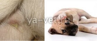
Symptoms and signs of hydrops in dogs that appear early are often confused with pregnancy and obesity. All these conditions are united by a rounded belly, which arouses suspicion only among the most attentive owners. For this reason, help is usually sought a little later - when the symptoms are complemented by the appearance of:
- poor appetite, which is not reflected in weight (despite the visible ribs and spine, the number on the scale does not change due to the amount of fluid);
- dull coat;
- shortness of breath during physical activity and at rest;
- extreme thirst;
- yellow or blue mucous membranes;
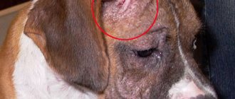
- swelling in the groin area, base of the ears, paws and chest;
- apathetic state with reduced activity (the animal is often in a sitting position);
- copious and frequent urination.
In the absence of recent mating, compliance with the feeding regime and sufficient mobility, veterinarians recommend conducting a special test. With its help, you can refute the imminent addition to the four-legged family and overeating.
To do this, you need to lift your pet by its front paws and watch its stomach. During stagnation, the round shape will change to a pear-shaped one, as all the liquid will rush down.
The clinical picture directly depends on the cause and stage of the pathology, therefore, in addition to the listed symptoms, diarrhea, vomiting, high fever, cough and severe pain may occur. To avoid complications, the animal should be shown to a doctor soon after a deterioration in appetite and loss of usual activity.
Laboratory examination of ascitic fluid
Certain information can be obtained by macroscopic assessment of ascitic fluid; however, the exact nature of its occurrence can only be determined through laboratory tests.
Classification of the causes of ascites in accordance with the composition of the fluid and the frequency of occurrence of each type of fluid. AB - arteriovenous; CVC - caudal vena cava; FIP - feline infectious peritonitis; GI - gastrointestinal; PSS - portosystemic shunt
Composition of the fluid - Diseases - Common causes - Rare causes
Transudate - hypoalbuminemia
- Liver failure
- Protein-losing enteropathy
- Protein-losing nephropathy
- None
- Inflammatory bowel diseases
- Glomerulonephritis
- Congenital pSS (rare)
- Cirrhosis
- Alimentary lymphosarcoma
- Lymphangiectasia
- Amyloidosis
- Subhepatic hypertension with presinusoidal block (ascitic fluid may be a transudate)
- Consequences of PSS ligation Portal vein thrombosis: occlusion of the vessel as a result of torsion.
- Vessel occlusion as a result of compression (hernia)
Modified transudate
- portal
- hypertension
- Intrahepatic portal hypertension
- Chronic hepatitis
- Cirrhosis
- Idiopathic liver fibrosis;
- AV fistula of the liver: liver neoplasms;
- fascioliasis (not found in the UK)
- Prehepatic portal hypertension with postsinusoidal blockade
- Compression/obstruction of the caudal vena cava
- No Obliterating endophlebitis of the hepatic veins:
- Badtz-Chiari syndrome; “tortuous” CPV (after injury); thrombosis of the CPV and the formation of fibrous fibers in its lumen; triatrial heart (cor triatrium dexter)
- Cardiac tamponade Idiopathic bleeding
- pericardial cavity
- Hemangiosarcoma of the right atrium
- Tumor of the base of the heart
- Restrictive pericarditis: mesothelioma; septic pericarditis
- Right-sided heart failure
- Damage to the valvular apparatus of the heart
- Dilated cardiomyopathy
- Neoplasms in the heart cavity
- Angiostrongylosis, dirofilariasis (not found in the UK)
- Septic (bacterial)
- Gastrointestinal perforation
- Migrating foreign body
- Penetrating wound
- Volvulus/infarction of the gastrointestinal tract
- Uterine rupture due to pyometra
- Mesocestoidosis (not found in the UK)
- Mycosis
- Nocardiosis or actinomycosis
- Exudate - inflammation
- Aseptic IPC
- Lymphocytic cholangiohepatitis in cats
- Pancreatitis
- Carcinomatosis
- Panniculitis
- Rupture of the diaphragm with organ entrapment
- Bile - leakage of bile
- Traumatic rupture of the gallbladder
- Necrotizing cholecystitis
- Perforation of the common bile duct (trauma, gallstones)
- Perforation of an ulcer in the proximal duodenum
- Blood - bleeding
- Systemic coagulation disorders
- Rupture of hematoma or hemangioma of the spleen
- Rupture of hemangiosarcoma of the liver or spleen
- Traumatic rupture of the liver or spleen
- Splenic torsion
- Torsion of the liver lobe
- Liver amyloidosis in cats
- Hepatic purpura in cats
- Lymph—lymphatic obstruction or leakage
- Cardiomyopathy
- Lymphangiectasia
- Bowel obstruction
- Neoplasms, in particular pheochromocytoma
- Urine - urinary tract rupture
- Bladder rupture
- Renal rupture
- Urethral rupture
- Diseases and changes that cause an increase in the volume of the abdominal cavity, which can be mistaken for ascites
- Neoplasms of the abdominal organs
- Diabetes
- Hyperadrenocorticism
- Obesity
- Ovarian cysts
- Paraprostatic cysts
- Pregnancy
- Pyometra
- Urinary retention (urethral obstruction or bladder atony)
With a gradient > 11 g/l, the effusion is the result of portal hypertension, with
In cats, a high protein content in a sample of exudative effusion is a characteristic sign of the development of lymphocytic cholangitis or, more often, the effusion form of feline infectious peritonitis (FIP). With IPC, the concentration of total protein in ascitic fluid usually exceeds 35 g/l and contains more globulins than albumins. An albumin:globulin ratio of 0.8 allows one to exclude IPC as a possible cause of effusion.
The detection of bacteria in ascitic fluid is a sign of contamination, unless the identified microorganisms are intracellular. If exudate is detected, a mandatory culture is required to isolate the bacterial culture.
Carrying out diagnostics
If a home test for ascites is positive, do not rush to self-medicate. Treatment methods directly depend on the cause of the disease, so a full examination cannot be avoided. The veterinary clinic will need:
- take urine and blood tests;
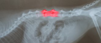
- undergo an ultrasound and x-ray;
- make a puncture.
Based on the results, the doctor determines the presence of diseases of the urinary system, injuries and tumors, the level of intoxication, the degree of organ damage, the exact amount and composition of the effusion. Next, the four-legged patient is given an individual treatment plan that takes into account the diagnosis.
Treatment
Before starting treatment for the underlying disease that caused the dropsy, supportive treatment is prescribed. They give medications that normalize the functioning of important organs: cardio medications for the heart and hepatoprotectors to improve liver function.
Another task is to empty the abdominal cavity of fluid.
This is done in two ways; the doctor will decide which one to use.
- Diuretics. There is no ideal medicine among them. Strong agents remove water completely, relieve swelling, but at the same time wash out too much potassium from the body. More gentle diuretics save some calcium, but have a side effect: dishormonal disorder. Light diuretics do not fully perform their function. Most of the liquid remains.
- Removal of fluid through a catheter inserted through the peritoneal wall. There are also important points here: the procedure must be performed slowly, and all the water cannot be removed completely at once.
With sudden elimination, kidney failure may occur as electrolytes, amino acids and proteins are lost. To avoid disruption of the metabolic process, drugs are administered intravenously to replenish the loss of these components.
If the results of the analysis show that the composition of the pumped out liquid is toxic, then antibiotics are prescribed.
After ascites is removed, treatment of the underlying disease begins. If treatment is not carried out, the dropsy will appear again.
Therapy methods
Treatment for ascites in dogs involves treating the underlying cause, relieving symptoms, and removing excess fluid. The first point is the most important, since without it symptomatic therapy gives only a temporary effect.
Medication
When sepsis and hypoxia are detected, blood transfusions and oxygen therapy are used, but most often treatment is limited to medication. Excess fluid is removed with diuretics, and all other drugs are selected individually:
- cardiotonics that stimulate the heart;
- antibiotics that destroy the causative agent of the detected infection;
- immunomodulators that prevent secondary infection by viruses or bacteria;
- analgesics that suppress attacks of pain;
- antiemetics, eliminating nausea and vomiting;
- sedatives and antipyretics;
- colloidal solutions that normalize protein levels.
Safe dosages and procedures for taking medications are determined by your veterinarian. They may differ from the instructions inside the package, since many medications are intended for human use.
Surgical intervention
Surgical treatment of dropsy in dogs is used when there is extremely high internal pressure and there is no effect from taking medications. In both cases, laparocentesis, or abdominal puncture, is used. In this procedure, effusion is drained through a small puncture in the abdomen. During the manipulations, the animal is under general anesthesia.
Also, indications for surgical intervention are necrosis, abscesses, internal bleeding and malignant neoplasms that do not have metastases. In these cases, treatment comes down to excision of dead tissue, cleansing of the purulent cavity, restoration of damaged tissue, or removal of the tumor.
Auxiliary folk remedies
Traditional medicine is used exclusively to eliminate symptoms. The recipes used are not able to affect the ascites itself, so in no case limit yourself to them.
Attention!
Many animals are prone to grass allergies. To be safe, use infusions and decoctions only under the supervision of a veterinarian.
Prevention
Prevention of ascites consists of general measures:
- balanced healthy diet;
- good living conditions;
- quarterly treatment for parasites (worms, fleas, ticks);
- routine vaccination;
- annual examinations by a doctor (especially for animals after 7 years).
Animal care and diet
During the treatment of ascites, it is recommended to reduce the duration of walking, increasing its frequency. Moderate activity and timely emptying will reduce the load on the heart, lungs and bladder. You will also need to put your pet on a temporary diet that includes:
- eating high-protein foods, boiled or steamed;
- reducing the amount of salt and water;
- taking vitamin and mineral complexes approved by a veterinarian.
With dry feeding, you will have to switch to a medicinal line designed for animals with sensitive digestion. To avoid allergies or diarrhea, it is better to choose food from a manufacturer you already know.
What to feed
If the appetite is preserved, then the dog is given food rich in protein. It could be chicken or turkey breast. They are pre-boiled or simply doused with boiling water. Carbohydrates and fats are reduced to a minimum. For porridges, you can add a little buckwheat or rice. Salt is completely excluded.
Appropriate industrial therapeutic diets are prescribed if the underlying disease is identified.
The dog's fluid intake should be reduced by pouring smaller amounts into the bowl and limiting access to water sources.
Good to know
- Sialocele
- Short bowel syndrome
- Irritable bowel syndrome
- Biliary vomiting syndrome
- Sclerotic encapsulating peritonitis
- Cececocolic intussusception
- Special feeding method
- Gut protectants
- Stomatitis
- Strongyloidiasis
- Tenesmus
- Digestibility and Absorption Tests
- Trichomoniasis
- Cytological analysis of stool
- Intestinal strangulation
- Congenital weakness of the esophagus
- Ulcers and erosions of the gastrointestinal tract
- Physical examination
- Hiatal hernia
- Chronic gastritis
- Septic peritonitis
- Schistosomiasis in dogs
- Sialadenitis, salivary gland necrosis
Further research
Further studies necessary to identify the cause of ascites are given in Further studies for ascites. APTT—activated partial thromboplastin time, AV—arteriovenous; CVC - caudal vena cava; DCM—dilated cardiomyopathy; CCA blood - general clinical blood test; BCh - biochemical analysis of blood serum; ECG - electrocardiogram; FRF—fibrin breakdown products; FIP - feline infectious peritonitis; OSPT is a one-step method for determining prothrombin time; Iα 1-P—α1-proteinase inhibitor; PIVKA, vitamin K deficiency-induced protein; PLE—protein-losing enteropathy; IPPL—pancreatic lipase immunoreactivity; NLP—protein-losing nephropathy; PSS—portosystemic shunt; TPI - trypsin-like immunoreactivity
Use of diuretics
Sometimes diuretics are prescribed to remove fluid. But potassium is removed in the urine, which is released in this way. To maintain it, you need to use special diuretics. But over time, they cause an imbalance in water and electrolyte metabolism. Therefore, do not abuse such drugs.
Good results can be achieved by using protectors for cardio and liver. They help these organs function normally.
The animal's diet should not contain salt, and fluid intake should be minimal.
Tactics
Before starting treatment, the doctor prescribes the following tests - ultrasound, x-ray, sampling of abdominal fluid for tests.
Based on the data obtained, a diagnosis is made and appropriate therapy is prescribed, which is aimed at eliminating the causes of ascites.
The dosage and methods of use of drugs that relieve the main symptoms of the disease are determined by the veterinarian.
Drugs
The most commonly prescribed medications are:
- Losartan;
- Veroshpiron;
- Furosemide;
- Eufillin.
In advanced cases of ascites, laparocentesis (pumping out fluid from the abdominal cavity) is prescribed. This procedure can significantly alleviate the dog’s condition and is carried out in parallel with taking medications. Additionally, the veterinarian may recommend taking antibiotics and protein drugs.
Methods
Depending on the disease causing ascites, the following treatment methods are also used:
- taking diuretics helps remove harmful substances from the animal’s body;
- taking cardioprotectors and hepaprotectors stabilizes the functioning of the heart muscle and liver;
- diet with limited salt and liquid.
How to protect your pet?
There are no preventive measures against abdominal hydrops. You should visit a veterinary hospital in a timely manner and get tested regularly. This will allow you to immediately identify diseases in your pet.
A sick dog needs extra attention. If surgical intervention is necessary, it is important to agree with the doctor so as not to risk the life of your pet. After surgery, active therapy is required.
The veterinarian will provide a list of necessary medications, give instructions, and nutritional recommendations. Owners must understand that the health of the animal depends on their care and love. Self-medication with human medications should not be practiced.
You might be interested
Diarrhea in a dog - how to treat it and why it occurs
Diarrhea or diarrhea in a dog is a very annoying phenomenon. On top of everything else, it can greatly …read more
What is a dog's normal body temperature and how to measure it
The easiest way to determine the well-being and health status of a dog is to measure its temperature. But you need to take into account that ...read more
The dog sheds a lot - should you worry and what to do?
When purchasing a dog for home, you should be prepared for any “surprises”, including periodic shedding ...read more