What is demodicosis in dogs?
Biologists have discovered more than a hundred species of Demodex; only three parasitize dogs:
- Demodex canis;
- Demodex cornei;
- Demodex injai.
Hundreds of studies have been published on these parasites, but questions still remain. In many publications, the demodex mite is described as a permanent inhabitant of the skin of all dogs, which multiplies when the immune system fails. This information is contradicted by a study of scrapings from healthy dogs without external signs of demodicosis. Of the 415 animals, only 36 were found to have ticks. The results of the study are described in a textbook on veterinary medicine.
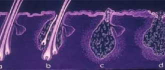
A subcutaneous mite cannot be seen without a microscope; its body length does not exceed 0.26 mm. Once on the skin, the parasite penetrates into the cavity of the hair follicle, sebaceous glands and excretory ducts. As it moves, it gnaws out paths in the epithelium, destroying the cells of the follicle, epidermis, and hair roots.
The life cycle of demodex lasts 25–30 days. During this time, the female lays dozens of eggs in the hair follicles. As the colony of parasites multiplies, it grows and conquers new living space. And the animal develops a demodectic focus.
Any dog can get demodicosis, regardless of gender, age and breed. But according to observations, the subcutaneous mite more often attacks short-haired animals:
- French, English, American bulldogs;
- Rottweilers;
- boxers;
- Dobermans;
- bull terriers;
- dachshund;
- German Shepherds and Great Danes;
- Staffordshire Terriers;
- Shar Pei.
Short hair does not protect well from external irritants, so dogs actively secrete sebum - a breeding ground for parasites.
Signs of the disease
Before looking at the signs of demodicosis, it is worth understanding what the disease is. This is an invasive disease, or in other words, a glandular disease. Its development is provoked by endoparasitic mites, which are located inside the sebaceous glands in the hair follicles. They can penetrate the tissues of internal organs.
Note! The main factors of this disease will help you understand that your dog has demodicosis. Typically, it causes dermatitis of the skin, manifestations of hyperkeratosis (thickening of the stratum corneum of epithelial tissue), damage to the tissues of internal organs, as well as depletion of the entire body.
The disease is usually detected in young dogs between 6 months and 2 years of age. But there are cases when demodicosis was detected in small puppies aged 2 months and older.
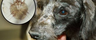
Typically, a subcutaneous tick in a dog causes many unpleasant signs that the owner can detect almost immediately. At first, they may look like pathologies with a fungal or bacterial nature.
The main symptoms include the following:
- feeling of itching occurs. The degree of itching can vary over a fairly wide range. In some cases it can be strong, the pet can scratch itself until wounds with blood appear;
- formation of redness of the skin;
- gradual loss of fur. If the parasitic organism has just penetrated the skin, then hair loss can be detected in areas with short hair - the stomach, ears, muzzle, and so on;
- The animal experiences changes in behavior and becomes restless. Sometimes outbursts of aggression may occur.
If a dog has had subcutaneous mites for quite a long time, then other manifestations may be detected in the pet:
- state of weakness, lethargy, general malaise;
- constant drowsiness;
- manifestations of anemia;
- the formation of secondary complications and pathological processes.
The above conditions usually occur as a result of the active development of subcutaneous mites. During its life, the parasitic organism releases toxic substances that spread throughout the animal’s body.
If the parasite dies, the animal begins to develop purulent processes. They may be accompanied by the formation of ulcers, pigment spots, and tubercles on the surface of the skin. All this leads to severe discomfort and complications for the pet’s health.
In any case, if similar symptoms and signs are detected, you should immediately contact a veterinarian. It is important to understand that the sooner appropriate treatment is provided, the sooner the unpleasant disease can be eliminated and serious problems with the animal’s health can be prevented.
Also learn about the symptoms, diagnosis and treatment of piroplasmosis in dogs.
Is canine demodicosis contagious to humans and other dogs?
People also suffer from demodicosis, but other species live on them: Demodex brevis and Demodex folliculorum. A dog tick can land on human skin, but will not be able to feed or reproduce and quickly dies in an alien environment.
With close, prolonged contact, adult ticks move from one dog to another. However, the likelihood of developing the disease depends on the state of the immune system. The risk group includes:
- weakened dogs;
- sick and recovering;
- elderly over 10 years old;
- puppies up to one year;
- animals with skin lesions.
A healthy dog with a normal immune system will not get demodicosis even after close contact with a sick animal.
Dog ticks are not dangerous for cats. They are parasitized by other species of Demodex - Demodex gatoi, Demodex cati.
How does infection occur?
The subcutaneous tick is transmitted from one animal to another. If the pet is contagious, and the owner neglects precautions, any, even short, contact with a healthy dog will cause infection. It doesn’t take long for parasites to move from one “victim” to another. They can jump over in a few seconds.
Representatives of no breed are immune from parasites. Knowing how dogs become infected with subcutaneous mites, you can try to prevent the disease as much as possible.
Many owners are interested in where ticks come from in a healthy dog. The reasons for the appearance are different, they are all associated with the rapid proliferation of Demodex in the skin. A favorable environment for them is increased sebum production. This is often due to deterioration of the skin condition that appears against the background of a weakened immune system.
When parasites are transmitted from one dog to another, dead skin particles and sebum are transferred. Along with them, a healthy dog receives parasites that quickly settle in a new place.
Forms of demodicosis in dogs
Based on the area of damage, there are 2 forms:
- Localized – up to 4 demodectic lesions up to 2.5 cm in size.
- Generalized – 5 or more scattered affected areas or extensive skin damage. This form occurs as a continuation of an untreated focal form or develops independently.
The division is conditional, since the ratio of the size of the dog and the affected areas is taken into account. For example, 3 lesions with a diameter of 2 cm for a Chihuahua are already generalized demodicosis, and 5 affected areas of 3 cm for a mastiff are a localized form of the disease.
Separately, pododemodecosis and otodemodecosis are distinguished. In the first case, lesions form on the paws and fingers. In the second, the parasite infects the surface of the ear.
Traditional treatment
Among folk remedies against demodicosis, birch tar is considered the most effective. For home treatments, you can use it in the form of soap. The dog is lathered with it in the affected areas and left for 3 hours. Then the soap should be washed off with warm water and the skin should be dried. Other effective recipes and folk methods against demodex in dogs:
- mix crushed celandine with vaseline in a ratio of 1:4;
- combine 1 part turpentine and 2 parts drying oil or animal fat;
- mix 5 tbsp. mustard oil with 1 tbsp. crushed garlic;
- prepare a mixture of 2 tsp. grated laundry soap, 1 tsp. birch tar, 2 tsp. melted fat and 1 tsp. sulfur.
Juvenile demodicosis in dogs under 1.5 years of age
Demodex mites are transmitted to puppies from their mother within 2–3 days after birth. Most kitties become asymptomatic carriers; the parasites do not affect health in any way. If the fragile immune system cannot cope and does not restrain the proliferation of parasites, by 3 months or later the puppy will develop demodicosis.
An additional burden in the 1st year of life is vaccinations, changing teeth, and cupping. These events can trigger stress, a dangerous enemy of the immune system. Focal juvenile demodicosis in 90% of cases goes away as the puppy grows up and develops immunity.
For the development of the generalized juvenile form, genetic prerequisites are needed. The reproduction of mites is inhibited by the T-link of the immune system, which recognizes and removes foreign agents. Dogs with defects in protective T cells are themselves prone to generalized demodicosis and pass this defect on to their offspring.
Causes of demodicosis in adult dogs
In animals older than 18 months, demodex mites more often “wake up” against the background of other disorders that reduce the overall immunity and barrier functions of the skin:
- bacterial, allergic dermatitis;
- hormonal imbalance;
- infection with worms;
- oncological diseases;
- autoimmune pathologies.
Dogs with impaired hair growth and decreased skin tone are predisposed to the development of demodicosis. It is easier for a subcutaneous mite to penetrate into the cavity of the follicle when the hair grows slowly after molting or the root does not adhere tightly to the walls of the hair follicle.
Often demodicosis begins when the hormonal balance changes. This happens during pregnancy, after estrus or long-term treatment with corticosteroids. Hormonal drugs save from severe inflammation, but lead to the death of lymphocytes and suppress the immune system.
During the treatment of demodicosis, corticosteroids are completely excluded.
Unbalanced nutrition, lack or excess of macro and micronutrients affects the condition of the skin and disrupts protective functions. The dog's hair falls out, bald areas appear, skin tone decreases, and dermatitis develops. As a result, favorable conditions are created for the spread of ticks.
Complications
Demodicosis in dogs is often complicated by the addition of secondary infections. When the body's defenses decrease, acariasis is accompanied by fungal (ringworm) or microbial infections. The course of the disease becomes noticeably more complicated. In this case, it takes a lot of effort to cure the animal.
Demodicosis of the eyes is not typical for dogs, but the underlying pathology is complicated by the development of inflammation of the mucous membrane of the organ of vision.
In the generalized course of the disease, pets experience damage to the digestive tract, hepatobiliary region, and problems with the endocrine glands appear.
Symptoms of demodicosis in dogs
The initial stage proceeds unnoticed. The fur is thinning in places. Skin color does not change. There is little or no itching. Well-being and behavior remain the same. As the tick spreads, the disease takes on a scaly (squamous), pustular or mixed clinical form.
At an early stage, a demodectic lesion seems harmless.
Signs of scaly demodicosis
The development of this form is indicated by the following symptoms:
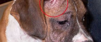
- The dog develops round bald areas with clear boundaries with a diameter of 1 to 20 mm. More often they appear on the face, around the eyes, on the forehead, and limbs.
The initial stage of scaly demodicosis.
- The skin on hairless areas turns slightly red, flakes, and becomes covered with scales.
- The fur at the border of the demodicosis lesion does not stay in the follicles and falls out.
- Gradually, the skin becomes rougher, cracks, and dense nodules appear on it.
- At a later stage, the color changes to bluish-gray with red inclusions.
In the scaly form, the animal does not suffer from itching. It begins when a secondary bacterial or fungal infection occurs. Body temperature often drops to 37℃.
Symptoms of pustular demodicosis
This form develops if pathogenic or conditionally pathogenic bacteria multiply in the demodicosis lesion:
- The skin turns red, swells, and becomes covered with nodules.
- Gradually they turn into yellow, brown, black bubbles with liquid.
- When the pustules open, pus flows out, sometimes with traces of blood. After drying, crusts form.
- The skin thickens, wrinkles, becomes cracked, and smells bad. Ulcers form at the site of burst pustules.
With otodemodecosis, the ears become swollen, hot, and painful. The inner surface turns red, sulfur is released intensely, and itching occurs. Demodicosis of the paws is painful. While walking, the dog rubs the injured skin. In addition to the standard symptoms, lameness occurs.
What are the causative agents of the disease?
Ticks are small, cigar-shaped organisms that resemble transparent worms with limbs and a jaw structure. They can only be seen under a microscope. Their main habitat is hair follicles. They bite along the hair, reaching the follicles, and continue to move throughout the epidermis, making “tunnels”. Along the way, eggs are laid at the roots of the hair, the number of which can reach 250-300 pieces.
As soon as the body weakens under the depressing effects of diseases or steroid drugs, active reproduction of worm-like mites begins.
It is worth noting that a sick dog cannot infect other animals or humans.
Analysis for demodicosis in dogs
It is impossible to make an accurate diagnosis based on external signs. Symptoms of demodicosis are similar to clinical signs of other diseases:
- Bacterial skin infections - folliculitis, pyoderma, impetigo, furunculosis.
- Fungal infections - trichophytosis, microsporia.
- Allergic diseases – flea infestation, atopic dermatitis, food sensitivity.
- Infection with scabies mites - sarcoptic mange.
- Thyroid dysfunction and hormonal imbalance – hypothyroidism.
The final diagnosis is made after microscopic analysis. The doctor makes a deep scraping from the lesion with a scalpel until a drop of blood appears. Then he transfers the materials onto glass smeared with Vaseline. Sexually mature individuals, larvae, and eggs are easily visible under a microscope. After counting the parasites at different stages, an acarogram is displayed.
Demodex mites under a microscope
If single adult individuals are found, then up to 5 repeated scrapings are made from the affected and healthy areas. In sensitive areas, such as around the eyes, instead of skin, a hair pluck is taken for analysis.
Additional studies will be needed to identify concomitant disorders:
- clinical urine test,
- blood biochemistry;
- coprogram;
- hormonal analysis of blood serum;
- bacterial culture to determine the sensitivity of the microflora if antibiotics are prescribed.
During treatment, scrapings are repeated every 3 to 4 weeks until not a single tick is found in at least two samples at a monthly interval.
Diagnostics
When a subcutaneous tick is detected in a dog, many owners panic and do not know how to help their pet. In these situations, you should pull yourself together and immediately contact a veterinarian; he will be able to conduct a full examination and prescribe the appropriate treatment.
To detect subcutaneous ticks in a dog, a veterinarian will prescribe a number of diagnostic procedures.
Carrying out a full diagnosis ensures the following:
- it reveals the main root cause of the pathological process;
- determines the severity of the disease;
- helps to choose the optimal treatment option.
Important! Before sending a dog for a full diagnosis, the veterinarian must conduct an external examination of the animal. Of course, it is impossible to visually detect the presence of demodicosis, because this parasitic organism has microscopic dimensions. But it’s still worth carefully examining your pet’s skin.
Often, with demodicosis, parasites penetrate into the deep layers of the skin, which is quite difficult to visually detect. But existing external symptoms in the form of redness, severe irritation, wounds from scratching may indicate the presence of a parasite in the animal’s body.
Every dog owner must take into account that the clinical manifestations of this disease may be similar to the symptoms of other infectious and non-infectious pathological processes. For this reason, if suspicious signs are detected, you should handle the animal carefully; the fact is that many diseases can be contagious and dangerous to humans.
Infectious diseases can be transmitted to humans in the initial stages of development. In this case, both the pathogen itself and the parasitic organism itself can be transmitted. In these situations, it is better to immediately contact a veterinarian who can conduct a full diagnosis of the animal.
Typically, a complete examination includes the following procedures:
- In order to detect demodicosis, the doctor first scrapes the dog’s skin. If this analysis is carried out by an experienced specialist, then he can almost immediately limit the search for identifying the type of parasitic organism - subcutaneous or intradermal;
- For an accurate diagnosis, a fragment of epithelial tissue is taken from the dog and examined under a microscope. To carry out this analysis, the animal must be injected with painkillers. Next, the veterinarian makes a small incision and takes a scraping. Performing this diagnosis helps determine the extent of the lesion;
- Additionally, tests of the animal’s urine, blood, and feces are prescribed.
Treatment of focal and generalized demodicosis in dogs
There is no general treatment regimen for all dogs. When choosing drugs, a lot of details are taken into account: age, heredity, area and shape of the lesion, test results, severity of symptoms, concomitant diseases.
According to the observations of veterinarians, focal uncomplicated demodicosis resolves spontaneously in 90% of dogs. But you should not hope for a successful outcome; the development of the disease cannot be predicted.
Treatment of the focal form includes the following areas:
- Cleansing demodicosis lesions.
- Treatment with acaricidal solutions, ointments or drops to reduce the population and destroy the tick.
- Treatment with ointments to restore skin, coat and prevent secondary infections.
In the generalized form the following are added:
- Systemic acaricidal drugs in the form of injection solutions or tablets.
- Antibiotics or external antiseptics to prevent and treat bacterial infections in the affected area.
- Antimycotics for suspected fungal development in a demodectic lesion.
Treatment should not be stopped until 2 negative laboratory tests are obtained, even if external symptoms subside. The drugs destroy adult ticks, but do not affect parasite eggs. When treatment is interrupted prematurely, they become more active, develop into sexually mature individuals, and demodicosis returns.
Shampoos
Before treatment, the hair around demodectic lesions is cut off. The dog is bathed to remove particles of damaged skin and excess fat. The effect of antiparasitic drugs is enhanced on a cleaned surface.
Here are examples of veterinary medicated shampoos with antibacterial and keratolytic properties:
- Peroxiderm. Benzoyl peroxide is added to the composition. This substance disinfects the affected area, regulates the secretion of skin secretions, and gently removes scales and crusts.
- Doctor - a line of shampoos with medicinal additives. The cleanser contains benzoyl peroxide. When pustules appear, use shampoo with chlorhexidine. When the lesions stop peeling, becoming covered with bubbles and crusts, they switch to restorative shampoos with birch tar or prebiotics and D-panthenol.
- DermaPet is a line of American dermatological shampoos. For demodicosis, use DermaBenSs. In addition to benzoyl peroxide, the composition contains 1% sulfuric and salicylic acids. These components disinfect and dry the skin. Sensitive dogs can be washed with the hypoallergenic DermaLyte shampoo from this range.
After bathing, the dog is not dried so as not to spread ticks. The wool is carefully blotted and dried with a hairdryer or allowed to dry naturally.
Acaricidal solutions for local treatment
All anti-tick medications contain toxic substances, so before treatment the dog is given a protective collar or the jaws are secured with a ribbon. Here are examples of solutions ready for application:
- Amitrazine – contains 3 main components. Amitraz causes paralysis and death of ticks. Dimexide relieves inflammation. Rapeseed oil restores skin. Amitrazine labeled “Plus” contains the antiseptic, antifungal substance decamethoxin. The solution is applied once every 5–7 days until recovery, Amitrazine Plus is applied every 3 days. To treat demodicosis, 5 to 8 procedures are usually enough.
- Prazicide complex is a solution based on a combination of ivermectin and praziquantel, which is harmful to demodex. The additional component levamisole stimulates local immunity by increasing the number of T-lymphocytes. The drug is applied to the affected areas at intervals of 10–14 days.
- Amit forte – combines 3 active substances. Fipronil destroys larvae and adult parasites. Diphenhydramine reduces itching and discomfort. Diflubenzuron interferes with the synthesis of chitin and disrupts the life cycle of the tick. The animal is treated 3–5 times with a time interval of 5–7 days.
- Decta - in addition to amitraz, contains chloramphenicol, which stops the growth of harmful bacteria. The auxiliary component propolis disinfects and accelerates skin restoration. The solution is used every 5 - 7 days until recovery.
Before treatment, scales and scabs are removed from the lesion with a cotton-gauze swab dipped in warm water or an antiseptic - chlorhexidine, miramistin. Antimicrobial solutions have no effect on ticks, but prevent the development of secondary infections.
The preparations are applied in a thin layer directly to the affected area from the borders to the center, covering 1 cm of healthy skin. Large areas are treated in 2 steps: first one half, then the other half a day later. After application, wait 20 minutes for the liquid to be absorbed, and only then remove the protective collar.
Acaricidal ointments
Instead of solutions, you can treat the lesions with ointments. They add glycerin, which softens and moisturizes the skin:
- Amidel gel - contains amitraz, lidocaine for local anesthesia, methyluracil to accelerate skin regeneration.
- Aversectin ointment is based on the aversectin C complex, which is destructive for larvae and mature individuals of Demodex canis.
The ointments are rubbed in every 5-7 days until complete recovery.
Acaricidal drops on the withers
The drugs are applied dropwise at the base of the skull and along the spine in 3 to 4 places. After treatment, the active ingredients are evenly distributed in the epidermis, follicles, and sebaceous glands:
- Advocate - the first licensed drops against demodex mites. The product contains a combination of imidacloprid and moxidectin. In mild cases, treatments are repeated every 3-4 weeks, in complicated cases - every 7 days.
- Promeris Duo - drops based on a combination of amitraz and metaflumizone. The drug effectively kills demodex mites, but often causes an allergic reaction.
- Dironet Spot it contains ivermectin and praziquantel. The dog is treated once every 10–14 days until recovery.
Drops on the withers work well with isolated lesions. In severe cases, they are included in complex therapy.
Restorative ointments
At the same time, they use products to reduce irritation, redness, discomfort, and prevent secondary infections:
- Sulfur ointment is a harmless remedy against inflammation, the formation of bacteria and fungus, and stops the spread of mites.
- Fir - contains pine resin, beeswax and foundation. The product improves blood circulation, resolves inflammation, disinfects and restores the skin.
- Vedinol is a line of ointments for the treatment of inflammation of the subcutaneous tissue and skin based on pine oil. An acaricidal phytocomplex has been added to Vedinol marked “plus”. The series includes an ointment specifically for paws. It helps with the appearance of redness and chafing in the affected area.
Acaricidal preparations for injections
For the generalized form, drugs based on avermectins are traditionally used. These are biopesticides that are harmful to ticks and are obtained from the decay products of the fungus Streptomyces avermitilis.
The avermectin series includes solutions containing ivermectin, aversectin, doramectin:
- Ivermek;
- Ivertin;
- Baymek;
- Aversect;
- Noromectin;
- Novomek;
- Ivomek;
- Dectomax.
The listed drugs are administered using intramuscular or subcutaneous injections or infused through the mouth according to the scheme. The safest from this list is Aversect. It is produced specifically for dogs and cats and has fewer side effects. The exact dosage, methods of administration, treatment regimen are prescribed in the instructions for Aversect for dogs.
The remaining solutions are intended for farm animals, although they cope with the canine type of demodex. Violation of the dosage results in severe complications, so the treatment regimen is prescribed only by the attending physician for a specific animal.
Avermectin drugs have been used to treat demodicosis for a long time, so they have become less effective against subcutaneous mites. Sometimes they turn out to be useless and have to be replaced with drugs from other groups.
Treatment of demodicosis in dogs with Bravecto and analogues
The active ingredients of Bravecto and similar tablets belong to the chemical class of isoxazolines. Substances of this group disrupt the connection between neurons, immobilize and destroy ticks. They have been used in veterinary medicine relatively recently; cases of Demodex resistance have not yet been identified.
Bravecto was invented to protect against ixodid ticks and fleas. Then it turned out that the drug copes with advanced demodicosis. Tablets are produced for 5 weight categories of animals from 2 to 56 kg. The active substance fluralaner enters the bloodstream and is slowly eliminated over 3 months. Tablets are given with food or after meals once every 12 weeks until recovery.
Other pharmaceutical companies have also released isoxazoline tablets:
- Frontline Nexgard . The active substance is afoxolaner. For demodicosis, during the first month, 2 single doses are fed at a two-week interval. Then they continue to give once a month. A detailed description of Frontline Nexgard is in the official instructions.
- Nexguard Spectra. The drug contains 2 active ingredients: afoxolaner and milbemycin oxime. In the first month, tablets are taken twice with an interval of 14 days, then once a month until clinical recovery.
- Simparica. Tablets are available in 6 dosages. The smallest is for small dogs with a body weight of 1.3 - 2.5 kg. Other drugs of the isoxazoline group are allowed to be used with a weight of 2 kg or more. For demodicosis, tablets are fed monthly, regardless of feeding.
The active ingredients are distributed unevenly in the tablets, so they are given whole and not divided into parts.
Dog breeders will find it useful: How to cure a dog from piroplasmosis and how to protect it from ixodid ticks.
Antibacterial tablets
Pathogenic and opportunistic bacteria quickly multiply on damaged skin. When pustules and ulcers appear, external antiseptics are added to the treatment regimen, and in advanced cases, antibacterial tablets, for example:
- Amoxicillin. Veterinary suspension for subcutaneous and intramuscular injections. A noticeable improvement occurs after 1 – 2 injections with an interval of 48 hours. The medicine is administered at the rate of 1 ml/10 kg of animal weight.
- Tsiprovet . 1 tablet is designed for 10 kg of body weight. The required dose of medication is given once a day for 5 to 7 days. In a preparation marked “for cats, small dogs and puppies,” 1 tablet is designed for 3 kg of body weight.
- Sinulox – veterinary tablets of 50, 250 and 500 mg. A single dose is 12.5 mg/1 kg of dog weight. The medicine is fed twice a day with an interval of 12 hours for 5 - 7 days. In advanced cases, it is allowed to increase the single dose to 25 mg/1 kg and extend treatment to 10–28 days. A complete analogue of Sinulox is veterinary tablets Xiclav.
The therapeutic effect of antibiotics is reduced if you interrupt the course ahead of time or skip a dose.
Immunity restoration
For any form of demodicosis, create the most favorable conditions for strengthening the dog’s immunity:
- The pet is examined and concomitant diseases are treated if they are detected.
- At home, the conditions of detention are analyzed. Demodex mites love warm, moist environments. The dog's place is located away from radiators. On the street they do not put you in a damp booth or enclosure. The bedding is kept clean, often washed in hot water or changed.
- Carefully protects from stress. Loud noises, unfamiliar people and new animals in the house, and harsh treatment become sources of nervous tension. Some dogs do not tolerate loneliness or travel well, and take a long time to get used to changes.
- Reviewing the diet. When eating natural food, do not overfeed with carbohydrates - cereals, pasta, bread. They do not give sweets, pastries, sausage, bones, or food from the common table. Avoid dry food with low protein content. Do not mix homemade products and industrial feed.
- To restore the skin and coat, it is useful to take a course of injections of the Catozal stimulating solution, add feed sulfur to food at the rate of 0.04 g per 1 kg of weight per day, but not more than 1 g per dog.
In the focal form, these measures are sometimes enough to restore the immune status and stop the spread of demodex. In difficult cases, Immunoparasitan, Fosprenil, Maxidin are included in complex treatment.
There is no consensus in the veterinary community regarding immunomodulators. Many do not see any benefit from them, since the effect of immunostimulants on the immune system that inhibits demodicosis has not been proven.
Forecast
Focal demodicosis disappears without a trace within 1 to 3 months. Treatment of the generalized form lasts from six months or longer. The indicator of clinical recovery is 2 negative tests of skin scrapings with an interval of 1 month. But a complete cure is stated when there are no relapses throughout the year.
With a generalized form in juvenile age, the risk of the disease returning in the future increases, since this often happens due to genetically determined immunity disorders. Such animals require increased attention and painstaking care.
Prevention of demodicosis in dogs
The main task is to eliminate conditions for the reproduction of ticks, which means maintaining the dog’s immunity:
- Once a quarter, the pet is taken for a preventive veterinary examination in order to identify hidden diseases at an early stage and treat them in a timely manner.
- Do not violate the vaccination schedule.
- The pet's area is kept clean and dry.
- Balanced food.
- Contact with obviously sick animals is limited.
- Regularly treated against external and internal parasites.
To prevent parasitic diseases, it is better to use universal remedies so as not to give several drugs. Bravecto, Simparica, and Frontline Nexgard tablets protect against ixodid, demodectic, ear, sarcoptic ticks and fleas. Nexgard Spectra, Advocate drops, in addition to the listed parasites, destroy roundworms and heartworm larvae.
Demodicosis in dogs is an unpleasant but curable disease, photos, treatment, symptoms depend on the clinical form and area of the lesion.
Clinical manifestations
Each form of demodicosis in dogs has its own characteristic symptoms. With otodemodecosis, the pet shakes its head and rubs it against various objects. In addition to clinical signs, several general symptoms of the disease can be identified:
- hair begins to fall out - on the chest, paws, muzzle, along the spine;
- comedones appear in areas of alopecia - acne, blackheads;
- if an animal has long hair, it begins to stick together - (follicular casts).
Symptoms of local skin lesions
Localized (focal or local) is characterized by damage to individual areas of the body. Areas of baldness are noted on the limbs and head. In the affected areas, the skin wrinkles and thickens. Its color changes to grayish or reddish-red. Dry scales appear on the skin. Other distinctive features of the localized form:
- pustules appear on the skin - light pink nodular rashes;
- then the pustules degenerate into abscesses, which then burst;
- the contents of the pustules dry out (females live in papules for up to 10 months), the skin becomes rough and red, becomes covered with folds, and acquires an unpleasant odor.
Signs of generalized demodicosis
With the generalized type, the mite penetrates deep into the layers of the skin, so it can even reach the internal organs. Clinical signs of this form of demodicosis are:
- foaming at the mouth;
- coordination problems;
- weakness;
- convulsions;
- muscle tremors;
- dyspepsia;
- increased salivation;
- vomit.