A healthy brain is the basis not only for the normal functioning of the body, but also for the socialization of animals, their upbringing and training. It is the parts of the brain that are responsible for the perception and processing of information. This is why brain diseases such as hydrocephalus are so dangerous, because a healthy and intelligent animal can turn into a disabled creature due to pathology.
Causes of the disease
Hydrocephalus is classified as a congenital disease, although acquired cases also occur in medical practice, but to a much lesser extent. Pathology occurs due to disturbances in the activity of the ventricles of the brain. CSF, a constantly circulating fluid, accumulates in them, which in turn leads to contraction of nerve cells.
To date, the exact causes of congenital hydrocephalus have not been established. If we are talking about an acquired pathology, then the provoking factors are, first of all, brain tumors and inflammatory processes in the body.
What is hydrocephalus?
Hydrocephalus most often occurs in puppies. Due to the rapid growth of the body, the cranial bones do not keep up with the brain, so an excessive amount of cerebrospinal fluid accumulates in their cavities. Gradually increasing pressure leads to the death of nerve cells and degradation of brain activity.
Mechanical damage and infections can cause a similar disorder, so adult animals are also not immune from the disease. Regardless of the cause, the pathology poses a serious threat to life and requires immediate treatment.
Which breeds are more susceptible
There is a group of dogs that are predisposed to congenital hydrocephalus. These are small decorative dogs: the so-called pocket breeds - Yorkshire terriers, Pekingese, Chihuahuas, etc.
This is due to the structure of the cranium, or more precisely, the size of the brain, which is very small. However, owners of such dogs should not immediately panic. The disease develops in puppies up to one year old. It appears within a few weeks after the baby is born.
At this age, dogs grow very quickly, and the increase in the bones of the skull simply does not keep pace with the growth of the brain. When the condition becomes critical, the pet develops hydrocephalus.
As for acquired pathology, it can develop in any dog, regardless of breed, age and gender.
Signs of hydrocephalus
The age at which symptoms first appear can vary, on average from 2 months to 5 years, but owners of animals under the age of 1 year most often seek help.
The most characteristic clinical signs of hydrocephalus include seizures and/or unusual behavior of the animal, for example:
- the dog runs in circles (as if chasing its tail);
- throws his head back;
- or tilts it to one side;
In addition, hydrocephalus is indicated by the characteristic appearance of the dog, which distinguishes it from its littermates:
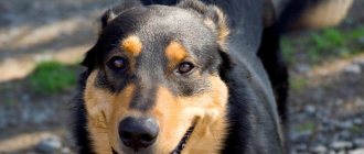
- a very large skull on a thin neck;
- strabismus of the eyeballs;
- behavioral disorders (aggression, bulimia, increased libido, difficulties in training).
Chihuahua with signs of hydrocephalus
If you notice such deviations, you urgently need to take the animal to a veterinary clinic for a comprehensive examination.
Main symptoms
It is very easy to determine hydrocephalus, since the disease has a pronounced clinical picture. The first signs appear in a puppy at 1-1.5 months; the most characteristic ones include:
- visual impairment visually resembling strabismus;
- increase in the shape and size of the head;
- seizures, convulsions;
- change in behavior (aggression or lethargy, apathy);
- unusual movements (rolling on the floor, throwing back the head, spinning around oneself).
The sick puppy stands out among the other babies in the litter: its head is much larger, its limbs are thin and thin.
Symptoms of the disease
The manifestation of the congenital disease, visible to the naked eye, can be detected within 2-3 weeks after the birth of the puppies. However, acquired hydrocephalus in dogs can manifest itself much later. The most striking signs of pathology, according to experts, include:
- inappropriate behavior, expressed in the fact that the dog can start circling in one place for no reason, unnaturally throw back its head and twist it in different directions;
- vision is impaired, the dog begins to clearly cross his eyes;
- seizures occur that are in many ways reminiscent of epileptic seizures;
- the shape and size of the skull changes, it becomes larger, acquires a spherical shape, cranial sutures are clearly visible under the skin;
- hydrocephalus causes the dog to become aggressive. When the owner tries to play with him, he may growl at him and even bite him.
The owner must be aware that skull transformation is only possible in representatives of “pocket” breeds; these symptoms will not affect a large dog. In dogs of medium and giant breeds, the disease manifests itself in that they lose their appetite, become apathetic and lethargic, sleep a lot, and when awake wander aimlessly around the house, bumping into pieces of furniture.
Diagnostics
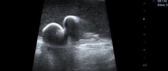
To make a diagnosis and prescribe treatment, the veterinarian carries out certain diagnostic measures. In addition to a visual examination of the animal, the specialist will need to take tests of urine, blood, and cerebrospinal fluid. If the fontanelle is open, an ultrasound examination is performed.
If we are talking about acquired hydrocephalus, encephalography and radiography are mandatory to establish the degree of brain damage and assess its functioning. In some cases, the computed tomography method is used. It is used in cases where it is not possible to accurately establish the picture of the disease.
Breeds at risk
The likelihood of developing the disease is influenced by breed. Most often, representatives of small breeds are affected, acquiring the disorder from birth. For representatives with larger dimensions, the situation is the opposite. In their case, the disease almost always turns out to be acquired.
Small breeds
Hydrocephalus occurs in Chihuahuas, Pekingese, miniature poodles and Yorkshire terriers - miniature dogs with small skulls. They are characterized by a congenital type of pathology associated with a discrepancy between the sizes of the brain and cranium. The first alarming symptoms appear 4-5 weeks after birth.
Large breeds
Pets with large dimensions get sick much less often. In their case, the cause of the disorder lies in concomitant diseases: infections or neoplasms. If preventive examinations and timely treatment are neglected, any pet can encounter pathology. Only the course of the disease will differ, excluding head deformation due to intracranial pressure.
Treatment method and prognosis
Once the diagnosis is made, the doctor prescribes treatment for hydrocephalus. It is carried out in a complex and includes taking diuretics, the action of which is aimed at removing excess fluid from the body. Prescribed:
- Prednisolone: 0.25-0.5 MK/1 kg of weight 2 times a day every 12 hours.
- Furosemide: 0.4-4 MK/1 kg 1 time per day.
- Acetazolamide: 10 MK/1 kg 4 times a day every 6 hours.
- Omeprazole: 10-20 MK/1 kg 1 time per day.
The dosage and course of taking medications are determined by the doctor depending on the weight of the animal, the condition and individual characteristics of the body. The owner must strictly comply with the treatment conditions, since uncontrolled use of medications can have a detrimental effect on the dog’s health and lead to numerous side effects.
Since convulsions are common in sick puppies, anticonvulsant medications are given. To strengthen the immune system, vitamin complexes are prescribed.
But conservative therapy is often ineffective, so there is a need for surgical intervention. The operation involves the introduction of a shunt for the outflow of cerebrospinal fluid.
After surgery, your veterinarian may prescribe corticosteroid hormone therapy.
Contraindications to surgical intervention include exhaustion of the body, advanced hydrocephalus, severe changes and deformation of the skull.
In most cases, the prognosis for hydrocephalus is unfavorable, since even with timely shunting it is impossible to completely get rid of the pathology. It is not always possible to restore vision and motor function. In addition, the risk of death during surgery is very high.
Literature
- Cerda-Gonzales S, Olby NJ, Pease TP: Morphology of the Caudal Fossa in Cavalier King Spaniels. Veterinary Radiology & Ultrasound, Vol. 50, N1, 2009, pp. 37-46.
- Rusbridge C, Knowler SP, Pieters L, McFadyen A. K: Chiari-like Malformation in the Griffon Bruxellois, Journal of Small Anima Practice Awaiting Publication, 2009.
- Rusbridge C, Caruthers H, Dube MP, Holmes M, Jeffery, ND Association Between Spinal Cord Dorsal Involvement and Pain in Syringomyelia Secondary to Canin Chiari Malformation. Journal of Small Anima Practice 48, 432-436, 2007.
- Dewey CW, Berg JM, Stefanacci JD, et al. Caudal Occipital Malformation Syndrome in Dogs. Compend Contin Educ Pract Vet 26: 886-896, 2004.
- Rusbridge C, Greitz D, Iskandar BJ. Syringomyelia: Current Concepts in Pathogenesis, Diagnosis, and Treatment. J Vet Intern Med 20:469-479, 2006.
- Garcia-Real I, Kass PH, Sturges BK, et al. Morphometric Analysis of the Cranial Fossa in the Dog: a Computerized Tomographic Study. Vet Radial Ultrasound 45: 38-45, 2004.
- Stalin CE, Rusbridge C, Grander N, et al. Radiographic Morphology of the Cranial Portion of the Cervical Vertebral Column in Cavalier Ring Charles Spaniels and Its Relationship to Syringomyelia. Am J Vet Res 69:89-93, 2006.
- Scrivani PV, Thompson MS, Winegardner KR, et al. Association between Frontal-Sinus Sire and Syringohydromyelia in SmallBreed Dogs. Am J Vet Res 68:610-613, 2007.
- Cerda-Gonzales S, Dewey CW, Scrivani PV, et al. Imaging Features of Atlanto – Occipital Overlapping in Dogs. Vet Radial Ultrasound 50: 264-268, 2009.
- Schmidt MJ, Wegger A, Jawinski S, et al. ultrasonographic appearance of the craniocervical Junstion in Normal Brachycephalic dogs and Dogs with Caudal Occipital (Chiari-like) Malformation. Vet Radial Ultrasound 49:477-476, 2008.
What to do at home
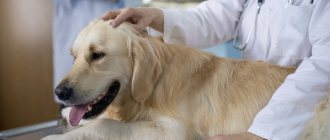
If owners really want to help their pets, they must understand that there is no question of any self-medication. The first thing to do if you notice characteristic symptoms is to contact your veterinarian. You cannot postpone your visit to the clinic - the fluid will compress the brain more and more, which will lead to death.
The operated dog needs to be provided with high-quality care to prevent displacement of the shunt, inflammation and suppuration of the wound. It is necessary to limit the puppy’s motor activity and its communication with other dogs. In the future, in order to prevent clogging of the shunt, a systematic examination in the clinic is recommended. The observation period is set by the doctor on an individual basis.
Veterinary clinic of Dr. Shubin
Definition
Hydrocephalus is an increase in the volume of cerebrospinal fluid (CSF) in the ventricular system or subarachnoid space of the brain.
Physiology of CSF circulation
The bulk of the CSF is produced by the choroid plexuses in the lateral, third and fourth ventricles, a small part is produced by the ependyma of the ventricles, the vessels of the pia and arachnoid membranes and the pia mater surrounding the brain and spinal cord.
CSF flows from the lateral ventricles into the third ventricle, then caudally through the mesencephalic duct to the fourth ventricle and then into the subarachnoid space. In the subarachnoid space, most of the fluid moves around the brainstem into the rostrotentorial region.
Most CSF absorption occurs in the arachnoid granulations in the dorsal sagittal sinus, with absorption likely through venous and lymphatic drainages around the spinal and cranial nerves.
Classification
Depending on the type of disturbance in the flow of cerebrospinal fluid, hydrocephalus is likely to be divided into the following types or categories: • Congenital and acquired hydrocephalus. • Internal hydrocephalus - accumulation of CSF in the ventricular system of the brain. • External hydrocephalus - accumulation of CSF in the subarachnoid space. • Obstructive or non-communicating hydrocephalus - obstruction of CSF outflow.• Communicating hydrocephalus - decreased absorption and/or increased production of CSF.• Ex vacuo hydrocephalus (secondary or compensatory hydrocephalus) - increased CSF volume with decreased brain volume.
Etiology
Obstructive hydrocephalus: • Congenital malformation of the mesencephalic duct (mild to absent stenosis) • Duct obstruction secondary to infection, inflammation or mass compression (eg brainstem neoplasm, abscess, granuloma).
Communicating hydrocephalus: • Malabsorption – inflammation of the meninges (eg canine distemper, CVC), subarachnoid hemorrhages, primary ciliary dyskinesia, neoplasia of arachnoid granulations. • Increased CSF production in neoplasms (extremely rare)
Compensatory hydrocephalus: • Developmental malformations (eg hypoplasia or aplasia of the brain) • Destruction of the brain parenchyma due to intrauterine viral infection (eg feline parvovirus) • Brain necrosis secondary to vascular disorders.
The most common type of hydrocephalus in dogs is congenital obstructive hydrocephalus due to various abnormalities of the mesencephalic duct.
Clinical signs
The main symptoms of congenital obstructive hydrocephalus are listed below; in other forms, clinical manifestations are weakly characteristic and may depend on the underlying disease.
Pedigree predisposition has been identified in the following dog breeds: Chihuahua, Toy Terrier, Yorkshire Terrier, Maltese, Toy Poodle, Pekingese, Pug, English Bulldog, Lhasa Apso, Pomeranian, Cairn Terrier, Boston Terrier.
Clinical signs are extremely variable; animals, even with significant ventricular dilatation and significant brain atrophy, may not show significant neurological impairment.
Most often, animals are brought to the clinic at the age of 2-3 months, the appearance of which can help to suspect hydrocephalus - a dome-shaped cranial vault, ventrolateral strabismus and the presence of an open fontanel. Puppies with hydrocephalus often lag behind their littermates in growth and development. Probable reasons for treatment are changes in consciousness (from lethargy to severe depression), increased drowsiness, hypoactivity, propulsive dizziness, drooping of the head, seizures, behavioral changes, dementia, visual deficit with normal pupillary response. A neurological examination is likely to reveal signs such as impaired consciousness, spastic paresis, cerebellar ataxia, sensory deficit with proprioceptive ataxia.
Diagnosis
Presumptive diagnosis based on physical and neurological examination. The final diagnosis is based on imaging findings. Expert method for assessing the volume of the cerebral ventricles - MRI and CT. It is possible to use ultrasound for open fontanel. Radiography may reveal a homogeneous haze of the calvarium and open suture lines.
Differential diagnosis
• Metabolic and toxic encephalopathies • Meningoencephalitis • Other congenital brain anomalies • Degenerative encephalopathies
Treatment
The choice of treatment depends on the cause and status of the animal. Acquired hydrocephalus requires treatment of initiating factors; for congenital hydrocephalus, therapy is carried out to reduce fluid formation or change outflow.
Based on clinical manifestations, animals can be divided into three categories: 1. Static clinical signs of moderate severity. 2. Acute and rapidly progressing clinical signs. 3. Chronic progressive signs.
If the signs are acute and rapidly progressing, emergency medical measures are taken to reduce the volume of CSF and stabilize the animal’s condition: • Osmotic and loop diuretics in various combinations. • Corticosteroids • Anticonvulsants
For chronic progressive symptoms, drug therapy is carried out aimed at reducing the volume of CSF: • Corticosteroids (prednisolone, dexamethasone) • Diuretics (furosemide, acetazolamide) • ± anticonvulsants (phenobarbital)
If the symptoms are static, periodic observation without treatment is likely.
Surgical treatment is used when conservative therapy is not effective - CSF is diverted through a shunt into the abdominal cavity. Shunt surgery has a number of disadvantages: high cost and the likelihood of various complications (shunt failure, infection).
Forecasts
Prognosis is variable and depends more on clinical manifestations. With moderate symptoms, long-term maintenance treatment is likely to be used. For severe lesions, the prognosis is unfavorable.
Valery Shubin, veterinarian, Balakovo