Dogs, like people, may well suffer from eye diseases - although they do not read, do not sit at the computer, and do not work in hazardous industries. However, inflammatory diseases of the organs of vision can appear regardless of the factors listed above. The most common inflammatory eye diseases in dogs include keratitis in dogs: with this pathology, the cornea of the eye suffers. In the article we will look at the features, causes and signs of this inflammatory process, and find out how keratitis is treated in dogs.
Causes of keratitis
Let us note several aspects that influence the occurrence of keratitis:
- injuries, burns, inflammation of the eye area;
- hereditary predisposition to inflammatory eye diseases;
- breed predisposition to mechanical eye damage (big-eyed, flat-faced breeds);
- metabolic disorders (enteritis, endocrine disorders, diabetes);
- weak immunity;
- allergies;
- young or old age;
- infectious agents;
- lack of vitamins (vitaminosis).
General information
The cornea is a relatively dense, transparent layer that protects the front, exposed surface of the eyeball. Both the shape of the cornea (it should be as flat, smooth, spherical as possible) and its transparency are of key importance, since any optical interference - turbidity, inhomogeneous inclusions, scars, etc. - distorts the normal refraction of light and/or reduces its intensity on the way to the retina inevitably affects the quality of the final visual image. Unlike some other inflammatory processes of the eye (blepharitis, conjunctivitis), which may not significantly affect visual acuity, with keratitis there is almost always a decrease in visual function. That is why it is extremely important to begin treatment as soon as possible, before irreversible scar changes form on the main optical axis of the eye.
Predisposition of different breeds
Some breeds develop keratitis more often. These include:
- brachycephalic breeds such as boxers, Boston terriers, bulldogs, Pekingese, pugs. They are characterized by pigmented, ulcerative keratitis;
- shepherds (German and East European shepherds and their mixed breeds), greyhounds, huskies, dachshunds, Dalmatians, etc. In shepherds, blood vessels often grow into the cornea and pigment is deposited, which makes vision difficult. This disease is autoimmune in nature and is called “shepherd pannus.” They are also characterized by superficial keratitis, which doctors call phlyctenulous.
Classification by manifestations and symptoms
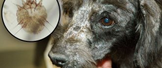
In the photo of dogs with keratitis, you can notice not only redness and dull mucous membranes, but also other signs. They depend on the mechanism of development of the disease, therefore, in accordance with the clinical picture, 8 more varieties are distinguished.
Purulent
Develops as a result of untreated catarrhal form. Accompanied by the release of purulent exudate and sticking of the eyes as a result of it drying out during sleep.
Catarrhal
The mildest form, characterized by very rapid (several hours) clouding of the cornea and a change in its surface to rough. When the course is prolonged, it is complicated by vascular ingrowth.
Vascular
Occurs when there is a pathological proliferation of blood vessels on the cornea, or neovascularization. Easily determined by visual inspection. A strong lumpiness will be noticeable on the mucous membrane. Due to a prolonged decrease in the transparency of the cornea, this type of pathology often leads to vision problems.
Parenchymatous
Most often it is formed as a result of infection, but it is also caused by injury. A distinctive feature of this variety is large spots and small dots on the cornea. May occur together with conjunctivitis and iritis.
Phlyctenulous
Develops due to chemical poisoning and allergies. Accompanied by the appearance of individual nodular areas with large bubbles filled with gray-white liquid. Thickening and tuberosity of the third eyelid is also noted. With a prolonged course, the bubbles burst and stain the mucous membrane with their contents, giving it a red-gray tint.
Ulcerative
With this type, the stroma is affected. The mucous membrane becomes cloudy, and one or several ulcers develop right on its surface. When they are opened, not only pus begins to be released from the eyes, but also exudate mixed with blood. May be complicated by necrosis of the affected surface.
Pigmentary
Pigmentary, or pigmentous keratitis in dogs develops under the influence of trauma and prolonged irritation of the mucous membrane. Its main symptom is a dark brown color of the cornea. If the course is prolonged, it becomes complicated by erosions and ulcers.
Spot
The rarest variety, the cause of which is still unknown. It can be easily identified by pearlescent cloudy spots on the mucous membrane. It occurs in a chronic form with a series of remissions and relapses.
Therapy of keratitis with Reparin-Helper®
The drug Reparin-Helper® heals and regenerates various damage to the eye area in dogs. The main active components in the drug Reparin-Helper® are cytokine proteins. Treatment of animals with cytokines activates the protective functions of the body itself in the damaged area. Thus, the healing process is much faster. Reparin-Helper® is especially effectively used for the treatment of ulcerative keratitis due to the good susceptibility of eye tissue to cytokines and rapid cell migration.
According to the instructions, the drug is used to treat:
- eye diseases (keratitis, conjunctivitis);
- all kinds of skin damage;
- after surgical operations;
- lesions of the oral cavity and in dental surgery.
Reparin-Helper® can be used not only for dogs, but also for horses, cats and other animals. The great advantage of the drug is that it can be used both in the clinic and at home. The main thing is to use it immediately after mechanical damage or detection of a disease - this will significantly speed up recovery.
Classification by degree of damage
Depending on the degree of damage to the cornea, pathology is divided into 2 types: superficial and deep. The first is characterized by a mild course, while the second is often accompanied by complications.
Surface
Affects the uppermost layer of the cornea or part of the stroma - the epithelium. Most often develops with mechanical damage to the mucous membrane. It is easily treatable at the initial stage, but if diagnosed too late it becomes more severe.
Deep
It affects not only the superficial (epithelial) layer, but also the deep layer (stroma, endothelium and boundary membranes). The main reason for the development of this variety is infection. It is accompanied by acute pain and does not respond to drug therapy. Scars often remain after surgery. There is also a high risk of vision impairment.
How does Reparin-Helper® work?
The drug acts in several directions.
- The drug has a local immunomodulatory effect due to the fact that it attracts immune cells (macrophages) to the site of damage.
- Normalizes the inflammatory reaction, which alleviates the animal’s condition and promotes recovery.
- Stimulates regeneration and collagen production, attracting and activating fibroblasts, which significantly accelerates the healing and rehabilitation of the eye. This is very important for eliminating ulcers, clouding, and also for restoring the cornea.
- Restores the transparency of the cornea and prevents the appearance of a scar (cataract).
Why is diagnosis in a veterinary clinic so important?
Self-diagnosis is complicated by the similarity with other diseases, so at the first signs of illness the animal should be shown to a doctor. The presence of inflammation is confirmed by checking the reaction to light, biomicroscopy, keratoscopy, measuring intraocular pressure and performing 2 tests: fluorescein and Schirmer. To determine the cause, standard laboratory and instrumental tests are used: blood tests, ultrasound and x-rays.
Prevention of keratitis
Keratitis, like many diseases, can be prevented. You just need to know about the right preventative measures and follow them.
- Daily hygiene, including eye hygiene. The eye area should be wiped with a cotton pad moistened with plain warm (boiled) water.
- Vaccinations. Vaccination prevents the occurrence of infectious diseases, which, in turn, cause keratitis.
- Balanced diet. The diet must be proper, rich in vitamins, because often four-legged animals that have a deficiency of microelements in their diet suffer from inflammation of the cornea. You can use high-quality industrial feed, or a natural menu, including meat, vegetables, cereals, dairy products, and eggs.
- Dogs are often injured in street fights; no one is immune from such actions. If the eye is damaged, antiseptic treatment is needed, after which you should immediately instill Reparin-Helper®. Be sure to show your four-legged friend to the doctor!
- If your eyes are inflamed, do not hesitate - go to the clinic, get tested, consult an ophthalmologist.
- If your dog is genetically predisposed to eye diseases or is in the risk age group, it is better to consult a veterinarian.
Features of the disease and similarities with other pathologies
Keratitis is an inflammation of the uppermost (outer) layer of the eye - the cornea. With pathology, the transparent surface turns red and becomes covered with a cloudy film. Turbidity occurs within a couple of hours or days, so it is recommended to focus on this sign first. With a longer course, the color of the cornea changes to gray or blue, and in the presence of pus - to yellowish.
The disease is very often confused with a mild allergic reaction, accompanied by similar redness and tearing. Conjunctivitis and iritis also have similar symptoms.
The first disease affects the conjunctiva, and the second - the iris. It is almost impossible to distinguish them from each other without special tests, so veterinarians strongly recommend not to engage in self-diagnosis.
Clinical picture
The clinical picture of keratitis is a series of manifestations specific to this disease, which have received a special name - corneal syndrome.
It includes symptoms:
- increased lacrimation;
- photophobia;
- narrowing of the palpebral fissure, it is impossible to open the eye completely;
- eye pain;
- sensation of a foreign object in the eye;
- redness of the eye.
In severe cases, the inflammatory process spreads to other parts of the eye and affects the sclera and iris. Another possible complication is ulceration at the site of inflammation. It can lead to perforation, in which infection enters the deep structures of the eye.