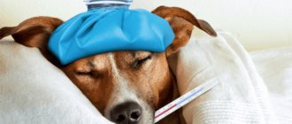
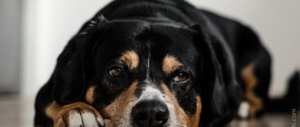
Pets require high-quality health care, and eye diseases account for most of the diseases. Glaucoma in dogs occupies a leading position among all eye diseases; about 80% of vision problems are caused by it. So, owners should pay special attention to the health of their pets’ eyes. If any abnormalities are diagnosed in time, it will be possible to return the dog to healthy vision with the least effort.
What is glaucoma in dogs?
This is a disease characterized by increased intraocular pressure. It is possible to determine the pressure level using a device - a tonometer. The cause of glaucoma is insufficient removal of fluid from the membrane of the eye. In this case, the secretion of fluid is not disturbed, that is, its amount is at the same level.
The eye is formed from various parts, but its shape, size and elasticity are directly controlled by the fluid contained within the eyeball. In this case, the liquid is under pressure, which in front is called intraocular. At the same time, it increases with a large accumulation of fluid, which causes the eye to bulge. Production occurs on an ongoing basis; the ciliary body is responsible for this function.
The fluid that is inside the eye contains the necessary nutrients for the membrane, as well as oxygen. When there is excess pressure, that is, a lot of fluid accumulates, it flows into the gap between the cornea and the iris. Constant pressure is maintained as long as fluid production and removal or drainage occur in equal proportions.
Diagnostics
Glaucoma is diagnosed using a visual examination, medical history (information about the animal’s life) and four main instrumental methods:
Tonometry. The method involves measuring intraocular pressure using a special device. In modern clinics, TonoPen and TonoVet are used for this purpose. This method is reliable and completely safe for the animal. Normally, IOP is 10 - 25 mmHg.
Gonioscopy is a method that allows you to maximally examine the main culprit of the disease - the iridocorneal angle of the eye. It is carried out using a special vacuum gonioscopic Barkan or Koepe lens. This is a completely safe procedure for the dog, causing nothing more than discomfort. Before starting the eyes, dogs are numbed with a mild anesthetic.
Ophthalmoscopy is a method of examining the eye using a special device - an ophthalmoscope. Such a device is certainly available in all veterinary clinics. An ophthalmoscope allows you to assess the condition of the fundus of the eye, this is very important, since it is the change in the fundus of the eye where the optic disc is located (glaucomatous excavation of the optic disc) that is the main diagnostic sign of glaucoma.
Ultrasound of the eyeball. Not every clinic can afford this diagnostic method, and it is not always necessary. This method is safe for dogs, as they use special sensors adapted for the eye. Ultrasound diagnostics is very informative - it allows you to determine the exact size of the eyeball, the position and shape of the lens, the condition of the retina and vitreous body.
Classification of glaucoma
Glaucoma is divided into several types: primary and secondary. Primary is characterized by the formation of high pressure, but the eye itself is healthy. However, certain breeds of dogs are more susceptible to the disease compared to others. The reasons are hereditary, as they contain certain anomalies in the structure of the iridocorneal angle, where excess fluid drains. So, at a narrow angle, the outflow of liquid becomes somewhat more difficult, which provokes an increase in pressure. Secondary glaucoma is caused by damage to the eye itself, which consequently led to the disease. This may include injury, illness, or damage.
The classification also includes another variety - according to the angle of the anterior chamber, which can be closed, open, or narrow. Moreover, both types of classification exist in parallel and occur in different forms.
Therefore, in the case of a hereditary predisposition to such a disease, you will have to especially carefully monitor the pet’s health. If the first signs are detected, examination and subsequent therapy will be required.
Primary open-angle glaucoma is considered a purely hereditary disease, which is more often observed in beagles and poodles. The disease is characterized by a chronic form, so the pressure increases gradually and takes a long time. Moreover, the dog is able to see even in the last stages of the disease.
Goniodysplasia is a variation of the form of the disease just described. It primarily affects Great Danes, Cocker Spaniels, Samoyeds, Beagles and Labradors. At the same time, it is typical for the disease to have a recurrent manifestation in the form of severe attacks. So the eye swells, the pupils tend to dilate due to a change in the angle of refraction of light, and hyperemia is noted. Sometimes it even leads to blindness in the pet.
How to check your pet's vision?
- The test can be done at home. Take the dog out of the room and close the door. Build an obstacle course from any objects in the place where the dog is expected to pass. Next, open the door, move behind the obstacle course and call the dog. If the dog walks around or jumps over objects without tripping over them, everything is fine with his vision.
- Test with an object. Take a piece of cotton wool, attract the dog's attention and throw it to the side. If the dog approaches the landed cotton wool, then its vision is fine. (The dog will focus only on sight, since it is unlikely to hear cotton wool falling)
- Bring your dog to a light source. Cover her eyes with your palm for a couple of minutes. Next, remove your palm and look at the pupils - they should react by narrowing.
Predisposition
The main difficulty in diagnosing and visually determining abnormalities in the eyes lies in their slow manifestation, while pronounced severity can only be acquired in the later stages. Due to the long period of development, glaucoma is rarely diagnosed in the early stages, since it does not bother the animal at all, and a slight increase in pressure is not visually noticeable.
- It will be interesting about seborrhea on the fur of dogs
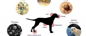
To prevent the disease, it is necessary to take into account the predisposition factors of the breed; if a dog has an increased risk of glaucoma, then it should undergo periodic examination. If at least one or more of the listed factors apply to your pet, you should especially carefully monitor the state of your eyesight and make an appointment with an ophthalmologist. Factors predisposing to glaucoma:
- Breed. Certain types of animals are more likely to encounter the disease than others; this must be taken into account when adopting Chihuahuas, beagles, divers, cocker spaniels, Dalmatians, huskies, poodles, etc.;
- Chronic eye diseases. If an animal has a seemingly harmless disease, perhaps the owner does not even know about it, the likelihood of glaucoma increases many times over. For example, conjunctivitis (inflammation of the mucous membrane) can provoke closure of the angle or thinning of the gap for fluid outflow. At the same time, improper treatment increases the risk of developing the disease; side effects from steroid drugs are especially dangerous. In this case, the disease progresses quite quickly and after 2 weeks the first manifestations may begin;
- Age. Age-related structural features of a dog’s body play a role, so animals over 6 years of age are much more likely to develop glaucoma;
- Hereditary predisposition. In addition to genetics associated with the breed, the disease can be passed on from parents. This factor can be determined by determining whether one of the parents had this disease. If the result is positive, you will have to more carefully monitor the pet’s health;
- Mechanical damage, eye injuries. This option plays a primary role; if the dog has received any damage, you should definitely contact a specialist. In the absence of timely assistance, there is a risk of developing a chronic, incurable disease;
- Other diseases. At risk are animals that have other types of illnesses: diabetes, cardiovascular diseases, they increase the risk of developing glaucoma.
Two tests for vision loss
For such cases there are two simple tests:
Labyrinth. Performed at home. It’s all very simple: build an “obstacle course” for your dog from available items, a so-called labyrinth, and call your pet to you from the other end of the room. Then repeat this test in a room with low lighting, not completely dark, but so that you can clearly see the outlines of objects. All you have to do is watch your four-legged friend. If he has no problems with his eyes, then it will not be difficult for him to come running to you.
Cotton wool test. Take a small piece of cotton wool and attract the dog's attention when he turns to you - throw the cotton wool to the side. A blind dog will not be able to observe this object, since cotton wool has practically no weight or smell, and the dog will not be able to notice it with any other sense organs except sight.
It is more difficult to notice when only one eye goes blind, then the dog’s behavior is unlikely to change. In this situation, the owner must be very attentive to notice the changes. You can confirm your doubts if you check the reaction of the pupil to light; the pupil of a completely blind eye will not change in size in bright light.
In addition to all of the above, a general deterioration in the pet’s condition will be noticeable.
Causes of glaucoma
From a medical point of view, there are some reasons that lead to the formation of diseases in dogs:
- Uveitis (inflammatory reaction in the inner lobe of the eye) - the cause of the disease is the penetration of infection into the intraocular fluid. In the presence of inflammation, the drainage channel is blocked, partially or completely, and leads to glaucoma;
- Subluxation in the lens of the eye is often a consequence of mechanical damage;
- The presence of benign or malignant eye tumors. This is how drainage is blocked due to an increase in the physical volume of the tissue;
- Intraocular bleeding - when blood clots, a clot is formed that can block the fluid outflow channel;
- Physical damage to the lens of the eye. Often this manifestation entails an inflammatory reaction, and, accordingly, swelling of the tissue and clogging of the canal;
- The occurrence of primary glaucoma is possible due to a defect associated with the anatomical structure of the iridocorneal angle. Moreover, it can be either narrow or closed due to the presence of parts of embryonic tissue.
Symptoms of glaucoma
Before moving on to treatment of the disease, you need to know for sure how cataracts manifest themselves and what are its main symptoms. If only a small part of the listed manifestations was identified, then it is likely that the disease is at an early stage or is not it at all. Nevertheless, this becomes a good reason to contact a specialist. Main symptoms of glaucoma:
- Increased tearing of the eyes;
- The eyeball increases in volume, but in the initial stages this is not very noticeable. The manifestation is popularly called “bull’s eye”;
- The vessels of the sclera protrude strongly, they become tortuous, with a pronounced red color;
- Pain in the eyes. This is evidenced by the dog’s behavior; it does not like it when the side of its head is touched, where painful sensations are observed;
- The pet eats much worse, the appetite disappears, or gets worse. This is accompanied by a depressed state and withdrawal from the society of animals or people;
- The dog has difficulty with orientation in space;
- Vulnerable to light, the pet tends to retreat to a dark corner, closes its eyes with the help of its paws or curls up into a ball, as with dropsy.
Symptoms
With the acute development of glaucoma, the animal suddenly loses its vision. But this process can be reversed, while with chronic slow development, vision is lost gradually and irreversibly.
Regardless of the cause, the manifestation of glaucoma is always the same. You may not notice everything at once, but if you notice anything from this list, then hurry up and seek help from a veterinarian. Symptoms of increased intraorbital pressure:
- Buphthalmos is an increase in size of the eyeball, this phenomenon is also called “bull’s eye.”
- Corneal edema.
- Redness.
- The vascular lines of the eye are visible.
- Soreness.
- Photophobia, the dog tries to hide in dark places.
- Mydriasis is dilation of the pupil.
Loss of vision. No matter how strange it may sound, you may not immediately notice that your pet’s vision has become much worse.
Glaucoma treatment
Treatment of glaucoma in a dog should be carried out upon examination by a doctor and determination of the cause or type of disease. So the information presented below should not replace a trip to a specialist, because self-medication can cause a lot of harm.
Treatment comes in many different forms and is highly dependent on the quality of the pet's vision when tested, the cost of the treatment needed, and the pet's temperament. If the dog can see, then it is necessary to reduce intraocular pressure, and it is important to do this as quickly as possible in order to preserve vision and also eliminate pain. So primary glaucoma in a dog should be treated in a timely manner. Emergency care may include:
- Osmotic diuretics:
- Glycerol. A fairly effective remedy, it should be administered intravenously in a volume of 1-2 ml per kilogram of animal weight at the beginning of 10-30 minutes and lasting 4-5 hours. The drug can be reused, but is contraindicated in cases of heart failure, renal failure, as well as systemic hypertension and dehydration. It is important that the animal does not inhale the substance;
- Mannitol. It is administered by intravenous injection at 1-2 g/kg body weight at regular intervals of 20 minutes. Initially, the cycle should be repeated every 30-60 minutes for 6 hours. The course can be reused. The drug is contraindicated in case of heart/renal failure and dehydration. After administration, you should refrain from drinking liquids, both orally and intravenously, for 2-4 hours.
1) Prostaglandins - they are great for monitoring the animal’s condition, but are expensive:
- The most common drugs in the group are Travoprost, Latanoprost, and Bimatoprost. Used topically, 1 drop every half day. Lead to the formation of strong mitosis.
2) Miotics - take special care in the presence of anterior uveitis, otherwise the formation of synechia, lens subluxation or exacerbation of glaucoma is possible:
- Pilocarpine – a 1-2% solution should be used 2-3 times a day;
- Dimecaine bromide – 0.125-0.25% solution instilled once a day.
3) Adrenergics:
- Levobunolol, Timolol (dosage 0.5%), Betaxolol - use 1 drop in the eye 2-3 times a day with a regularity of 8-12 hours. For mild severity, they are prescribed 2 times, in complex forms: three.
4) Neuroprotective agents:
- Amlodipine - taken orally in a volume of 0.625 mg once a day. It has a protective effect for ganglion cells.
5) Carbonic anhydrase inhibitors - effective for short-term or long-term blood pressure control. They also have a kaliuretic effect, so drugs containing potassium should be added to treatment if it is long-term:
- Dorzolamide, brinzolamide - used topically at 8-hour intervals;
- Acetazolamide - administered orally at 10 mg/kg twice a day;
- Methazolamide - dosage 5 mg/kg twice daily;
- Dichlorphenamide – dosage 2.5 mg/kg 2-3 times a day
Treatment without examination and diagnosis in a veterinary clinic is prohibited, as there is a high probability of side effects and worsening of the disease.
Currently reading:
- Diagnosis and treatment of liver cholestasis in dogs
- A warning sign for a dog with cloudy eyes
- Review for choosing carriers for small and large dogs
- Thyroid dysfunction in dogs (hypothyroidism)