In this article I will talk about cataracts in dogs, the causes of the disease, symptoms, methods of diagnosis and treatment. I will describe its stages and possible preventive measures to prevent the disease.
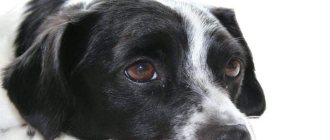
Cataract is clouding of the lens of the eye. Develops from the age of 8 years and progresses over the years.
Causes of cataracts in dogs
Cataract is a hereditary disease. When buying a pet, it is advisable to ask the breeder whether the offspring suffered from this disease. If yes, then you should think about changing the kennel to buy a dog.
The main reasons for the development of the disease:
- diseases of the visual system (glaucoma, uveitis);
- diseases of the endocrine system (diabetes mellitus);
- decreased immunity;
- mechanical eye injuries;
- consequences of drug treatment;
- age over 8 years.
The following breeds are more susceptible to the disease:
- golden retriever;
- Boston Terrier;
- poodle;
- Cocker Spaniel;
- Yorkshire Terrier.
The disease is age-related, i.e. occurs in adult animals over 8 years of age.
In rare cases, cataracts are congenital.
Cataracts develop in dogs after years
Kinds
By type, cataracts are divided into congenital and acquired.
- Congenital lens opacification is a rare phenomenon and is associated with concomitant chronic diseases such as diabetes mellitus. It also occurs with intrauterine injury to the eyeball, underdevelopment.
- The acquired form is the most common type and occurs at a certain age due to genetic inheritance. It occurs more often in dogs over eight years of age, because by this age the condition is aggravated by chronic illnesses.
Cataract stages
The rate of development of the disease and the transition to the next stage depends on the individual characteristics of the animal.
There are four stages:
- Initial - partial clouding of the lens, occurs in the peripheral zone. Vision decreases slightly. The pet may not be able to separate individual parts of objects.
- Immature - partial clouding of the lens in the central optical zone. Vision is greatly impaired. A dog can only distinguish the outlines of objects.
- Mature – complete turbidity. The animal experiences a loss of orientation in space; only light is felt.
- Overripe - the fibers of the lens liquefy and it begins to disintegrate. Light perception is impaired.
- The last stage is characterized by complete destruction of the lens, the eye looks like it is covered with a white veil. Diseases develop: glaucoma, phacolytic uveitis, iridocyclitis.
The initial stage of cataracts in a dog
Last stage of cataract
Symptoms
To begin treating cataracts, you need to know how to determine the presence of this disease. In the early stages, correct diagnosis is more difficult, sometimes almost impossible.
Stages of development and their distinctive features
How difficult it will be to cure depends on the development of the disease. As the disease process progresses, the symptoms become more pronounced. Cataracts are divided into several stages:
initial stage
The dog does not suffer from severe visual impairment. Cloudiness of the lens is almost invisible and appears along the periphery.
Immature
The animal sees only vague outlines of objects. Turbidity becomes noticeable in the central part of the lens, as in the photo.
Mature
The pet only notices light fluctuations. Cloudiness is distributed throughout the lens.
Overripe
The dog is completely blind. The lens is white.
Treatment Options
Conservative treatment
At the initial stage, this type of treatment is aimed at slowing the progression of the disease. Therapy is carried out using drops of Smirnov, Katahrom, Vittaiodurol, Vitafacol, Vicein, etc.
This type of treatment is more suitable for eliminating the root cause of the disease, i.e. disease that led to cataracts. For example, if the disease develops in one eye, eliminating the disease that led to the clouding will help prevent the disease from developing in the other eye.
Conservative treatment of cataracts is possible only at the initial stage
Surgery
The only effective treatment is surgery. Eye surgery is complicated. Effectiveness depends on the stage of development at which it is carried out.
The essence of the operation is to replace the biological lens with an artificial or intraocular lens.
At the initial stage of cataract, surgical intervention is minimally traumatic and in most cases successful.
The clouded lens at this stage is still soft and is removed through a puncture without causing serious injury to the eye tissues.
The operation occurs as follows:
In later stages, cataracts can only be treated with surgery.
Prevention
No preventive measures have been developed to completely prevent a dog from developing cataracts. Owners are advised to monitor their pet’s diet and daily routine, take frequent walks, and enjoy active rest. All this will help reduce the risk of developing chronic diseases, and they serve as an impetus for clouding of the lens. Since genetic predisposition plays a big role, when purchasing a puppy, do not be lazy to ask the breeders about the presence of this disease in the parents or members of his family.
Visit your veterinarian regularly for preventive examinations, because early detection of the disease makes the treatment process easier. Add vitamin complexes to your pet's diet. Pay attention to changes in your dog's habits. Examine the condition of your eyes yourself periodically. If you notice a bluish tint, seek medical help.
Cataracts are an insidious disease, the development of which is difficult to predict. Do not be indifferent to your pet, regularly monitor its condition and do not delay visiting the veterinary clinic. Remember that we are responsible for those we have tamed.
Symptoms and diagnosis of lens opacities
Symptoms are usually related to the degree of visual impairment. For example, dogs with less than 30% pupillary opacity are asymptomatic, whereas patients with more than 60% pupillary opacity may experience vision loss or difficulty in dimly lit areas.
Cataracts are often discovered by chance when examining a dog's eyes. This is due to the fact that clouding of the lens does not always lead to blindness. If the area of opacity is small or shifted to the periphery of the lens, then the dog’s vision is preserved. Vision problems are observed in pets with damage to the central zone of the lens.
Meanwhile, if your dog has diabetes-related cataracts, you may also see increased thirst, increased frequency of urination, and weight loss in your dog, as well as symptoms of visual impairment.
Visual acuity decreases, the degree of which depends on the intensity and location of the clouding. On examination, the lens is blue (true cataract); the accumulation of white strands on the lens indicates a false cataract.
Classification of cataracts in animals:
- Initial : localized clouding and vision is not impaired. Up to 15% of the surface of the pupil is affected.
- Mature : The opacification is more noticeable and the fundus may be partially hidden during fundus examination. In dogs with bilateral damage, vision may be impaired. This is an ideal time to take your pet to an ophthalmologist, as a fundus examination can still be performed.
- Overmature : The lens begins to lyse and some vision may return. The operation must be performed before this stage, since lens resorption leads to the release of protein structures of the lens into the anterior chamber, causing the development of anterior uveitis and possibly glaucoma, which reduce the success rate of the operation. In cases where the nucleus lyses and sinks to the bottom of the lens, the term Morganian cataract is used.
The maturity of the cataract is determined using a slit lamp. Ultrasound diagnostics and measurement of intraocular pressure are often used, especially if there are suspicions of other eye diseases.
In severe cases of the disease, the diagnosis can be established based on a simple examination with the naked eye. However, with weak opacities, to establish the position of the opacities and its shape, it is necessary to use lateral or focal lighting and use ophthalmoscopy. To facilitate examination, it is recommended to dilate the pupil with atropine. With complete and dense opacities, the pupil is dilated.
Video of diagnosing bilateral cataracts in a Yorkshire Terrier
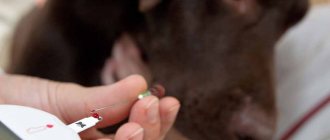
What medications are prescribed for cataracts?
To slow down the process of clouding of the lens, ophthalmologists prescribe medications - eye drops with various active ingredients. Oral medications may be taken.
List of drugs that are most often used in modern medicine to treat cataracts:
- "Vicein." A combined product that contains cysteine, glutamic and nicotinic acids, sodium salt, calcium chloride and other substances. Doctors usually prescribe two drops of solution daily. Treatment must be continued for a long time: usually at least a year;
- "Quinax." This solution has an antioxidant effect. It protects the eyes from the negative effects of free radicals and activates proteolytic enzymes. The latter dissolve opaque protein compounds in the lens and thereby restore transparency;
- "Taufon". Drops with the active ingredient taurine. Activate the regeneration and repair of eye tissue, have a beneficial effect on metabolic processes. Thanks to the effects of taurine, cell membranes are stabilized and the electrolyte composition of the cell cytoplasm is restored. The drug is used for degeneration of eye tissue, including cataracts;
- "Oftan Katahrom". The main active ingredient is cytochrome C. The drug also contains adenosine and nicotinamide. These substances have an antioxidant effect, stimulate metabolic reactions in the cells of the lens and improve its trophism. At the same time, the solution inhibits clouding in the transparent structures of the organs of vision, restores cells that have already become cloudy, and improves blood circulation. "Oftan Katahrom" relieves inflammation and improves the condition;
- "Vita-Iodurol". Drops used to prevent the accumulation of protein deposits in the lens. Improves nutrition and metabolic processes in it.
Patients with cataracts are also prescribed solutions containing riboflavin, insulin, calcium, magnesium and other substances that normalize the condition of the lens. An aqueous solution of zinc restores epithelial cells.
Among oral medications, Lutein-complex is considered the most effective. It contains blueberry extract, lutein and taurine. The drug has a strong antioxidant effect. It improves microcirculation in the eye tissues and thereby slows down age-related changes.
The drugs “Vitalux Plus” and “Okuwait Lutein” have a similar effect. They are prescribed orally when it is necessary to slow down the process of clouding of the lens.
The appearance of complications
There is a complication called lens-induced uveitis. This complication appears at the moment when the cataract matures, and the amount of proteins in the lens increases, which have very strong antigenicity, as well as toxicity to the inner membranes of the eye. Most often, these proteins are dangerous to the retina. Due to the action of these proteins, prostaglandins are released and accumulated. In addition, certain types of cells migrate into the structures of the eye that surround the lens. Moisture appears in the anterior chamber, in the iris, as well as in other areas. Among the symptoms of this disease, one can distinguish that the color of the iris changes, posterior synechiae, as well as pod membranes, are formed. If this disease is started, the animal’s vision will become much worse over time, secondary glaucoma appears, as well as subatrophy of the eyeball.
Intumescent cataract. This disease, as well as an acute attack of glaucoma in an animal, is associated with the fact that the processes of clouding of both translucent and cloudy lenses begin. Due to the fact that fibers that are not interconnected penetrate into the lens, they begin to swell very much and become opaque over time. As a result, first the outer and then the deep layer of the lens is damaged. Due to swelling cataracts, visual functions deteriorate greatly and intraocular pressure increases.
In this case, secondary glaucoma may develop, this is due to the fact that a mechanical blockade of the anterior chamber angle occurs, and the circulation of intraocular fluid, which also enters the anterior chamber, deteriorates. Because of this, a strong pressure gradient appears; it increases in the back of the eye from volumetric indicators. Pressure decreases in the anterior chamber of the eye, causing the diaphragm to move. As a result, the animal's intraocular pressure increases greatly and can be critical. All this happens in just a few hours. As a result, the optic nerve completely atrophies in a very short time.
If an animal has a swelling cataract, then the lens must be urgently removed. In general, at any stage of cataract, urgent surgical intervention is necessary. If this disease occurs, subsequent removal of the entire eyeball may be necessary.
All autoimmune panophthalmitis mentioned above appear due to the release of lens masses into both the anterior and posterior chambers. The result is autoimmune toxicity. This process develops very quickly, the animal’s condition will be acute, the disease develops very aggressively and progresses greatly. Often, if this disease is neglected, the eyeball will simply die. If you conduct an external examination of the eyes, you may notice swelling on the eyelids, the eyelids will be very thick, and there will be problems with the conjunctiva. In subsequent stages of the disease, the cornea will swell very much and also become cloudy, and folds of the membrane can be seen. At the moment when eye pressure increases, the disease is much more difficult to cure. If you conduct an ultrasound, you can see a very strong, rough cloudiness in the vitreous body, which consists of molten tissue.
Antibacterial and corticosteroid therapies for autoimmune panophthalmitis are often simply ineffective. In addition, in some patients, the procedure of vitreous lavage with solutions of amikacin and ceftazidime is useless. If autoimmune panophthalmitis occurs, then implantation of an intraocular prosthesis is necessary.
After the operation, will I be able to see perfectly at any distance?
The patient immediately notices a significant improvement. With multifocal intraocular lenses, most patients do not need to wear glasses for 90-95% of their daily activities. This means that most patients with multifocal intraocular lenses can read, drive a car, watch TV, work on a computer, do makeup, and play sports without glasses. They are advised to have only one pair of glasses for small text or for reading in low light.
Pankov glasses for the treatment of cataracts
This is an effective device for treating the initial stages. The glasses regenerate light pulses using LED emitters. They affect the eye muscles, causing them to contract rhythmically. As a result, the pupil reflexively contracts and expands under the influence of light waves. This promotes the outflow of intraocular moisture, improves lymphatic drainage and activates blood circulation. The lens receives the necessary nutrition. To achieve the maximum therapeutic effect, it is recommended to simultaneously use Pankov glasses and eye drops, which contain vitamins and minerals.
Achieving a therapeutic effect is possible only with long-term use of this device. Sessions must be conducted regularly. The interval between them should be no more than three days. When using the device for the first time, the session lasts no more than three minutes. Then the duration can be gradually increased. The maximum duration of one session is 15 minutes. The course of treatment usually consists of fifteen sessions. Therapy can be repeated at intervals of one month.
Table of contents:
What are senile cataracts Symptoms of age-related cataracts Methods of treating senile cataracts Features of surgery to remove senile cataracts in the elderly Cost of treatment In people after 50 years of age, age-related changes occur in the organs of vision. One of the most common pathologies is senile cataract, which is also called senile cataract. The lens becomes less transparent, the person complains of blurred vision. If left untreated, the situation worsens over time, and the person risks going blind. To prevent this from happening, it is important to consult an ophthalmologist in a timely manner. The Santa Clinic is staffed by experienced specialists who will accept patients at any time.
Diagnosis of the disease
Diagnosis of this disease takes place in three stages:
- General examination of the dog. The veterinarian generally checks the pet’s health and activity.
- Examination of the eye from the outside. At this stage, the veterinarian evaluates the external condition of the lens.
- If at the second stage the specialist suspects the development of cataracts, he conducts an electroretinogram (ERG - the study of electrical phenomena in the retina and optic nerve).
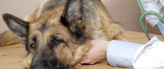
An accurate diagnosis can only be made by a veterinarian.
Based on the data obtained, the veterinarian can make an accurate diagnosis and determine the stage of development of the disease.
In pregnant women, lactating women and puppies
Cataracts can be found both in puppy dogs and in nursing dogs and puppies. During diagnosis, it is noticed that the transparent layers of the lens are slowly interspersed with cloudy layers. The dog often turns its head so as to catch an object in focus and examine what interests it.
Pregnant bitches may suffer from cataracts if they have an infectious disease. If a dog with this disease, which she received as a result of an injury, gave birth and is nursing puppies, then cataracts are not inherited by her offspring.
A puppy with congenital cataracts will often show signs such as a white pupil and squinting. He does not show any reaction to silent objects and those that do not smell.
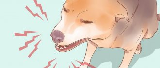
Such a puppy is inactive, hardly plays, and reluctantly leaves its mother. Animals suffering from cataracts become nervous and sometimes aggressive. They are insecure, inactive and have difficulty allowing a person or other animal to approach them.
Prevention of eye disease involves good maintenance of the pregnant dog. To feed a puppy, you should enrich her diet with vitamins A, C, B, E, PP. It should also be vaccinated on time to protect against infection.
Puppies, after starting complementary feeding and throughout their lives, are given multivitamin complex preparations to maintain eye health and immunity in general.