Icterius or jaundice in dogs is not a disease, but a dangerous symptom indicating the development of a fairly large number of pathologies in the body. Jaundice occurs due to inflammation of liver structures, various etiologies, blood-parasitic diseases, and intoxication of the body with hemolytic poisons.
Icterius is diagnosed in animals with pathological conditions of the gallbladder and the development of stones. Jaundice is a serious symptom associated with serious changes in the body and requires immediate attention from a veterinarian. Any delay can cost your pet's life.
Description
Leptospirosis (other names: jaundice, Stuttgart disease, Weill-Vasiliev disease) is a severe infectious disease that affects many species of mammals, including dogs, as well as humans.
The cause of leptospirosis is exposure to a pathogen, which is bacteria of the genus Leptospira. These bacteria thrive and can multiply in stagnant warm water or in moist soil, where they come mainly from the urine and excretions of sick animals. Sun rays or strong heat kill them, and various disinfectant solutions also have a detrimental effect.
In Russia, the disease in animals is caused by leptospira from six serological groups, which are divided into three independent groups: L. Icterohaemorrhagiae, L. canicolau, L. grippotyphosa. In dogs, L. icterohaemorrhagiae and L. canicolau are more often isolated.
Jaundice in dogs
The risk group for leptospirosis includes urban dogs, especially at an early age, as well as hunting dogs. An increase in the number of cases of the disease usually occurs in the summer, although infection with leptospirosis is not excluded in the cold season.
Males are more often affected by leptospirosis, as they show the greatest interest (sniffing, licking) in dog marks. The main route of entry of the pathogen into the body is through mucous membranes; transmission through open wounds directly into the blood is possible. A dog that has swum in a stagnant pond may well develop leptospirosis. It is also possible to transmit the pathogen through household contacts, through a common collar, when dogs “get to know each other” on the street, or through tick bites. Dogs can also become infected with leptospirosis by eating rats, mice, raw meat, or from sick pets.
The outcome of the disease in unvaccinated dogs is fatal in most cases (up to 90%). Unfortunately, vaccinated dogs also have a risk of becoming infected under unfavorable epidemiological conditions. Recovered dogs remain leptospiron carriers for three years and are a source of the pathogen for animals and humans.
Standard vaccination regimens (once a year) are ineffective in unfavorable conditions. In such cases, vaccination twice a year is recommended. Animals with leptospirosis are potentially dangerous to humans.
Causes of the disease
There are various causes of the disease. The main one may include damage to the liver or spleen
. Veterinarians often encounter acute hepatitis. If this is an old individual, then chronic inflammation of the organ may occur. It also affects the pancreas and small intestine.
fungal infections can also cause illness.
, which include coccidioidomycosis, histoplasmosis. This option is very dangerous, since fungi are not afraid of drugs and do not die from them. It is necessary to use stronger substances that can have a detrimental effect on the animal.
The cause of the disease is also elementary obesity. This pathology is typical for dogs that constantly eat low-quality food. Pathology can manifest itself against the background of hormonal disorders.
Inappropriate or incorrect use of medications also causes this problem. Acetaminophen (Tylenol), phenobarbital or primidone, and metronidazole should be used carefully. Antibiotics, which owners often use to treat their pets themselves, can also cause a similar reaction. It is always better to ask an experienced veterinarian for advice so as not to harm your pet.
The dog could simply get poisoned. The poison initially enters the liver structure. Herbicides, rodent poisons, zinc-based compounds and others are harmful to pets. Less commonly, the disease is associated with liver failure or any genetic diseases of the breed.
“ Sometimes we are talking about problems with the bile ducts and the gallbladder itself.
The cause of the disease can also be worms that live in the bile ducts. The hemolytic form can develop after a tick bite
.
Liver amyloidosis often occurs in some dogs. Breeds at risk include the boxer, hounds, sharpei, and bedlington terrier. A separate group consists of hunting species and pets that live on the street.
Unfortunately, no animal is immune from this disease.
Development of leptospirosis disease
Having penetrated the body, leptospira actively move, multiply, and secrete toxins. In response to this, the temperature rises and antibodies are produced, which cause the destruction of microbial bodies. This is accompanied by the release of endotoxins, which in turn cause hemolysis of red blood cells and the release of hemoglobin. Hemoglobin is excreted in large quantities in the urine, turning it red (hemoglobinuria). In addition, hemoglobin colors the skin, mucous membranes and sclera yellow-red. Blood coagulation systems are activated. Blood capillaries become clogged with blood clots, which leads to necrosis of large areas of tissues and organs. Depletion of coagulation systems leads to bleeding and hemorrhage. Microbial toxins and cell breakdown products cause damage and inflammation to the liver, intestines and other organs. Having penetrated the kidneys, leptospira are inaccessible to immunoglobulins and macrophages. Acute renal failure may develop.
Clinical signs of leptospirosis
The disease occurs acutely and chronically. Acute leptospirosis occurs in two clinical forms: hemorrhagic and icteric. The incubation period for canine leptospirosis ranges from 2 to 15 days.
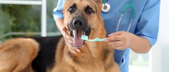
The hemorrhagic form is characterized by a sharp increase in temperature to 40.5-41.1°C, loss of appetite, dry cracked nose, and bad breath. The oral mucosa is bright red, burgundy, and later bleeding ulcers appear on the mucous membranes of the oral cavity. This is accompanied by vomiting and bloody diarrhea. Sometimes there was spontaneous bleeding of blood and ichor from the nose and anus. The animal quickly loses weight, the eyes are sunken, the lymph nodes are enlarged.
In the later stages of the disease, muscle tremors, vomiting with blood, and nosebleeds are noted. The animal loses weight sharply, body temperature drops below normal (to 36-36.5 degrees). Urine is excreted in small quantities and contains bile and protein.
The death of animals with this form of the disease can occur in 65-90% of cases. Animals die while in a coma or with convulsions. The illness with this form of the disease lasts 2 - 3 days, sometimes 5 - 10.
Reviews from owners and veterinarians
Julia: “The dog is 12 years old. On Saturday afternoon she was bitten by a tick, and on Sunday we noticed that she began to turn yellow. The mucous membrane in the mouth, the skin is all yellow. Doesn't stand on its paws, temperature is low. On Monday, the vet said it was due to a bite. No tests were done.
He prescribed Karsil and Ursoferran for 3 days. At the hospital I was injected with forticarb. Last week we were at the lake, the dog drank water from there. Tell me, is it just jaundice? Do I need to inject antibiotics?
Veterinarian: “Your dog has piroplasmosis. The mucous membranes and skin have turned yellow due to the process of destruction of red blood cells and the level of bilirubin has increased.
To stabilize the condition, the dog needs injections of Prednisolone, Derinat, Epocrine and drips with saline solutions. Antibiotics are completely useless, because piroplasmosis is not a bacterial disease.”
Oleg: “Laika dog, age 1.5 years. Suddenly vomiting and diarrhea began, the dog stopped drinking and eating, and its hair began to fall out. We went to the vet and the diagnosis was hepatitis. We have been undergoing treatment for a whole month, but there is no improvement. After the IV, everything seemed to be fine, but a few days later it all happened again.
Prescribed: folic acid, Noshpa, Dexamethasone, Allochol, Essentiale in capsules. Food options include rice and beef. An ultrasound showed an inflamed liver, a round gallbladder, but nothing about the pancreas. What are we doing wrong?
Veterinarian: “Essentiale needs to be administered intravenously and very slowly. Do not feed your dog anything other than medicated food. Take Royal Canin Hepatic. When feeding such food from a well-known brand, additional enzymes are not needed.
Before going for a walk, put a muzzle on your dog so that he doesn’t eat any herbs! The fact that dogs “heal” on their own is nothing more than a common story.
And in case of pancreatitis, enzymes should generally be used with great caution. They have many categorical contraindications. Pancreatic necrosis may occur. In Russian, the pancreas will fall off.”
Pavel: “Our three-year-old St. Bernard fell ill. He lay down and doesn’t get up, doesn’t eat, very weak. They called the veterinarian, and on his advice they gave him an injection of bicillin, noshpa, gamavit and 2 doses of glucose. The next day we took him to the doctor. By this time, yellowness appeared on the skin and in the mouth.
Twice a day at the clinic we place a drip of saline and glucose. There was vomiting with blood. The dog urinates frequently and constantly asks for water. We force feed him chicken broth. Dehydration is already severe, it was difficult to get an IV.
Tell me, why did this hepatitis appear? We can’t take a blood test because the clinic doesn’t have a laboratory.”
Veterinarian: “In your case, jaundice may be a sign of hemolytic anemia or liver damage. Blood must be donated in any way - both a general analysis and biochemistry. Only this option will help you understand what’s wrong with your dog.
Another very informative option is an ultrasound of the entire abdominal cavity. A peripheral blood smear from the claw is needed to rule out piroplasmosis. But you should not treat a dog for piroplasmosis without an accurate diagnosis. The treatment there is very specific.”
Symptoms
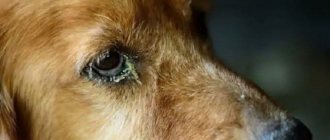
The icteric form is most often recorded in puppies, but can also develop in adult animals. The disease may develop gradually or go undetected until severe jaundice appears. At the onset of the disease, clinical signs often resemble parvovirus enteritis: vomiting, diarrhea or constipation, dull fur, often with dandruff, gelatinous discharge from the eyes, and a sharp unpleasant odor emanates from the dog. The liver and lymph nodes are enlarged.
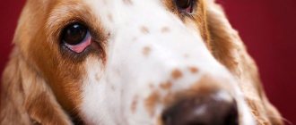
The icteric coloration of the mucous membranes of the mouth, eyes, and individual areas of the skin becomes clearly visible a day or two after the disease. Urine is dark yellow, high in protein. Inflammation of the conjunctiva of the eyes and itching often occur. Depending on the course of this form of the disease, it lasts from 2 to 10 days.
The mortality rate of dogs with this form of the disease reaches 40-60%. Chronic leptospirosis is often asymptomatic.
Mechanical jaundice. Pancreas cancer
Return to section:
Oncology
Obstructive jaundice - what is it?
Rice. 1 - The anatomical structure of the biliary tract is normal |
First, let me remind you: bile is formed in the liver, after which it enters the duodenum through the bile duct system, where it helps digest food. The gallbladder is a kind of “reservoir” where bile “accumulates” between meals (Fig. 1).
So, any obstacle to the “movement” of bile into the intestine forms a pathological condition called “obstructive jaundice.” Bile begins to accumulate in the bile ducts and gallbladder, enlarging and expanding them. Excess bile is released into the blood, and the body removes it through other routes, including the kidneys and skin. Therefore, one of the initial signs of obstructive jaundice is an increase in bilirubin in the blood, a bile pigment that is the main component of bile. Since bile does not enter the intestines and does not color the stool, it becomes light, even gray. The urine becomes very dark due to increased secretion of bilirubin by the kidneys. The skin turns yellow, and due to an excess of bile salts in the blood, severe skin “itching” may begin.
Thus, “obstructive jaundice” is a pathological syndrome associated with a violation of the outflow of bile, which is externally manifested by yellowing of the sclera and skin, lightening of the stool and darkening of the urine, and can cause weakness, drowsiness, abdominal discomfort and itching. Quickly identifying the cause of this syndrome (that is, making a diagnosis) is the main task of the doctor in this situation.
Obstructive jaundice: causes.
| Rice. 2 The most common causes of obstructive jaundice. |
There are many diseases that cause disruption of the flow of bile.
The main ones are listed in Figure 2.
The method of treatment, its result, as well as the prognosis of the disease (i.e., the expected duration and quality of life with this disease and its "curability").
Unfortunately, in 40-67% of cases, the cause of obstructive jaundice is tumors, and they are benign only in 2-3% of cases.
The most common cause that doctors have to deal with is cancer of the head of the pancreas.
The tumor compresses the duct from the outside, disrupting the outflow of bile. (Fig. 3)
Obstructive jaundice in pancreatic cancer - treatment methods
| Rice. 3 Compression of the common bile duct due to a tumor of the head of the pancreas. |
It is impossible to consider all possible types of treatment for various causes of obstructive jaundice in one article. Therefore, I will dwell in more detail on treatment options for the most difficult category of patients - those with malignant tumors that cause compression of the bile ducts.
The only chance for patients with a malignant tumor causing obstructive jaundice is radical surgery (complete removal or resection of part of the organ), but this is possible in less than 30% of cases. This happens because the disease develops very slowly and begins to “manifest” already at an advanced stage.
In each case, the question of the possibility of complete tumor removal is decided individually; it depends on many factors: the extent of the process, the age of the patient, the presence of concomitant diseases, etc. These operations are considered one of the most difficult in modern abdominal surgery and are performed, as a rule, in specialized departments by experienced oncologist surgeons.
All other methods of treatment - radiation, chemotherapy (so-called dietary supplements, phyto- and homeopathic therapy, I do not consider “treatment” in principle) are ineffective and are aimed only at slowing down the growth of the tumor and improving the “quality of life” of the patient.
Why is obstructive jaundice dangerous?
As I already mentioned, the main parameter that assesses the severity of obstructive jaundice is the level of total bilirubin in the blood. All of the above treatment methods, including radical surgery, with rare exceptions, are possible when the level of total blood bilirubin is below 50-90 µmol/l (normal 3-17 µmol/l) due to the high risk of complications. However, visible jaundice of the sclera and skin occurs, as a rule, when the bilirubin level is above 100-120 µmol/l. At levels above 300-350 µmol/l, bilirubin begins to penetrate the blood-brain barrier, i.e. enter the brain and, with further growth, cause severe intoxication, even death.
According to the literature, in conditions of biliary tract obstruction and inflammation, surgical treatment is risky, accompanied by a large number of complications, and mortality reaches 10-34%, which is 4 times higher than in cases where obstructive jaundice can be eliminated before surgery.
Therefore, one of the first tasks in the treatment of obstructive jaundice is to reduce the level of bilirubin in the blood - to treat intoxication and prepare the patient for one or another type of specialized medical care (surgery, chemotherapy or radiation therapy).
Choosing a treatment method for obstructive jaundice
Conservative therapy (intravenous infusions of drugs) in patients with obstructive jaundice of tumor origin is rarely effective. of decompression of the bile ducts come to the fore - i.e. methods aimed at restoring the outflow of bile from the bile ducts into the digestive tract. Among the surgical methods for treating obstructive jaundice, three main areas can be distinguished:
decompression of the bile ducts – i.e. methods aimed at restoring the outflow of bile from the bile ducts into the digestive tract. Among the surgical methods for treating obstructive jaundice, three main areas can be distinguished:
- Endoscopic techniques for drainage and stenting of the bile ducts.
- Percutaneous transhepatic techniques under ultrasound and fluoroscopy control.
- Open surgery is the creation of an “anastomosis” (i.e., “connection”) between the bile ducts and the intestine, bypassing the tumor.
The latter method is used quite rarely today, as it is associated with a greater number of complications. It is used when it is technically impossible to perform the operation using the first two methods or when there are no specialists of the required profile in the hospital.
The choice between endoscopic (“retrograde”) (Fig. 4) or percutaneous transhepatic (“antegrade”) (Fig. 5) techniques, all other things being equal, largely depends on the specific situation.
| Rice. 4 “Retrograde” stenting of the common bile duct using an endoscope for the treatment of obstructive jaundice caused by compression of the common bile duct by a pancreatic tumor. | Rice. 5. “Antegrade” - percutaneous transhepatic drainage of the bile ducts for obstructive jaundice caused by Klatskin’s tumor |
Thus, for the technical feasibility of percutaneous puncture under ultrasound control, a necessary condition is the expansion of the intrahepatic bile ducts. At the same time, the use of endoscopic techniques in patients who have previously undergone surgery on the stomach or duodenum, as well as with obstructive jaundice caused by a tumor in the “gate” of the liver, is difficult and, at times, impossible.
In 70-80% of patients with obstructive jaundice, it is possible to use both methods of decompression, and then the choice largely depends on how common one or another method is in a particular hospital (technical equipment, experience of a particular specialist, on whom it largely depends percentage of successful interventions and number of complications).
Obstructive jaundice - surgical treatment in St. Petersburg
The State Budgetary Healthcare Institution “City Hospital No. 40” has implemented the ability to provide urgent and emergency medical care to patients with obstructive jaundice of any etiology using all of the listed methods. The availability of the latest equipment and experienced specialists allows us to provide timely, highly qualified medical care to this complex category of patients.
More information about minimally invasive methods of percutaneous transhepatic drainage and stenting of the bile ducts for relieving obstructive jaundice can be found in the second part of the article.
Percutaneous transhepatic drainage and stenting of the bile ducts for the treatment of obstructive jaundice.
As a specialist x-ray surgeon, I would like to dwell in more detail on the technique of percutaneous transhepatic decompression of the bile ducts for the malignant nature of obstructive jaundice.
Percutaneous transhepatic cholangiography – conditions for implementation, advantages and disadvantages of the method.
A necessary condition for performing percutaneous puncture is the expansion of the intrahepatic bile ducts to 3-5 mm. With obstructive jaundice of any etiology, this phenomenon is quite common; if the outflow of bile is disrupted, it begins to accumulate primarily in the ducts, gradually expanding them. If an obstacle (stone or tumor) does not completely compress the common bile duct, i.e. Some bile still flows into the intestine, but this process may take some time.
Fig.1. X-ray operating room.
Advantages of the method:
- Performed under local anesthesia (i.e. does not require general anesthesia)
- In experienced hands, the rate of successful drainage is 98-100% (which exceeds the technical success of endoscopic methods).
- Fewer complications (if the necessary equipment and experienced specialists are available).
Disadvantages of the method:
- It is performed under the control of fluoroscopy (although modern equipment makes it possible to reduce the radiation dose to minimal figures - less than during computed tomography).
- When installing external or external-internal cholangiodrainage, part of the bile flows into a special plastic container, which must be carried with you for 3 to 14 days, which worsens the patient’s quality of life.
In the hospital, patients with obstructive jaundice are admitted to the surgery/oncology departments. As a rule, operations aimed at decompressing the bile ducts are urgent - i.e. urgent enough to avoid complications associated with bilirubin intoxication, but at the same time not performed immediately upon admission of the patient. Usually doctors have 1-3 days to further examine the patient - to determine the cause of jaundice (stone, tumor, stricture), determine the level of bilirubin in the blood, and other tests that need to be taken into account when preparing for surgery.
The purpose of the operation, its risks and possible complications are explained to the patient, and voluntary informed consent to the procedure is signed. A light dinner is allowed the day before, and fasting on the day of intervention.
Percutaneous transhepatic drainage for pancreatic and bile duct cancer.
Rice. 2. Puncture of the bile ducts under ultrasound control on the right in the 8th intercostal space.
Percutaneous transhepatic cholangiodrainage (PTCHD) and stenting operations are performed in a specially equipped cath lab.
The intervention is performed under local anesthesia, usually 20-30 ml of a 1% lidocaine solution. In our hospital, an anesthesiologist-resuscitator is always present in the operating room, who, if necessary, provides intravenous anesthesia.
The puncture site is selected individually, depending on the anatomical structure and location of the obstacle. As a rule, access to the ducts of the right lobe of the liver is carried out from the 7-8 intercostal space along a line drawn perpendicular to the anterior angle of the axilla. Access to the ducts of the left lobe is from under the xiphoid process.
The correct choice of access has the greatest impact on the safety of the technique.
How is bile duct drainage surgery performed?
After treating the skin with an antiseptic solution and anesthesia, the skin at the puncture site is incised with a scalpel to facilitate insertion of the puncture needle. The needle itself has a diameter of less than 1 mm. Under ultrasound or fluoroscopy control, it is carried out to a depth of 5-10 cm until it enters the dilated bile duct.
Several milliliters of a non-ionic iodine-containing contrast agent (Omnipaque, Optireum) are injected through a needle. This is done to ensure that it enters the bile duct and not the liver vessels. A thin soft conductor with a diameter of up to 0.3 mm is inserted through the lumen of the needle, the needle is removed, and a thin plastic catheter (diameter less than 2 mm) is inserted along the installed conductor. Through it, 20-30 ml of contrast agent is injected - the so-called. Cholangiography.
| Rice. 3. Percutaneous transhepatic cholangiography. | Percutaneous transhepatic cholangiography. The following is determined: a) pronounced expansion of the intrahepatic bile ducts; b) complete block in the distal third of the common bile duct (compression of the head of the pancreas by the tumor) |
| Rice. 4. Cholangiography for obstructive jaundice caused by Klatskin tumor. | Cholangiography for obstructive jaundice caused by Klatskin tumor. A pronounced narrowing of the right (a) and left (b) lobar bile ducts is determined due to the germination of cholangiocarcinoma. Tight filling of the bile ducts allows you to accurately determine the level and degree of blockage of the bile ducts, the degree of their expansion, defects in their filling (large stones and intraluminal tumors are visible), as well as determine the tactics and method of further treatment - decompression of the bile ducts. |
| Rice. 5. Cholangiography for intrahepatic cholangiolithiasis | Cholangiography for intrahepatic cholangiolithiasis: a) multiple small concretions (stones) up to 2-3 mm in size inside the dilated bile ducts of the right lobe of the liver; b) benign (post-inflammatory) stricture of the terminal part of the common bile duct; c) entry of the contrast agent into the duodenum through the installed percutaneous transhepatic drainage. Bile obtained during primary puncture of the bile ducts is often taken for culture and determination of sensitivity to antibiotics. This greatly helps to combat such a common complication of obstructive jaundice as cholangitis - i.e. inflammation of the wall of the bile duct. |
Rice. 6 - Drainage tube for percutaneous transhepatic drainage of the bile ducts. | After determining the level of the block, the doctor, using catheters of various shapes and conductors of different stiffness, performs recanalization of the obstacle (the conductor is passed through the stricture or the common bile duct compressed from the outside into the small intestine). A plastic tube with a diameter of about 3 mm with a large number of holes is inserted into the intestine along the guide to restore the outflow of bile - drainage. It is positioned so that the drainage holes are located both before and after the obstacle. Thus, bile enters the drainage tube before the obstruction and exits the openings into the intestine after it. |
For the first 2-3 days, a plastic bag is connected to the outer end of the drainage (in the intercostal space). This allows you to eliminate excess bile located in the ducts and control (in time identify) possible complications, such as hemobilia - bleeding into the bile ducts.
If the obstacle cannot be passed, then drainage is left only for external outflow in order to reduce the level of bilirubin in the blood and its toxic effects. In such cases, the patient has to drink bile (along with juice or water), since with it the necessary liquid and microelements that are necessary for an already exhausted body are lost. After a few days, when the inflammation and swelling of the wall of the bile ducts passes, as a rule, a second attempt is made to pass the obstacle. After installing the drainage in the desired position, it is fixed to the skin with a suture, which reduces the risk of its displacement.
Treatment after a decrease in bilirubin levels. Caring for bile duct drainage.
Rice. 7. External-internal percutaneous transhepatic cholangiodrainage.
The success of drainage largely depends on the availability in the hospital of the entire range of instruments and the experience of the surgeon performing the intervention. In our department, the success of external-internal drainage with restoration of normal passage of bile into the intestine is 98-99%.
In the case where, after a decrease in the level of bilirubin in the blood, radical surgery is possible (i.e., completely eliminating the root cause of obstructive jaundice), the drainage is removed during or after this operation. In cases where the process is inoperable, the drainage is closed after a few days and remains with the patient on an ongoing basis. It must be washed - once a day, by injecting 20 ml of saline solution into the drainage. This is done in order to avoid its rapid “clogging” with bile salts or so-called “sludge” - thick stagnant bile. Patients are prescribed drugs that “thin” bile, such as Ursosan. Despite all these measures, the drainage has to be changed every 4-6 months. This happens quite quickly, since there is no need for repeated puncture of the bile ducts and the drainage channel has already been formed.
However, even the presence of a foreign body in a patient for a long time, even a thin plastic tube without a bag, causes psychological discomfort and reduces the quality of life. By itself, the drainage can shift and cause inflammation when food enters the bile ducts through its openings from the intestine; Bile may leak through the external drainage channel and stain clothing.
Stenting of the bile ducts for obstructive jaundice.
In order to avoid these complications, bile duct stenting surgery was developed in patients with inoperable malignant process (in some cases with other causes of obstructive jaundice). In fact, it is a logical continuation of the drainage operation, and, if possible, is performed on a stable patient with a satisfactory prognosis for survival.
Stenting of the bile ducts is usually performed 1-4 weeks after drainage surgery, after assessing the dynamics of the decrease in bilirubin levels and preparing the patient. It is performed through the same access - a thin conductor is inserted into the intestine through the existing drainage, after which the drainage tube is removed. A special balloon is inserted along this guide, which is positioned inside the stricture (benign or malignant), and opened for a minute for “plasticity” of the common bile duct - i.e. expanding it to allow insertion of a mesh metal structure – a stent – into it.
The diameter of the opened balloon is 6-8mm. The balloon is deflated and removed, and a stent is inserted along the same guidewire.
| Rice. 8. Bile duct stent | Rice. 9. Balloon catheter for “plasty” of common bile duct strictures. | Rice. 10. Balloon plasty of malignant common bile duct stricture before stent placement. A balloon with a diameter of 6 mm (a) is opened on a guide (b) in the area of the stricture. |
The size of the stent is determined in advance, according to the data of cholangiography. Most modern stents are coated with a special material (looks like fabric on the outside). Such stents are called “grafts” and have a much lower percentage of “tumor growth” through it - and therefore relapse of obstructive jaundice.
The stent (like the balloon) is rolled up on a special delivery system, which is quite thin and does not require additional expansion of the channel in which the drainage was previously located.
The stent is inserted and opened in such a way as to block the stricture, but not block the remaining bile ducts.
| Rice. 11. Stent-graft with polytetrafluoroethylene coating for stenting of bile ducts. | Rice. 12. Stent-graft installed in the common bile duct from the confluence of the lobar bile ducts (a) to the duodenum (b) for the treatment of obstructive jaundice caused by metastases of colon cancer to the porta hepatis. |
If necessary, intravenous anesthesia is added at the time of balloon and stent deployment. After installation of the stent, the patient is observed for several days in the surgical department, then, after making sure that there are no complications, he is discharged to continue treatment (chemotherapy, radiation therapy, photodynamic therapy) in a specialized institution or at the place of residence (symptomatic therapy).
Obstructive jaundice is the cost of an operation where drainage and stenting of the bile ducts are performed.
In most hospitals in St. Petersburg, minimally invasive operations are performed for a fee, because require quite expensive consumables and the presence of experienced specialists.
St. Petersburg State Budgetary Healthcare Institution “City Hospital No. 40” provides residents of St. Petersburg with the opportunity to perform such operations free of charge, using coupons for the provision of high-tech specialized medical care within the framework of the compulsory medical insurance program.
Doctor on X-ray endovascular methods of diagnosis and treatment, oncologist Popov V.V.
← Back
Diagnosis of leptospirosis
To make a diagnosis, microscopic examinations are carried out in a dark field microscope. Leptospires have the appearance of thin silvery threads, bent at one or both ends, and are mobile.
- Bacteriological studies, isolation and identification of pure culture in the studied material.
- Serological - detection of specific antibodies in the blood,
- Clinical - based on clinical signs.
- Pathoanatomical – autopsy of dead animals.
It is not Leptospira that is found in the blood of animals, but antibodies to this disease. Antibodies are produced by the animal's immune system in response to the penetration of a pathogenic microorganism, in this case, the penetration of Leptospira into the body. Therefore, for the diagnosis to be final, it is necessary to repeat the study a week later. With leptospirosis, there will be an increase in antibody titer several times.
The material for intravital diagnostics is blood and urine, post-mortem - the corpses of small breeds of dogs, from large dogs they take the heart, kidney, bladder with contents, pieces of parenchymal organs.
Differential diagnosis
Differentiate leptospirosis from canine distemper. Distemper is more often diagnosed in young dogs, and leptospirosis in adults.
Leptospirosis can be distinguished from plague by the presence of bloody vomiting, an increased amount of protein in the urine, and the presence of bile pigments in it. Often, at an early stage of the disease, it is possible to differentiate plague from leptospirosis only with the help of laboratory tests.
It is also necessary to exclude piroplasmosis, a dangerous disease in dogs - the blood is examined for the presence of blood parasites, food poisoning, immune hemolytic anemia, viral hepatitis, herpes virus infections, brucellosis, canine distemper, toxoplasmosis, liver and kidney neoplasms, urolithiasis.
What is this?
Jaundice is one of the manifestations of some dangerous diseases, in which a massive accumulation of bilirubin occurs in the blood and tissues. As a result, the bile pigment colors the sclera, skin and mucous membranes with a yellow tint of varying intensity.
Bilirubin is essential for the breakdown of free radicals and metabolism in general. Its abnormal excess is formed for various reasons.
This can occur when the liver cells lose the ability to utilize it and remove it through the intestines. As a result, the gallbladder and ducts become clogged, and the concentration of bilirubin in the blood steadily increases.
The progressive breakdown of red blood cells, due to which bilirubin remains in a free form without the possibility of binding and transporting to the liver, is another common cause of excess pigment.
In addition, incomplete removal of bound bilirubin through the intestines and its subsequent partial return to the vessels are similarly manifested by an icteric tint on the skin and mucous membranes.
Is it transmitted to humans?
Jaundice is quite common in dogs. If we are not talking about leptospirosis (infectious jaundice) and primary infection with helminths, in itself, as a symptom, it is not dangerous for other animals or people.
Treatment of leptospirosis
It is important to remember that any treatment must be carried out under the supervision of a veterinary specialist. This is especially true for diseases with severe forms. For any serious illness, timely initiation of appropriate treatment is very important. In most cases, independent attempts at therapeutic assistance provided at home lead to disastrous results. When such animals reach a veterinary clinic, unfortunately, they cannot be cured. The disease progresses quite quickly, destroying other vital organs, so every missed day, unfortunately, turns out to be of no benefit to your pet.
The best therapeutic effect for leptospirosis is achieved by a combination of specific and symptomatic therapy.
Prevention
A balanced, nutritious diet is certainly the most important preventive factor. But this is still not enough to protect the dog from one of the root causes of jaundice 100%.
Regular timely deworming will help to avoid blockage of the bile ducts by worms. Any chemicals and poisons must be completely out of reach of the animal.
And the most guaranteed protection is systematic vaccination of the dog with a mono- or polycomponent vaccine against infectious hepatitis, leptospirosis and enteritis. To create a vaccination schedule and choose a vaccine, you just need to visit with your pet any veterinary clinic that inspires the greatest degree of trust in you.
Specific therapy
Specific therapy - this type of therapy is designed to eliminate the pathogen from the dog’s body. Their use is most effective at the acute initial stage of infection, until the diseased animal develops severe lesions in its organs and tissues. Hyperimmune serum against leptospirosis is administered, optimally no later than 4-6 days from the onset of clinical signs of the disease.
During the period of Leptospira circulation in the blood, penicillin antibiotics, dihydrostreptomycin and streptomycin are used.
Is jaundice transmitted to humans?
It must be taken into account that yellowing of the mucous membranes, skin and other integuments is a consequence, and not the root cause. Therefore, you cannot become infected with jaundice as such, but you can get infected with the disease that caused it. These include helminthiases, that is, helminthic infestations, and leptospirosis. The latter is a rather dangerous disease, which, however, is curable.
To protect yourself and other animals in the house from the root causes of jaundice, you need to follow hygiene measures and the veterinarian's instructions. If a sick dog lives in an area with small children or people in poor health, it is better to ensure that their mutual contact is kept to a minimum.
Symptomatic therapy
Symptomatic therapy is aimed at eliminating certain symptoms of the disease and alleviating the general condition of the animal: it includes the use of antispasmodics, drugs that protect the liver, antiemetics, anticonvulsants and cardiac drugs. The dog is placed in a warm room, and if dehydration develops, balanced solutions (lactated Ringer's solutions, glucose solutions), and vitamins are administered intravenously.
When treating leptospirosis, diet therapy is no less important. During the period of illness and after it, dogs should be fed food with a low protein content, and in the case of icteric form - with a low fat content.