Tracheal collapse is a genetically determined degenerative disease associated with flattening of the tracheal rings. These changes in the structure of the tracheal rings are associated with their softening. Thus, the tracheal rings, instead of being O-shaped, become C-shaped and the trachea loses its rigidity, becoming lunate. This is accompanied by friction of the upper wall of the trachea relative to the lower one, irritation of the receptors occurs, and this in turn leads to a cough, sometimes suffocating and debilitating. Depending on the area of localization of tracheal collapse, there are 2 types: cervical and thoracic.
Changing the shape of the trachea during collapse
What is tracheal collapse
To understand the real danger, it is necessary to understand the structure of the trachea. It is located at the intersection of the larynx and bronchi and performs 2 functions: conductive and protective.
Anatomical features
The breathing tube is hollow inside, and outside it is surrounded by rigid half rings that form its frame. Their open side faces the spine, where it is covered by a special membrane. It is very mobile and consists of muscle fibers and connective tissue. It makes the rings look complete.
Such a well-thought-out structure does not restrict the mobility of the neck and facilitates the passage of food and air currents. The flexible organ constantly contracts and expands, regulating inhalation and exhalation during the breathing process.
No less interesting is what is inside. The walls of the hollow tube are covered with ciliated epithelium, containing a huge number of special cilia. It is responsible for the production of mucus, which traps harmful microorganisms. Bacteria stuck in it are coughed up along with sputum or spat out with saliva.
Collapse and what it can be confused with
As a result of tissue softening, the tracheal half-rings are flattened and deformed. The membrane covering them sags into the tube, narrowing the existing lumen. The walls of the deformed organ begin to come into contact with each other, irritating the nerve endings.
In response to these changes, the body launches an inflammatory reaction, accompanied by a debilitating cough. In this way, he tries to clear the clogged passage, as the epithelial cells cease to cope with their work. Gradually they are replaced by fibrous tissue.
Due to lack of oxygen, internal pressure also changes. It increases in the pulmonary artery, causing right ventricular failure. Without timely medical care, the animal may die not only due to hypoxia, but also due to cardiac arrest.
The symptoms of the pathology are similar to most respiratory diseases. For this reason, it is necessary to be confident in the diagnosis and not self-medicate before contacting a veterinarian.
General information about the disease
This is a little-known disease, although it occurs quite frequently in some dog breeds. This term refers to airway obstruction. It is worth mentioning that the trachea is a special tube consisting of fairly strong cartilage rings. This organ performs a simple function - through it, air enters and leaves the lungs during breathing.
Sometimes, for some reason, the cartilage rings begin to collapse, and then the breathing process becomes noticeably more difficult. In such situations, the cartilage rings are not even destroyed, they are simply greatly flattened, and this is quite enough to cause significant difficulty breathing. The respiratory muscles begin to constantly be under serious tension, and an inflammatory process may even begin in these tissues.
Main causes of attacks
The wear and tear of cartilage tissue that causes tracheal collapse in dogs occurs due to a genetic predisposition or as a complication of a number of diseases. In the first case, the disorder is often diagnosed 6 months after birth.
Adverse factors and health problems
Tissue deformation develops against the background of acute glucosamine deficiency. This substance is part of the fluid that lubricates joints. Its deficiency results in faster wear of the articular surfaces and loss of shock-absorbing function.
Unfavorable factors that accelerate the process of compression of the tracheal tube include:
- excess weight;
- frequent colds;
- allergic reactions and regular inhalation of aggressive substances that irritate the nasopharyngeal mucosa (household chemicals, tobacco smoke, perfume);
- indoor air is too dry;
- prolonged stress or excessive emotionality;
- mechanical damage to the neck;
- reverse sneezing syndrome;
- an incorrectly selected collar that compresses the trachea.
The secondary type of pathology is diagnosed in the presence of cardiovascular diseases, extensive neoplasms, respiratory infections and inflammation of the respiratory system. In these cases, the main treatment is aimed at eliminating the underlying cause.
Breed predisposition
The risk group includes decorative dog breeds that suffer from a lack of physical activity. Miniature Poodles, Chihuahuas, Pomeranians, Pekingese and Yorkies spend most of their time on the couch. If you don’t monitor their weight, it will go up very quickly.
But the mentioned factor is not fundamental. It only exacerbates a problem that has been present since birth. The fact is that all of the listed breeds have much softer tracheal rings than those of shepherd dogs, Labradors and other large dogs. Signs of a congenital anomaly worsen with age, reaching their peak by 6-7 years.
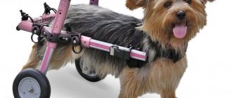
Postoperative care
We are often asked questions about what actions owners should take to alleviate the pet’s condition. In fact, everything is quite simple, but important points are still worth examining in detail. Pay special attention to the dog's nutrition; it should be subject to a diet, the choice of which in most situations becomes the task of the doctor.
You can't keep your dog hungry, but it should eat little food. Start walking your pet, and the more often you do this, the better the effect will be. It is important not to overdo it, and also to walk where the air is as clean as possible. Avoid active games, as this can worsen the condition!
If the animal has been operated on, then the same advice must be followed, but it is important to devote the maximum amount of time to this. Do not overload the diet under any circumstances, especially at first. Only easily digestible foods are allowed. Give your pet plenty of clean water, and carefully monitor his health. If necessary, call your veterinarian or take your dog for an examination!
Complications after surgery
Complications do not always arise, but if treatment was not started on time or was incorrect, then problems will arise with many systems of the dog’s body, various inflammatory processes may begin in the trachea, which most often threatens infection. A large number of other problems may arise, related not only to the disease itself, but also to side effects from the drugs.
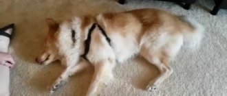
Symptoms and manifestations
When the trachea collapses, the dog develops a chronic cough. This is the main symptom, the severity of which can be used to judge the advanced stage of the disease.
Other symptoms include the following:
- cyanosis of the mucous membranes, paw pads and ears on the inside;
- gurgling wheezing and shortness of breath;
- fatigue and lethargy;
- attacks of suffocation and short-term loss of consciousness.
Breathing problems can occur when you inhale and exhale simultaneously, or when you inhale air exclusively. The first option is noted when the thoracic vertebrae are affected, and the second option when the cervical vertebrae are affected.
The latent period of the disease, accompanied by rare coughing, often drags on, preventing timely diagnosis. Despite this, distinguishing pathology from a common cold is not so difficult. When the tracheal rings are damaged, most attacks occur during physical exertion, accidental pulling on the leash, and excessive expression of emotions at the sight of their beloved owner.
Prevention
If your dog has a genetic predisposition to problems with the trachea, then if you follow the rules of prevention, you can significantly delay the development of the disease. Here are the main measures that are necessary for prevention:
- refusal of a collar, as its harness can put a lot of pressure on the neck;
- maintaining a balanced diet throughout life;
- an attempt to avoid various drafts, as well as places where there is smoke or a lot of dirt, so
- how any of these factors can provoke the development of one of the diseases of the respiratory system, and they can lead to deformation of the trachea.
If you do everything correctly, you will significantly improve your dog’s life, because you will avoid a treatment process that is almost unpredictable.
Currently reading:
- Thyroid dysfunction in dogs (hypothyroidism)
- Seven Signs and Remedies for Getting Rid of Fleas in Dogs
- Pinched nerves and subtle symptoms in dogs
- What to do if your dog has an abnormal bite
Severity of the problem
The method of treatment depends not only on the cause, but also on the severity of the disease. Depending on the nature and scale of the deformation, there are 4 degrees of severity:
- The tracheal lumen is narrowed within 25%
. The sagging of the membrane at this stage is insignificant, so the rings operate as usual without losing their functionality.
- The lumen narrows by half
. The membrane membrane sags heavily, and the cartilage becomes denser. There is a partial loss of function.
- The percentage of narrowing increases to 75%
. The sagging membrane almost touches the rings, which take the shape of an ellipse.
- The lumen narrows to 95-100%
completely blocking breathing. The membrane shell fits closely to the rings. At this stage, suffocation occurs even with complete rest and can result in the death of the animal.
It is important to note that the recommended rate of full tube dilatation is not typical for Yorkies. The tracheal cartilages of these dogs have an ellipsoidal shape from birth.
Symptoms of the disease
Symptoms of the disease vary from mild (but persistent) coughing to "full-blown" respiratory failure in which the animal appears to be about to suffocate (and often does). The cough during collapse is very specific - it is hard, dry, and a “resonating barrel” effect is possible. The cough sharply intensifies and gets worse with emotional excitement, physical activity, while eating and drinking.
In severe cases, it is enough to stroke the dog on the chest so that it immediately begins to choke on a cough. Allergies, tobacco smoke, dust, a tight collar - all these factors aggravate the course of the disease. Some breeders write that the cough during collapse is similar to the “wheezing of a dying goose.” At the third and fourth stages, the dog’s condition is so severe that it does not want to move too much and constantly lies down.
When making a diagnosis, you need to remember that tracheal collapse may be accompanied by other diseases of the respiratory system, “supplementing” and aggravating the collapse, and also greatly complicating the formulation of an accurate diagnosis. We are talking about pathologies of the bronchi and lungs, as well as various heart diseases.
How is diagnostics carried out?
It is possible to understand how to treat tracheal collapse in a dog only after a complete diagnosis. To do this, you will have to make an appointment at the veterinary clinic.
The condition of the affected organ is examined using:
- palpation, revealing noticeable deformation of the walls of the tracheal tube;
- X-ray, which determines the size of the lumen and the presence of foreign bodies;
- fluoroscopy, which allows you to observe a specific area in dynamics;
- endoscopy, necessary to take mucus samples and assess the actual extent of the lesion;
- Ultrasound to check for tumors and associated diseases;
- clinical and biochemical blood test confirming pathological deviations in basic parameters.
X-rays and endoscopy are performed under sedation, since the dog’s movement may interfere with taking a picture or taking a piece of biomaterial. For other studies, the administration of anesthetic is not practiced.
Fluoroscopy is considered the most effective diagnostic method. Despite the high radiation dose to everyone present, the data obtained turns out to be 100% correct. This avoids the false positive and false negative results that are common with x-rays.
Rehabilitation rules
After the operation, the veterinary specialist gives a list of rehabilitation rules, which includes:
- use of antimicrobial and corticosteroid drugs;
- care of the seam area;
- administration of antitussives;
- measures to evacuate mucus if it is impossible for animals to cough it up on their own.
It is also necessary to strictly observe the timing of diagnostic x-ray and endoscopic examination, which allows monitoring the condition of the built-in devices and the nature of changes in the walls of the trachea.
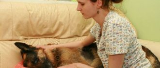
How to help your pet
Emergency assistance from the owner is permissible only in case of a severe attack. In all other cases, the animal must be treated by a veterinarian.
Conservative therapy is effective for disease of 1st and 2nd severity. Four-legged patients with a more severe diagnosis are required to undergo mandatory surgery.
First aid for an attack
First aid is aimed only at temporarily improving the condition, so the victim will still have to be shown to a veterinarian. If you experience an attack of suffocation, follow these steps:
- Calm your pet with an approving voice and strokes. Try to remain calm, as your excitement will certainly be transmitted to the patient.
- Try to eliminate the provoking factor. To do this, you need to remove the collar and ventilate the room if you are not outside.
- Wet your dog's head with cool water and apply a cold compress wrapped in a thick cloth to his chest.
- Contact your veterinary clinic by phone to receive personalized advice from your veterinarian.
An oxygen cylinder, which can be purchased at a regular pharmacy, will help normalize breathing. Codeine, which blocks the cough center, also copes well with the problem. If you don’t have any of these remedies on hand, don’t waste precious time looking for them and go to the veterinary clinic.
Drug therapy
To stimulate respiratory function, glucocorticosteroids (Prednisolone) are used, and to suppress spasm and cough - bronchodilators (Albuterol, Terbutaline, Theophylline), antitussives (Butamirate) and expectorants (Mukaltin, Bronchipret) drugs. The list of other medications depends on the cause of the disease. It may include:
- cardiotonics and vasopressors that lower blood pressure and normalize the force of heart contraction;
- antibiotics (Baytril, Amoxicillin, Tsiprovet) and antiviral (Anandin, Neotime, Fosprenil), eliminating the causative agent of infection;
- anti-inflammatory, dulling the body's response to irritation;
- immunomodulators that restore defenses;
- sedatives that have a calming effect.
Thanks to drug therapy, stabilization of the condition is observed in 71-93% of four-legged patients, depending on the severity of their disease. In other cases, surgical intervention is resorted to.
If a large amount of mucus is detected, the respiratory tract is sanitized. The frequency of procedures depends on the rate of sputum formation and the characteristics of the cough, since when it is dry, the natural discharge of pathological secretions is difficult.
Surgical intervention
The operation carries many risks, so it is used only when the threat to life outweighs the possible complications. Its main task is to correct the narrowed lumen using prosthetics. To do this, use one of two methods:
- Installation of external implants
, replacing damaged areas. Suitable exclusively for pathologies of the cervical spine and is often accompanied by rejection of the material.
- Installation of a special metal stent
, performing a frame function, from the inside. It is used for dysfunction of both the cervical and thoracic vertebrae, but does not solve the problem of constant coughing and copious sputum discharge.
In addition to possible rejection, the disadvantages of implants include the high likelihood of damage to the laryngeal nerves during surgery, which can lead to paralysis of the larynx. For this reason, many owners agree to stenting, which is attractive not only due to lower risks, but also due to more gentle anesthesia.
The persistence of chronic cough during such an operation is explained by the atrophy of the cilia on the mucous membrane, which are forced to constantly contact the tube inserted into the lumen. Despite this, stenting does make breathing easier, so with stable use of symptomatic medications, the patient’s condition is successfully stabilized.
Cough
The most characteristic syndrome of this disease is cough. Appears unexpectedly, intensifies when the leash is pulled or when the trachea is irritated. As the pathology progresses, it intensifies and other symptoms are added to the cough: difficulty breathing, severe lethargy.
In this case, a cough usually appears during feeding, when drinking water, when the dog experiences a strong emotional reaction.
In case of pathology, veterinarians usually prescribe sedative antitussive narcotic analgesics, which allows them to control the functioning of the cough center. The most popular drug in this group is Butorphanol. Doctors can prescribe Hydrokadone; in Russia, the circulation of this medicine is limited, so problems arise with the purchase of the drug.
The dosage is selected individually, based on the pet’s condition and the degree of the disease.
Are there any complications?
As a result of deformation of the tracheal tube, not only the organs of the respiratory system, but also the heart and brain are affected. Without timely treatment, the disease provokes the development of the following disorders:
- pulmonary hypertension, characterized by an increase in blood pressure due to narrowing of blood vessels;
- right ventricular heart failure, which can lead to death due to gradual heart failure;
- death of brain neurons suffering from prolonged lack of oxygen.
Long-term use of antibiotics can also cause complications. These drugs destroy not only pathogenic microorganisms, but also their own immune cells. As a result of such therapy, the existing disease is often complicated by a secondary infection.