Dirofilariasis is the only helminthiasis in the temperate climate zone that is transmitted transmissibly through mosquito bites. The disease is recorded in warm-blooded animals and humans. In this article I will tell you how dirofilariasis develops in dogs, I will describe treatment, diagnostic methods, drugs for prevention based on the latest developments of parasitologists and infectious disease specialists.
- Laboratory diagnosis of dirofilariasis in dogs.
- Means for the destruction of adult heartworms.
- Drops.
What is dirofilariasis in dogs?
The disease is caused by round filamentous worms of the genus Dirofilaria. The name is translated from Latin as “evil thread”. Helminths reach a length of 15–30 cm and a diameter of 1.3 mm.
In the temperate climate zone, 2 types of heartworms are recorded:
- Dirofilaria immitis. Adult worms settle on the right side of the heart - in the ventricle, atrium, and lumen of the pulmonary artery.
- Dirofilaria repens. Sexually mature helminths live under the skin - in fiber or muscles.
With severe infestation, worms sometimes migrate to the abdominal cavity, brain, spinal cord, settle under the eyelids and even in the eyeball.
Dogs are very susceptible to heartworms, easily become infected and become the main carriers of helminths. One animal can be simultaneously parasitized by 1 to 250 mature worms. Intermediate hosts are mosquitoes of the genus Anopheles, Aedes, Culex. Insects carry larvae from sick animals to healthy dogs and humans.
Cats and wild canines are less susceptible to heartworms. They are less likely to have the most dangerous form of cardiopulmonary disease.
Life cycle of heartworm
The development cycle of helminths begins in the body of an infected dog. Viviparous female adult heartworms produce stage 1 larvae, microfilariae, daily. They circulate in the bloodstream of an infected animal before meeting a mosquito and live for 2–3 years.
Video. Microfilaria Dirofilaria repens in the blood of a dog.
At the moment of the bite, the insect swallows the larvae along with blood. Inside the mosquito, the parasites mature to stages 2 and 3. They gradually migrate to the head section of the insect and become infectious to animals and humans.
The duration of maturation of larvae in the body of a mosquito depends on the temperature and humidity of the environment. The average period is 14 days. At 20 – 28℃ and above, the time is reduced to a week, at 18℃ and below it increases to a month.
When the temperature drops to 14℃, the development of microfilariae stops; when the air warms up, it resumes.
When an infected mosquito bites a healthy dog, stage 3 larvae burrow into the skin and penetrate the subcutaneous fat, where they remain for up to 21 days. The cardionematode Dirofilaria immitis then enters the bloodstream and reaches the heart 3 to 4 months after infection. Subcutaneous parasites Dirofilaria repens remain within the subcutaneous fat.
In the dog's body, the larvae molt twice. At the fourth stage they increase to 1.8 cm, at the fifth stage - to 12 - 16 cm. After 6 - 7 months from the beginning of infection, mature females begin to produce microfilariae in the blood. Adult heartworms live 5–7 years.
Is canine dirofilariasis dangerous for humans?
Humans are not typical carriers of heartworms. In Russia, 30–40 cases of infection are registered every year. Helminths settle under the skin, often localized in the area of the eyelids and eyes. They do not mature to the sexually mature stage. Throughout history, only 4 cases have been recorded when worms reached the heart.
The larvae become invasive to humans at stage 3, when they develop in the mosquito's body. Therefore, the only method of transmission is transmissible, through an insect bite. The owner cannot become infected directly from his dog. But animals influence the frequency of diseases in the region: the more dogs with dirofilariasis, the higher the risk of human infection.
How does the invasion develop?
The main carriers of heartworms are mosquitoes. Infection with helminths occurs through the bite of a dog or other animal. Less commonly, other insects become pathogens - lice, ticks, horseflies and even fleas. However, symptoms of the disease do not appear immediately. This is due to the fact that nematodes must go through a full life cycle - from penetration into the animal’s body to the reproduction of adult individuals and distribution through the circulatory system.
Unfortunately, dirofilariasis is a very common disease. Outbreaks of disease among dogs occur during the hot season, since mosquitoes are the most common carrier of helminths.
Dirofilariasis in Russia – statistics
The incidence of dirofilariasis depends on the number of days per year with an average daily temperature above 14℃. The screenshot shows a fragment of the guidelines on dirofilariasis of the Federal Service for Sanitary and Epidemiological Surveillance of the Russian Federation (2018):
Traditionally, foci of dirofilariasis are recorded in the south of Russia:
- in the Krasnodar and Stavropol Territories;
- Crimea;
- Rostov, Astrakhan, Volgograd regions;
- Republic of Adygea.
In 2022 - 2022, the study was carried out by employees of the Novosibirsk Institute of Animal Systematics and Ecology and the Moscow veterinary laboratory Vet Union under the leadership of the famous parasitologist Sergei Konyaev.
The research is based on an analysis of hundreds of publications from 1910 to 2019 and the results of a survey of practicing veterinarians. To collect information, a questionnaire was published on the parasitology ru website and on social networks, which was filled out by 1039 doctors:
- 71.6% of veterinarians encountered the cardiopulmonary form of dirofilariasis;
- 36.4% rate it as a frequent disease - 10 or more cases per year;
- 10.8% were treated only with the subcutaneous form;
- 5.3% were diagnosed with cardiopulmonary dirofilariasis at least once.
Over the past decade, the range has expanded, the disease is recorded in the following areas:
- Vladimirskaya;
- Nizhny Novgorod;
- Ivanovskaya;
- Tula;
- Lipetskaya;
- Tyumen;
- Belgorodskaya;
- Tambovskaya;
- Kurganskaya;
- Tula;
- Sverdlovskaya;
- Jewish Autonomous Region;
- Kabardino-Balkaria;
- Tatarstan.
Full research results are published on the VKontakte page.
Symptoms of infection
Dirofilariasis does not have clear clinical signs that clearly signal infection and help to immediately make a diagnosis. The most common symptoms - increased fatigue, weight loss, weakness - accompany a lot of diseases.
During walks and training, the dog is reluctant to move, stop, sit down, or lie down. After physical exertion he breathes heavily and recovers more slowly.
If helminths settle in the heart, pulmonary arteries, symptoms of respiratory and heart failure occur:
- Impaired capillary refilling. When you press on the gums, the normal pink color returns within 2 seconds.
- Pallor or cyanosis of the mucous membranes.
- Coughing.
- Foamy or bloody sputum.
- Dyspnea.
- Noisy breathing.
- Cardiopalmus.
During the examination, the veterinarian notices pathological changes in the heart: abnormal murmurs, rhythm disturbances, tachycardia.
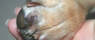
Dirofilaria repens under the skin of a dog
Subcutaneous heartworms do not affect internal organs, but cause anxiety to the animal. In places where parasites are localized, the following occurs:
- seals;
- movable tubercles;
- rashes;
- sores;
- itching
Scratching causes fur to fall out and wounds to appear. If bacteria enter them, purulent inflammation develops. Indirectly, infection is signaled by symptoms of complications of dirofilariasis - an enlarged liver, free fluid in the abdominal cavity.
Complications
Severe consequences occur in the cardiopulmonary form:
- Thromboembolism of pulmonary vessels. After natural death and during treatment, helminths disintegrate into fragments. Decaying parasites partially or completely clog the lumens of blood vessels and block the blood flow to the internal organs. Thromboembolism is the main cause of death of an animal due to dirofilariasis.
- Eosinophilic pneumonia. Occurs in dogs with a hypersensitivity reaction as an immune response to antigens of microfilariae circulating in the blood.
- Interstitial pneumonia is inflammation of the walls of the alveoli and connective tissue of the lungs.
- Ascites is an accumulation of free fluid in the abdomen.
- Glomerulonephritis is an immunoinflammatory lesion of the renal glomeruli. The complication arises as a reaction to the symbiont bacteria dirofilaria - Wolbachia. They accompany helminths, maintaining vitality and reproductive functions.
- Hepatomegaly is an enlargement of the liver due to circulatory problems.
- Respiratory distress syndrome is acute respiratory failure.
- Cardiogenic shock is an extreme degree of heart failure.
In the subcutaneous form, pathogenic microflora sometimes spreads at the site of worm localization and an abscess develops.
How to make a diagnosis
To develop a treatment strategy, it is necessary to identify the pathogen and determine the form and severity of the disease. Your veterinarian will order the following tests:
- immunodiagnostics;
- for antigen;
- on microfilaria;
- for antibodies.
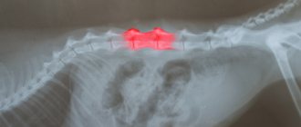
Instrumental studies include chest x-ray and echocardiography.
It is preferable to collect blood for microfilaria in the evening. Parasites are waiting for mosquitoes to appear, so their concentration in the skin capillaries at this time of day is highest. The diagnosis is made when the following conditions are met:
| Diagnostic test | Result | |
| Heart shape | Cutaneous form | |
| Somatic antigen | + | – |
| Presence of microfilariae | + | + |
| X-ray revealed pulmonary vascular lesions | + | – |
| ECHO detected helminths | + | – |
The cutaneous form of the parasite can be diagnosed by its movements under the skin. It happens that the inflamed area opens up and the parasite crawls out. Making an accurate diagnosis is possible no earlier than six months after infection.
Forms of cardiopulmonary dirofilariasis
The severity of a dog’s condition is determined by 3 factors:
- Duration of carriage of helminths: the more time passes from the moment of infection, the higher the risk of complications.
- Intensity of infestation: the more worms settle in the heart, the more severe the disease.
- Individual resistance of the animal.
According to the symptoms, cardiopulmonary dirofilariasis occurs in three forms:
- Latent. The disease develops without clinical signs. The dog feels fine, and the owner does not even suspect the disease. Infection is detected accidentally when microfilariae are detected in a general blood test.
- Chronic. The disease develops slowly, with increasing symptoms. First, 1–2 clinical signs are noticed, and others gradually appear.
- Heavy. This form is diagnosed when a whole ball of worms accumulates in the heart, and the dog remains without treatment for a long time. The animal develops signs of cardiac and respiratory failure.
The course of the disease is influenced by the dog’s lifestyle. With dirofilariasis, the animal needs complete rest. If the disease is hidden, the owner is unaware of the infection and does not reduce physical activity. Against the background of an asymptomatic course, complications suddenly develop in the animal, and the condition sharply worsens.
How is heart dirofilariasis spread?
The disease does not spread directly from dog to dog.
Because transmission of the disease requires a mosquito as an intermediate host, the disease cannot be transmitted directly from dog to dog. Consequently, the spread of the disease occurs during the mosquito season, which in some regions can last year-round. The number of infected dogs and the length of the mosquito season directly correlate with the incidence of heartworm disease in any region.
The mosquito usually bites the dog in those places where the coat is thinnest. However, having long hair cannot prevent infection.
Diagnostic methods
If dirofilariasis is suspected, it is necessary to identify the pathogen, determine the form of invasion, analyze the risk of complications, and exclude concomitant diseases. Therefore, a complex of studies will be required.
Laboratory diagnosis of dirofilariasis in dogs
In veterinary clinics, helminths are detected in three ways:
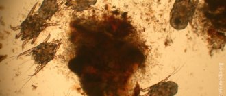
- Native smear method. Drops of fresh blood are placed between glass slides and examined under a microscope. When infected, mobile translucent filamentous microfilariae are visible. This is a simple, accessible and at the same time inaccurate method. If the degree of infestation is low, the larvae do not get into the samples, and the test shows a false negative result.
- Knott's method. Blood and 2% formalin are mixed in a test tube and rotated in a centrifuge. Then the supernatant liquid is drained and a dye, for example methylene blue, is added to the precipitate. This method is more accurate than the previous one. Microfilariae are stained and clearly visible under a microscope. The Knott method does not determine the type of pathogen. If there are few microfilariae, they may not be included in the samples at all.
Close-up of a stained microfilaria.
- Polymerase chain reaction (PCR) method. Enzymes are added to the venous blood that copy fragments of the isolated DNA of microfilariae. The laboratory technician then checks the results against the database. Larvae are easily detected due to the multiple increase in pathogen DNA in samples. The PCR diagnostic method is the most sensitive and accurate. Dirofilariasis is detected even if 1 microfilaria circulates in the blood, and the type of larvae is determined.
If the results are negative, but suspicions remain, then the studies are repeated up to 3 times at weekly intervals.
Express test
To quickly identify heartworms, an immunochromatographic analysis is performed. It detects an antigen protein in the blood that is synthesized by adult female heartworms.
The analysis is carried out using a test cassette. Blood is dripped onto designated areas. Then the result is assessed by the number of colored stripes: 1 line – positive, 2 – negative, no stripes – errors.
The studies take 5–15 minutes; dirofilariasis is detected with high accuracy only during intensive and long-term infection. False negative results occur in the following situations:
- less than 7 months have passed since infection;
- Only males are parasitic in the body;
- less than 5 females are localized in the heart;
- The dog receives preventive medications against helminths.
If microfilariae are detected in the laboratory using the Knott method or a native smear, and the tests show a negative result, then the subcutaneous form of dirofilariasis is diagnosed.
Additional Research
If infection with Dirofilaria immitis worms is confirmed, the heart and pulmonary arteries are examined to assess the risk of complications:
- Echocardiography – reveals pathological changes in the heart. Sometimes adult heartworms are visualized during the study.
- Electrocardiography - shows heart rate, rhythm disturbance.
- Chest X-ray - determines enlargement of the right atrium, ventricle, abnormal changes in the pulmonary arteries.
To assess the general condition, a clinical blood test is prescribed.
Treatment of dirofilariasis in dogs, drugs
Treatment of the cardiopulmonary form includes 3 areas:
- Destruction of adult helminths and larvae.
- Prevention of vascular blockage.
- Correction of cardiac disorders, prevention and treatment of complications.
There is no general scheme. Specific medications are prescribed only by the attending veterinarian after a comprehensive diagnosis, assessment of the animal’s condition and possible risks.
A drug for the destruction of adult heartworms
Now the only remedy prescribed against sexually mature helminths is melarsomin, trade name Immiticide. The drug was developed by American pharmacists in 1995. It is approved by the FDA, but is not licensed in Russia and is sold only in online pharmacies.
Before the advent of melarsomin, the drug thiacetarsamide was used to treat dirofilariasis. The drug caused severe complications from the liver and kidneys, and has now been abandoned.
Melarsomin is a toxic arsenic compound. It destroys sexually mature worms and does not affect larvae less than 4 months old. It is relatively safe for dogs in a therapeutic dose.
According to the instructions, the solution is injected deep into the lumbar muscles 2 times with an interval of 24 hours. A single dose of melarsomine is 2.5 mg per 1 kg of body weight. A total dose of 5 mg per 1 kg contains 0.75 mg/1 kg of arsenic.
Practicing veterinarians have developed their own administration regimens depending on the intensity of infection:
- 2 injections with an interval of 1 month.
- 1 injection, after 1 – 3 months 2 more at daily intervals.
With such schemes, the helminths die in parts, reducing the risk of blockage of the pulmonary artery due to the rapid mass death of parasites. Melarsomin is contraindicated in animals with renal or liver failure. The price of Immiticide is 24 thousand rubles.
After each injection, physical activity is completely excluded for 6-8 weeks, the space for movement is limited to the point of being kept in a cage, and the dog is walked on a short leash.
Drugs against microfilariae
The larvae are not as destructive as adult worms, but they also cause harm. They disrupt blood microcirculation and cause an allergic reaction in sensitive animals. To destroy microfilariae, macrocyclic lactones obtained from soil fungi are used:
- ivermectin
- milbemycin;
- moxidectin;
- doramectin;
- aversectin.
They do not destroy adult individuals, but reduce viability and suppress reproductive functions.
To destroy microfilariae, injection solutions with ivermectin and aversectin are often prescribed:
- Ivermek;
- Baymek;
- Novomek;
- Otodectin;
- Eprimek;
- Aversect K&S.
The solution is administered intramuscularly 1 to 5 times with an interval of 7 to 10 days. The exact regimen and dose are determined by the attending physician based on the condition of the animal. After the injection, microfilariae die within 4 to 24 hours.
Adjuvant therapy
Maintenance medications are prescribed before the administration of melarsomin and after the destruction of helminths:
- Doxycycline - reduces the population of Wolbachia bacteria, which complicate the course of the invasion.
- Alteplase - dissolves fibrin clots in the lumen of blood vessels. They are formed due to damage to the heart and poor circulation. Alteplase is administered once by intravenous drip.
- Prednisolone or Prednivet - suppresses the activity of the immune system against pathogen antigens, inhibits the allergic reaction.
- Heparin – inhibits blood clotting and prevents thrombus formation.
- Curantil - improves venous outflow, blood microcirculation in the heart, kidneys, stimulates the synthesis of interferon.
Previously, aspirin was prescribed for dirophyriasis to thin the blood and prevent the formation of fibrin clots. Now many veterinarians have abandoned it, considering the benefits to be insignificant and unproven.
Surgical treatment of cardiopulmonary dirofilariasis in dogs
Surgery is prescribed if the dog is brought to the clinic too late. When too many worms accumulate in the heart, the animal may not tolerate drug treatment. Due to the massive death of worms after administration of the drug, there is a high risk of complete blockage of the blood vessels of the heart and lungs.
During the operation, helminths are removed from areas accessible for extraction. The outcome depends on the degree of damage to the heart, lungs and arteries by parasites, the equipment of the clinic and the skill of veterinarians. If it is not possible to remove all the worms, drug treatment is prescribed after the operation.
Treatment of subcutaneous dirofilariasis in dogs
Dirofilaria repens does not threaten the life of the animal, so potent melarsomine is not used. More often, the dog is prescribed drugs to kill microfilariae. The drugs are prescribed in prophylactic doses for a course of 8 months to several years - until the natural death of the worms. Sometimes the worms are removed through cuts in the skin.
How is heart dirofilariasis treated?
There is some risk when treating a dog with heartworms, although deaths are quite rare.
In the past, deworming medications contained high levels of arsenic.
Previously, deworming medications contained high levels of arsenic, which caused toxic side effects. Now there are new drugs that do not cause side effects, which can successfully treat more than 95% of sick dogs.
Typically, when a dog is diagnosed with heartworm disease, it means the disease is already advanced. This means that the worms are present long enough to cause significant damage to the heart, lungs, blood vessels, kidneys and liver. Some cases are so advanced that it would be safer to treat the organs themselves than to deal with the risky removal of worms. Dogs in this condition are unlikely to live more than a few weeks or months. Your veterinarian will advise you on the best treatment for heartworm disease.
Methods for treating dirofilariasis of the heart:
There are injectable medications that can kill helminths.
There are injectable medications that can kill adult heartworms. They kill adult helminths in the heart and adjacent vessels. Injections are carried out within 30 days. Your veterinarian will be able to determine the dosage and injection schedule based on your dog's condition. Many dogs are also prescribed antibiotics to combat potential infection from the bacteria (wolbachia) that inhabit the worm.
After treatment, complete rest is essential.
After treatment, complete rest is essential. Adult worms die within a few days and begin to decompose. When they decompose, they are transported to the lungs, where they settle in small blood vessels and are eventually processed by the body. Processing can take weeks or months, and often dead worm fragments can cause complications. This can be a dangerous period, so it is absolutely necessary that the dog is kept at rest and does not engage in physical activity for one month after treatment. The first week after injections is critical because this is the period when the worms die. Many heavily infected dogs will have a noticeable cough for seven to eight weeks after treatment. If the cough seems severe, notify your veterinarian.
Surgery to treat complications is necessary if the dog has a significant reaction within a few weeks of starting treatment for heartworm disease, although such reactions are rare. If your dog has loss of appetite, shortness of breath, severe coughing, coughing up blood, fever or depression, you should tell your veterinarian. Treatment is with anti-inflammatory drugs and antibiotics, replacement therapy and intravenous fluids.
Getting rid of microfilariae. About a month after treatment for the adults, the dog should return to the veterinarian to receive treatment for heartworm microfilariae. The dog may have to stay in the clinic for the whole day. After treatment, preventive measures will be applied.
New treatment protocols use different drugs to kill microfilariae.
New treatment protocols use different drugs to kill microfilariae. Your veterinarian will select the appropriate medications and create a medication schedule based on your pet's condition.
Preparations for the prevention of dirofilariasis
To protect against heartworm larvae, anthelmintic drops, tablets or solutions are used. Repellent preparations do not always protect against mosquitoes; they are used as an additional remedy.
In regions where dirofilariasis is intense, prevention begins in March-April, 1 month before the start of the mosquito season. Finish in October-November, a month after the end of insect activity.
Drops
The easiest way to protect your dog from infection is to apply drops on the withers. The following drugs have been approved for the prevention of dirofilariasis:
- Advocate. The drops contain a combination of moxidectin and imidacloprid. The first component enters the bloodstream and is excreted within 30 days, the second is distributed in the epidermis. Advocate destroys heartworm larvae at stages 3 and 4, when they penetrate the skin and subcutaneous tissue after the bite of an invasive mosquito. The animal is treated once a month.
- Stronghold. The drops contain selamectin and are available in a concentration of 6% for puppies, 12% for adult dogs. Dogs tolerate Stronghold safely; it is prescribed even to infected animals to reduce the concentration of microfilariae circulating in the bloodstream. Drops are applied every 30 days. The dog will have to be treated additionally for ixodid ticks; Stronghold cannot cope with them. In veterinary pharmacies you can find similar drops with selamectin - Selafort.
- IN-AP complex. The drug contains aversectin C1, which is active against heartworm larvae. The interval between treatments is longer than with previous drops - 6 weeks. IN-AP complex contains 2 more antiparasitic components: against ticks, fleas, tapeworms and roundworms.
If basement mosquitoes live in the house, then the dog is treated all year round.
Pills
To prevent dirofilariasis, tablets are given once a month.
- Nexguard Spectra. The drug consists of 2 active components: afoxolaner and milbemycin. The first protects against external parasites, the second protects against nematodes, including heartworm larvae. Tablets are available in different dosages for 5 weight categories of animals. The drug is contraindicated in puppies up to 8 weeks old and adult dogs lighter than 2 kg.
- Milbemax. The tablets contain a combination of praziquantel and milbemycin. There are 2 types available: for puppies and small dogs weighing up to 5 kg and for medium and large breeds. The prophylactic dose of milbemycin to protect against heartworms is 0.5 mg/kg. Milbemax can be given to puppies from 2 weeks of age.
- Helmimax. The tablets contain a combination of praziquantel and moxidectin. There are 3 types of tablets sold in pharmacies: Helmimax 4, 10 and 20. They are intended for small, medium and large breeds, respectively. Gelmimax can be given to puppies from 3 weeks.
- Dironet. The drug consists of 3 components: ivermectin, pyrantel and praziquantel. Puppies are allowed from 3 weeks. Contraindicated for pregnant dogs. The tablets are also available in 3 versions for small, medium and large dogs. Dironet contains a prophylactic dose of ivermectin: 0.006 mg/1 kg.
Solutions
For prevention, drugs with ivermectin are used, as for treatment, but in microscopic doses. Solutions are administered intramuscularly or taken once a month.
Most medications are difficult to dose. The prophylactic dose of ivermectin is only 6 mcg, and 1 ml of solution often contains 10 mg. It’s easier to calculate and fill the syringe with Otodectin. It contains 1 mg of ivermectin per 1 ml of liquid.
Measures to eliminate dirofilariasis
4.1. State laboratory of veterinary medicine when confirming the diagnosis of dirofilariasis
urgently notifies the chief state inspector of veterinary medicine.
4.2. The Chief State Inspector of Veterinary Medicine, in accordance with the expert opinion of the state laboratory, issues an order to impose quarantine, prior to the meeting of the local state anti-epizootic commission, but for no more than 72 hours.
4.3. Upon the recommendation of the relevant chief state inspector of veterinary medicine, the local emergency anti-epizootic commission must, within 24 hours, make a decision on introducing a quarantine restriction on the spread of dirofilariasis in accordance with the veterinary legislation of the state.
4.4. During the first 24 hours from the moment the decision is made to introduce quarantine for dirofilariasis, the local emergency anti-epizootic commission takes all required measures to inform about this all persons who are in the quarantine zone, and local government bodies and local executive authorities of adjacent administrative-territorial units. An information message on the introduction of animal quarantine for dirofilariasis must be published in the official publication and distributed throughout the relevant administrative-territorial zone. To ensure prompt notification of persons about the introduction of quarantine for dirofilariasis of dogs, cats and other species of animals, the local State Emergency Anti-Epizootic Commission uses the media, television and radio broadcasting.
4.5. During quarantine:
4.5.1. The boundaries of the invaded (5 km, in cities 500 m) and buffer (10 km, in cities 1000 m) zones are determined.
4.5.2. In the infested area:
- All animals susceptible to dirofilariasis are registered;
- Conduct diagnostic tests on all animals susceptible to dirofilariasis using the method specified in paragraph 2.3.2. of these Instructions for diagnosing and carrying out preventive measures and improving the health of animals from dirofilariasis and repeat such studies on the 60th day;
- The import and export of susceptible animals is prohibited;
- Take measures to reduce the number of stray animals;
- In areas of mass breeding of mosquitoes (areas with high humidity, near swamps, rivers, lakes, etc.), delavation of reservoirs is carried out using insecticidal preparations;
- Dogs that are in the infected area are treated with insecticidal preparations throughout the entire mosquito activity season (from the beginning of April-May to the end of October-November).
4.5.3 In the buffer zone:
- All susceptible animals are registered;
- Conduct diagnostic tests on all susceptible animals using the method specified in paragraph 2.3.2. these Instructions for diagnosing and carrying out preventive measures and improving the health of animals from dirofilariasis;
- The import and export of susceptible animals is prohibited;
- Measures are being taken to reduce the number of stray animals.
4.5.4. Animals infested with microdirofilaria are isolated and given a full course of microfilaricidal therapy, which includes the use of drugs from the group of macrocyclic lactones: abamectin (duotin), ivermectin (Baymek, biomectin, brovermectin, ivermik, ivermectin, ivomec, intermectin), doramectin (dectomax), moxidectin (Cydectin, Advocate), selamectin (Stronghold) and closantel (Closantel, Brontelu, Santel), etc., in recommended therapeutic doses.
4.5.5. Animals that have completed the full course of treatment are laboratory examined on the 60th and 90th days.
4.5.6. If necessary, patients with dirofilariasis are euthanized, and their corpse is destroyed by burning (as a corresponding act is drawn up) or disposed of in a biothermal pit (Beccari pit, Czech pit).
4.5.7 Quarantine is removed after receiving a negative laboratory test result on day 90 (as defined in clause 4.5.5) of the last infected animal, or after the destruction of the last infested animal.
Forecast
The success of treatment is determined by 4 factors:
- number of sexually mature helminths;
- degree of damage to the heart, arteries, lungs;
- period from the moment of infection;
- restriction of animal mobility.
The most favorable outcome is with early diagnosis, when at the time of detection the disease is asymptomatic or with 1–2 clinical signs. In this case, dirofilariasis passes without a trace without complications. With delayed treatment and severe infestation, the dog develops irreversible changes in the lungs and heart, which shorten its life expectancy.
The article used materials from webinars of Doctor of Veterinary Sciences A.N. Shinkarenko, articles and recommendations on forums for dog owners by parasitologist S. Konyaev.
Can a person get sick?
Because a dog or cat is the definitive host; the likelihood of a person becoming infected with heartworms from a sick animal is minimal. But mosquitoes, being intermediate hosts, can lead to infection of humans with worms.
People more often become ill with helminths that affect the skin, eyeballs and eyelids of a person.
To avoid possible infection, you need to use repellents in the warm season, treat not only clothing, but also especially exposed areas of the body.