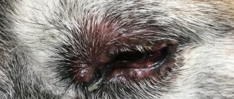
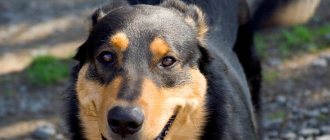
Eye diseases have not spared our smaller brothers either. Blepharitis in dogs is quite common. The disease is an inflammation of the ciliary edge of the eyelids and is accompanied by swelling, redness and itching. It can be complicated by inflammation of the so-called meibomian glands - small glands on the edges of the eyelids that produce an oily secretion. This fluid serves to lubricate the surface of the sclera and protects the eyes from drying out. Blepharitis disrupts this process, causing eye irritation. The dog feels a constant burning sensation, squints in bright light and often rubs his eyes, which further worsens his condition.
The disease can be complicated by conjunctivitis, keratitis, and blurred vision. And if unfavorable, it leads to complete blindness. In this article we will look at the causes, types, main symptoms, as well as methods of treatment and prevention of this disease.
Causes of the disease
There are many reasons for the development of blepharitis. They are divided into two large groups:
- Infectious blepharitis is an inflammation of the eyelids caused by a bacterial infection, as well as fungal or mite-borne microorganisms.
- Non-infectious - can be congenital, traumatic, allergic. It may have another origin.
Infectious blepharitis
- Bacterial blepharitis is the most common. Their main cause is a staphylococcal or streptococcal infection that affects the eyelids. Inflammation of the eyelids can be primary or secondary if its source is not in the eye itself, but somewhere nearby (ears, oral cavity, skin). This type of blepharitis is dangerous due to the occurrence of diffuse abscesses, which are very difficult to treat.
- The source of tick-borne blepharitis is parasitic infection of the skin, hair follicles or sebaceous glands. A typical example of such a disease is demodicosis or red scabies.
- Fungal blepharitis or mycosis of the eyelids is quite common. It may be caused by long-term antibiotic therapy, the use of corticosteroids, as well as autoimmune disorders. This type of disease is very difficult to diagnose and equally difficult to treat.
Non-infectious blepharitis
- Traumatic blepharitis. This type of inflammation of the eyelids occurs against the background of mechanical damage to the eyes (cuts or bruises of the eyes, dust) or eye burns (chemical or thermal).
- Congenital blepharitis. They are caused by congenital pathologies of the development of the eyelids (entropion of the eyelids, the presence of an extra row of eyelashes growing inside the eye).
- The disease can occur in dogs with a breed predisposition. Dogs with an unusual type of muzzle are most predisposed to the disease: short (brachycephalic) or elongated (dolichocephalic). The first group includes bulldogs, mastiffs, Pekingese, and the second, respectively, hounds, collies, pinschers and others. This is something to consider when purchasing a puppy.
- Allergic blepharitis. If a dog's eyes are very sensitive to various types of irritants (food, medications, insect bites, flowering plants), then the allergic reaction is expressed in the form of inflammation of the eyelids. Endogenous allergization of the body due to various problems of the gastrointestinal tract or kidneys can lead to a change in the quality of lubrication produced by the meibomian glands, which will increase the risk of meibomian blepharitis.
- Benign or malignant neoplasms localized in the eyelid area are the most dangerous cause of blepharitis. In this case, there is often a need for a radical solution, since only by removing the eye can the dog’s life be saved.
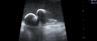
What is the third eyelid?
The popular name “third eyelid”, scientifically in veterinary medicine is called the crescent-shaped fold of the conjunctiva; it is located in the corner of the eyeball.
The absence of pigmentation on the free edge of the third eyelid is not a pathology, but only a feature of some animals.
The semilunar fold protects the eye from various injuries. If you press lightly on your dog's eye, it will immediately appear and instantly cover the surface of the cornea. The second feature is that in the thickness of the third eyelid there is an additional gland, which ensures the production of about 30% of tears. Also, when the third eyelid moves, tears are evenly distributed over the surface of the cornea, which helps remove or wash away foreign particles and bacteria.
Types of disease
Blepharitis is classified according to several criteria.
Based on the localization of the inflammatory process, we can distinguish:
- Anterior edge - when only the ciliary edge of the eyelid is involved in the painful process.
- Posterior marginal is said to occur when the meibomian glands and surrounding tissues are inflamed.
- Angular or angular - indicates the localization of inflammation mainly in the corners of the eyes.
- Mixed involves all the tissues of the eye, as well as the skin and surrounding muscles.
Based on the characteristics of clinical signs, the following forms of the disease are distinguished:
- Simple. It has mild symptoms - moderate redness of the dog's eyelids, thickened edges, whitish secretion in the corners of the eye.
- Scaly. The edges of the eyelids are thicker. It can be recognized by the characteristic scales of the epidermis, which are located at the base of the eyelashes.
- Ulcerative. This is a purulent form of the disease. Crusts of dried pus on the eyelids form painful ulcers after removal. Inflammation is accompanied by loss of eyelashes, severe itching and pain. After the ulcerations heal, scars form on the eyelids, interfering with the normal growth of eyelashes.
- Meibomian. It occurs as a consequence of hyperfunction of the meibomian glands. Excess secretion becomes a breeding ground for the development of pathogenic microflora. The disease can be complicated by a purulent course.
- Furunculous or phlegmous also has a name - barley. The most characteristic signs are the appearance of one or more abscesses along the edge of the eyelids, on the eyelash line, which open over time. This is the most dangerous type of disease, as it can have a dangerous life-threatening complication - sepsis.
Purpose of eye drops
"Diamond Eyes"
Drops are recommended for daily care of your pet's eyes. They have a slight bacteriostatic effect and can help relieve redness, itching, and reduce lacrimation. They contain taurine and antioxidants. They are a first aid remedy in case of foreign body penetration or minor injuries, as well as for the prevention of age-related changes.
"Leopard".
This product as an active ingredient contains 2 components - furatsilin and chloramphenicol, therefore it is active against a wide range of pathogenic microflora. The drops also have anesthetic and anti-inflammatory properties. They are used for the prevention and treatment of keratitis, all types of conjunctivitis, and blepharitis. Bars drops are also used in the treatment of eye injuries.
"Iris".
The active ingredient gentamicin sulfate is active against a wide range of pathogenic microflora. Penetrates through the cornea and maintains optimal concentration of the drug inside the eye. The ophthalmic drug has proven itself in the treatment of keratitis, conjunctivitis, ulcers and erosions of the cornea, as well as blepharitis and uveitis. Particularly active against pyogenic microflora.
"Tsiprovet."
Characterized by a wide spectrum of antimicrobial action. It is used to treat conjunctivitis, blepharitis, septic iridocyclitis, and corneal ulcers. It is also used by veterinary ophthalmologists as a prophylactic agent in the postoperative period and to prepare the visual organs before surgery. In addition, it is a means of prevention in case of foreign body penetration or corneal injuries. "Tsiprovet" even affects microorganisms that are resistant to most antibiotics, such as chlamydia and mycoplasma.
It should be understood that eye diseases should be treated by a specialist, and not by the owner, breeder or pharmacist. If eye drops eliminated, for example, purulent discharge from your dog, this does not mean that they will help an animal with a completely different problem. Therefore, please contact your veterinarian for a prescription.
Main symptoms
Each type of blepharitis has its own set of characteristic symptoms. But there are signs that are inherent in any form of the disease. These include the following:
- swelling and redness of the eyelids;
- severe itching and burning;
- photophobia;
- lacrimation;
- thickening of the diseased eyelid.
Sometimes these symptoms are accompanied by so-called blepharospasm. This is a reflex spasm or painful tension in the muscles surrounding the inflamed eye.
Purulent and ulcerative blepharitis is characterized by discharge with purulent contents and accumulation of purulent exudate in the corners of the eyes.
Animals at risk
The risk group includes animals that are most vulnerable to the listed disorders. These include:
- brachycephals with bulging eyes (Japanese chins, Shih Tzus, Pekingese, pugs);
- breeds with folded skin (chow-chow, sharpei, English bulldogs);
- pets with long hair (Yorkshire terriers, Afghan hounds, lapdogs).
In the first case, vulnerability to external factors is explained by the visual organ being too large. In the second - skin folds on the face, increasing the likelihood of inflammatory processes due to accumulating dirt and dust. In the third - long hair, fraught with frequent irritation and injury.
Treatment of the disease
The choice of treatment regimen for the disease depends on the cause that caused it. It is important to correctly diagnose and treat the primary disease. Otherwise, relapses will follow one after another. A veterinarian-ophthalmologist prescribes a course of treatment and monitors its progress.
Therapy includes symptomatic treatment aimed at relieving inflammation and special treatment designed to eliminate the cause of the disease.
Symptomatic treatment of blepharitis in dogs consists of the following steps:
- It is necessary to cleanse the eye of dried purulent crusts and scales. To do this, they are soaked and carefully removed with a cotton swab soaked in a solution of furatsilin or saline.
- In order to suppress inflammation, anti-inflammatory eye ointments (hydrocortisone or dexamethasone) are applied to the affected area.
The choice of primary treatment methods depends on the causes of the disease:
- For infectious blepharitis, antibiotics and sulfonamides are used for general and local use. The most popular are Tetracycline eye ointment, Albucid eye drops with sodium sulfacyl and others.
- Tick-borne blepharitis necessarily requires the use of anti-tick drugs (Metronidazole).
- Allergic blepharitis can be treated by eliminating the allergen. Additionally, antihistamines (Tavegil or Suprastin) are prescribed.
- If the disease has developed against the background of low immunity, the use of immunomodulatory drugs should be combined with antibiotic therapy, hardening, and physical activity that increases resistance.
- There can be many reasons why the disease has developed, so it is necessary to examine the dog, sanitize chronic sources of infection, and deworm it. It is important to pay attention to how the hygienic conditions of keeping the animal are maintained.
- If the disease was caused by a tumor or congenital pathology of the eyelids, then the problem can only be solved surgically.
- To prevent the dog from scratching the sore eye and further infecting it, a special protective membrane (the so-called Elizabethan collar) is placed around the neck during the illness.
Typically, the dog is treated at home.
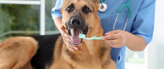
How to give eye drops to a dog
- Wash your hands thoroughly to prevent further infection.
- With the help of an assistant, secure the dog using a collar or muzzle (for aggressive dogs).
- Carefully remove all contaminants (dried exudate, dust, etc.). To do this, use gauze napkins or thick cotton pads moistened with warm boiled water or a hygienic eye care product.
- While holding the dog's face in place with one hand, gently pull back the lower eyelid and apply the medication into the conjunctival sac or directly onto the eyeball.
- Hold for 2-3 seconds to evenly distribute the drug over the mucous membrane.
If the owner does not have the necessary experience, then you should ask the veterinarian to show you how to properly drop the medicine into the pet’s eyes.
Prevention of the disease
Measures to prevent blepharitis are aimed at maintaining the dog’s general condition at a high level. They consist of regular diagnosis and timely elimination of all sources of inflammation in the body. Equally important are proper nutrition, proper rest, and sufficient physical activity. It is necessary to monitor the hygiene of the dog’s resting place and wash its dishes in a timely manner. It is important to rid your dog of helminths and skin parasites in a timely manner.
With good and timely treatment, the prognosis of the disease is favorable. In most cases, vision can be preserved. But sometimes, with a prolonged chronic course and repeated relapses, a deterioration in visual function can occur.
Dangers and complications
Timely therapy allows you to avoid many complications: atrophy, eye loss, partial or complete blindness. Due to the variability of possible causes, in addition to vision loss, it is worth considering other dangers associated with the original pathology or associated infection.
The possibility of disruption of internal organs and systems, which could lead to the death of the pet, should not be ruled out. It is much safer to try to prevent the disease by avoiding its occurrence.
Entropion of the third century
This type of third eyelid prolapse most often occurs in the youngest dogs. The appearance of the pathology is associated with an increase in the diameter of the eyeball as the dog grows older. This provokes the process of lengthening the cartilage stalk, which is attached to the ligamentous apparatus of the organ. If the leg lengthens excessively, the cartilage breaks over time, causing it to lose its flexibility and cease to perform supporting functions. As a result, the eyelid begins to bulge, disturbing the animal, and attempts to insert it yourself are doomed to failure.
A cartilage fracture interferes with the normal functioning of the eyeball, which becomes extremely vulnerable to the harmful influence of microbes; infection can easily get there. This will inevitably lead to inflammatory processes. Dog breeds such as Newfoundland and Great Dane are at risk. Unfortunately, the animal cannot be cured without surgery. Only a qualified veterinarian will be able to properly suturing the cartilage so that the eyelid returns to its place.
First aid
An immediate visit to a veterinary clinic is recommended if there is a sudden increase in growth, the appearance of ulcerations and severe pain, as well as a change in the color and consistency of the discharge. All these signs accompany a severe course of the disease, so any delay is very dangerous.
To prevent scratching, be sure to wear a protective collar on your pet. To alleviate the condition, you can wash your eyes with warm chamomile infusion using cotton pads.
Many owners relieve inflammation with Dexamethasone (glucocorticosteroid) and Ciprovet (antibiotic). It is better to consult a veterinarian on this issue, since the first drug is not recommended for purulent processes, and the second for cerebral circulatory disorders.
Lagophthalmos
Another disease associated with problems with the innervation of the eye. Clinically, lagophthalmos is manifested by the inability to close the eyelids. The cause is paralysis of the facial or trigeminal nerves. Treatment of the eye in dogs with this pathology is aimed at eliminating the inflammatory process of nerve fibers and restoring the physiological innervation of the eye. As an auxiliary therapy when such a condition occurs, agents are used that moisturize the cornea and also prevent bacterial microflora and mechanical irritants from entering the eye.
Forms of manifestation
- Scaly - characterized by redness and thickening of the edges of the eyelids with the formation of purulent crusts;
- Ulcerative - accompanied by swelling of the edges of the eyelids, the formation of ulcers and bleeding ulcers, profuse lacrimation, eversion and inversion of the eyelids;
- Meibomian - has a pronounced thickening and purulent inflammation of the eyelid in a dog;
- Furunculous - manifested by general purulent inflammation of the glands and hair papillae of the entire century (barley);
- Phlegmonous - characterized by the formation of a local abscess that breaks through the eyelid (abscess).
Along the way, acute and chronic blepharitis is distinguished; the chronic form is difficult to treat and leaves irreversible consequences on the animal’s eyelids. The acute form, with timely consultation with a specialist and the appointment of effective treatment, can be completely eliminated without consequences for the body.